
Radiobiological motivation for Lh ARA Dr Jason Parsons
Radiobiological motivation for Lh. ARA Dr Jason Parsons Cancer Research Centre Department of Molecular and Clinical Cancer Medicine
Radiotherapy in cancer treatment • ~50 % of all cancer patients will receive radiotherapy. • Conventional (photon) radiotherapy is the still the most effective treatment for solid tumours (e. g. head and neck). • Dose rates of ~1 -5 Gy/min utilised. • Significant irradiation of normal tissues and organs at risk in proximity to the tumour being treated. • Biological factors including oxygen (hypoxia) and inherent radioresistance of tumours (e. g. glioblastoma) are a barrier to effective treatment.
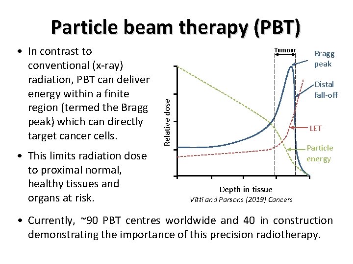
Particle beam therapy (PBT) • This limits radiation dose to proximal normal, healthy tissues and organs at risk. Tumour Bragg peak Distal fall-off Relative dose • In contrast to conventional (x-ray) radiation, PBT can deliver energy within a finite region (termed the Bragg peak) which can directly target cancer cells. LET Particle energy Depth in tissue Vitti and Parsons (2019) Cancers • Currently, ~90 PBT centres worldwide and 40 in construction demonstrating the importance of this precision radiotherapy.
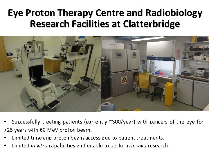
Eye Proton Therapy Centre and Radiobiology Research Facilities at Clatterbridge • Successfully treating patients (currently ~300/year) with cancers of the eye for >25 years with 60 Me. V proton beam. • Limited time and proton beam access due to patient treatments. • Limited in vitro capabilities and unable to perform in vivo research.
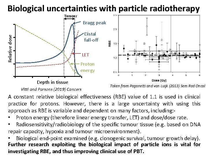
Biological uncertainties with particle radiotherapy Tumour Bragg peak Relative dose Distal fall-off LET Proton energy Depth in tissue Vitti and Parsons (2019) Cancers Taken from Paganetti and van Luijk (2013) Sem Rad Oncol A constant relative biological effectiveness (RBE) value of 1. 1 is used in clinical practice for protons. However, there is a large uncertainty with using this approach as RBE is variable and dependent on many factors, including: • Proton energy (therefore linear energy transfer, LET) and dose/dose rate. • Radiosensitivity/radiobiology of the specific tumour tissue (e. g. based on DNA repair capacity, hypoxia and tumour microenvironment). • Biological end-point examined (e. g. clonogenic survival, tumour growth delay). Further research exploiting the biological impact of particle ions is vital for investigating RBE, and thus improving clinical use of PBT.
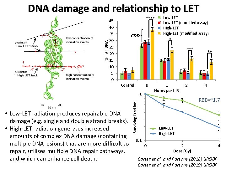
% Tail DNA damage and relationship to LET **** 45 40 35 30 25 20 15 10 5 0 CDD Proton-IR Low-LET (58 Me. V) Proton-IR (58 Me. V; modified) Low-LET (modified assay) High-LET (11 Me. V) Proton-IR High-LET (11 (modified assay) Proton-IR Me. V; modified) * *** Control 0 1 2 4 RBE=~1. 7 Surviving fraction • Low-LET radiation produces repairable DNA damage (e. g. single and double strand breaks). • High-LET radiation generates increased amounts of complex DNA damage (containing multiple DNA lesions) that are more difficult to repair, utilises multiple DNA repair pathways, and which can enhance cell death. 1 Hours post-IR ** Low-LET 58 Me. V High-LET 11 Me. V 0. 1 0 2 Dose (Gy) Carter et al, and Parsons (2018) IJROBP Carter et al, and Parsons (2019) IJROBP 4
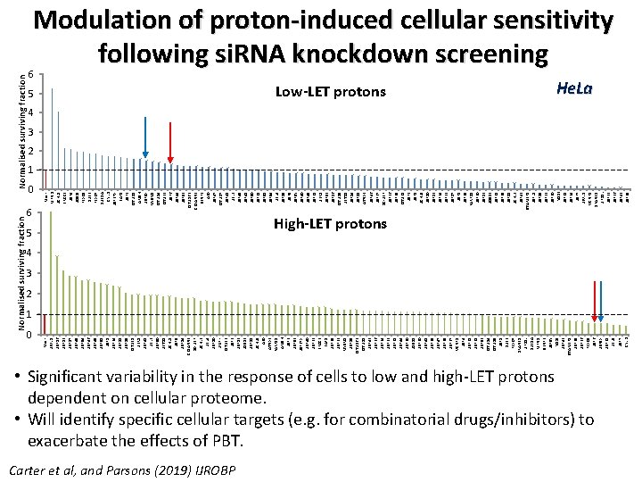
Normalised surviving fraction 6 0 Mock MPND UCHL 1 BRCC 3 USP 4 JOSDI FBX 08 DUB 3 FBX 07 DUB 1 A CYLD USP 27 X BAP 1 USP 3 OTUD 1 AMSH USP 9 X MYSM 1 OTUD 4 OTUB 2 USP 6 USP 44 USP 52 OTUD 6 B CEZANNE SENP 2 A 20 USP 47 OTUD 7 USP 41 UBL 3 USP 45 USP 48 USP 50 USP 28 USP 51 USP 54 UBL 4 USP 15 USP 9 Y USP 40 USP 49 USP 21 UBR 1 USP 53 USP 16 OTUD 5 UBTD 1 USP 14 USP 18 ATNX 3 USP 46 USP 37 DC-UBP USP 26 USP 25 OTUB 1 USP 2 USP 1 UCHL 5 USP 10 USP 13 USP 24 USP 22 USP 17 USP 8 USP 35 PARP 11 USP 20 USP 12 JOSD 2 USP 31 USP 29 USP 11 UCHL 3 USP 42 STAMBP 1 USPL 1 USP 19 USP 32 USP 30 YODI USP 38 USP 34 USP 7 UEVLD VCPIP 1 ZRANB 1 UFD 1 L USP 33 USP 36 USP 43 USP 39 0 Mock UEVLD USP 16 USP 13 USP 17 USP 45 USP 54 USP 46 USP 49 USP 51 USP 34 USP 11 USP 19 OTUB 1 UBR 1 USP 48 UBL 3 USP 50 UBTD 1 UCHL 1 USP 5 USP 14 CEZANNE DC-UBP UCHL 3 UBL 4 USP 10 UMPK OTUB 2 USP 12 JOSD 2 USP 18 UCHL 5 A 20 ATNX 3 PARP 11 AMSH USP 3 USP 52 USP 27 X USP 40 USP 22 BRCC 3 BAP 1 USP 25 USP 33 MYSM 1 USP 15 OTUD 6 B OTUD 1 USP 42 USP 26 USP 24 USP 32 USP 28 USP 44 USP 20 JOSD 1 USP 30 USP 41 USP 35 USP 47 USP 29 USP 37 VCPIP 1 USP 4 USP 21 USP 38 USP 53 OTUD 4 OTUD 5 USP 8 DUB 3 FBX 07 ZRANB 1 UFD 1 L DUB 1 A MPND SENP 2 USP 9 Y YODI USP 43 STAMBP 1 USP 39 USP 36 FBX 08 USP 6 USP 9 X USP 31 UBL 5 USP 7 CYLD Normalised surviving fraction Modulation of proton-induced cellular sensitivity following si. RNA knockdown screening 6 5 Low-LET protons 5 Carter et al, and Parsons (2019) IJROBP He. La 4 3 2 1 High-LET protons 4 3 2 1 • Significant variability in the response of cells to low and high-LET protons dependent on cellular proteome. • Will identify specific cellular targets (e. g. for combinatorial drugs/inhibitors) to exacerbate the effects of PBT.
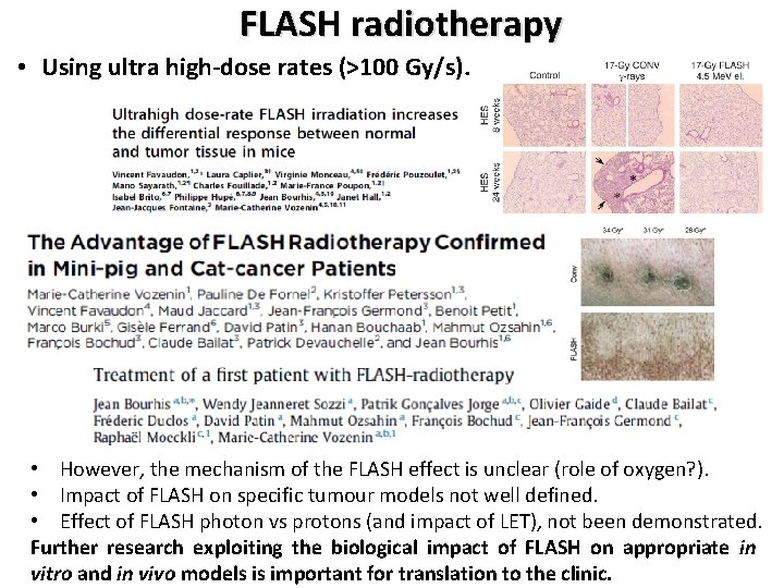
FLASH radiotherapy • Using ultra high-dose rates (>100 Gy/s). • However, the mechanism of the FLASH effect is unclear (role of oxygen? ). • Impact of FLASH on specific tumour models not well defined. • Effect of FLASH photon vs protons (and impact of LET), not been demonstrated. Further research exploiting the biological impact of FLASH on appropriate in vitro and in vivo models is important for translation to the clinic.
Challenges and opportunities for PBT radiobiology research Challenges • The radiobiology of PBT at the molecular and cellular level is still not entirely understood (e. g. impact of LET, dose rate delivery). • Other factors that impact on RBE of PBT not well defined (e. g. hypoxia, tumour microenvironment, drug-IR combinations, fractionated doses, FLASH). • More research required using specific and validated cancer models, plus relevant normal tissue models, in vitro (e. g. 3 D spheroids/organoids) and in vivo. • Significant lack of access to PBT facilities for radiobiology research. • Current facilities not fully equipped for in vitro, but more so in vivo experiments. Opportunities with Lh. ARA • Highly accessible facility for both in vitro and in vivo particle ion radiobiology research, capable of analysing a multitude of biological end-points. • Enhance our understanding of the radiobiological effects of particle ions (protons and carbon ions), including examining dose delivery (FLASH) and combinatorial treatments through high-throughput screening. • Significant opportunity to develop research that will have a major Worldwide impact through the optimisation and personalisation of cancer treatments using PBT in the clinic.
Feedback from Yolanda Prezado • Lh. ARA is a very promising facility which would offer unique beam features and infrastructure for radiobiology research. The main characteristics to be highlighted are: Flexibility Flexible temporal and spatial beam structure allowing exhaustive investigations of beam parameters (pulse length, repetition rate, instantaneous dose, FLASH) on biological response utilising a stable beam. Different beam species One of the few places in the World to offer the evaluation of different particle ions (protons to heavier ions) within the same facility. Accessibility Greater accessibility of Lh. ARA in comparison to clinical facilities, with greater flexibility (e. g. in vitro and in vivo experiments; dose fractionation; more complex biological end-points; immunotherapy/chemotherapy combinations). • Lh. ARA has the potential to drive a change in current clinical practice by increasing the wealth of radiobiological knowledge.
- Slides: 10