RADIATION IN MEDICINE Uses of Radiation in Medicine


 or oral administration of a radioactive material into the")


")
- Slides: 14
RADIATION IN MEDICINE
Uses of Radiation in Medicine What is Radiation? 1. Radiology 2. Nuclear Medicine 3. Cardiology 4. Radiation Oncology
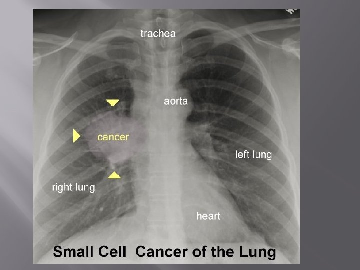
Radiology: Bombard tissues with x-rays: the thicker and denser tissues will absorb the x-rays and thinner, less dense tissues, will allow the x-rays to pass through and hit a photographic film causing an exposure 1.
Diagnostic Applications: 1. Emphysema 2. Pneumonia 3. Lung cancer 4. Broken bones How X-Rays Work
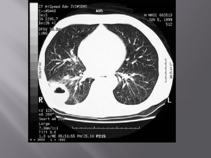
2. CT Scans: Computerized tomography uses x-rays from multiple angles to build an image of inside of the body by making images of “slices” of the body May utilize a contrast dye such as iodine or barium or both - ingested by mouth or enema - injected by vein Enhances the image by highlighting different regions
Diagnostic Applications: 1. Detect abnormal growths and cancer 2. Provide information about the stage of a cancer, response to treatment, or reoccurrence 3. Guide a procedure such as a biopsy or cancer treatments
3. Mammography: x-ray of breast tissue to look for breast cancer 4. Barium Enema: patients drink a milkshake made with barium sulfate which passes through the digestive system and makes the large intestine visible – allows to see if there are blockages or polyps in the intestine
5. Nuclear Medicine: Injection (IV) or oral administration of a radioactive material into the body and tracing the material as it travels through the body to examine how well organs are functioning. As the materials pass through the body the medicine emits positrons which can be detected making a PET scan – Positron Emission Tomography How Does a PET Scan Work?
Diagnostic Applications: 1. Inspect the blood flow, oxygen intake, and metabolism of your organs and tissues at the cellular level. 2. Detect cancer, heart problems, brain disorders, and problems with the central nervous system 3. See how cancer metabolizes, has metastasized, or is responding to chemotherapy.
Non-Ionizing Radiation � MRI – magnetic resonance imaging � How an MRI works – a bit more detail �
Tissues are hit with a strong magnetic pulse which aligns the hydrogen atoms (protons) in their molecules. The protons are hit with pulses of various radio frequencies. This excites the protons to a higher energy state which causes them to give off energy signatures that are translated to images of the soft tissue.