RADIATION DETECTORS USED IN NUCLEAR MEDICINE IMAGING Submitted

RADIATION DETECTORS USED IN NUCLEAR MEDICINE IMAGING Submitted by : Miss. Ruqiya Ramzan Assistant Professor in the Department of Paramedical Sciences. Msc. RIT (RIMT University Punjab)

INTRODUCTION When radiation from a radioactive material pass through matter, they interact with atoms and molecules and transfer energy to them. The transfer of energy has two effects: ionization and excitation. Ionization occurs when the energy transferred is sufficient to cause an orbital electron to be removed away from its parent atom or molecule, thus creating an ion pair. Excitation occurs when electrons are perturbed from their normal arrangement in an atom or molecule, thus creating an atom or molecule in an excited state.

USES Measuring doses of radiopharmaceuticals Performing radiotracer bioassays Monitoring and controlling radiation risk in clinical environment ABSOLUTE EFFICIENCY Ø Absolute efficiency is defined as the number of radiation events detected in a given interval divided by the number of radiation quanta (particles or photons) emitted by a radiation source in the same interval.

INTRINSIC EFFICIENCY Intrinsic efficiency is defined as the number of events detected in a given interval divided by the number of radiation quanta (particles or photons) incident on the detector in the same interval. The intrinsic efficiency is determined by the stopping power of the transducer. The stopping power for a particular radiation is determined by the thickness, density, and atomic number of the detector.

GEOMETRIC EFFICIENCY Geometric efficiency is defined as the number of radiation quanta (particles or photons) incident on the detector in a given interval divided by the number emitted by the radiation source in the same interval. For a point source detected by a simple cylindrical detector , the geometric efficiency decreases with increasing source-to-detector distance in accordance with the inverse square law.

TYPES gas-filled detectors scintillation detectors, and semiconductor detectors. The ionization detector Geiger-Müller detector extremity and area monitor dose calibrator Gamma ray spectrometer solid-state personnel dosimeter, and intraoperative probes

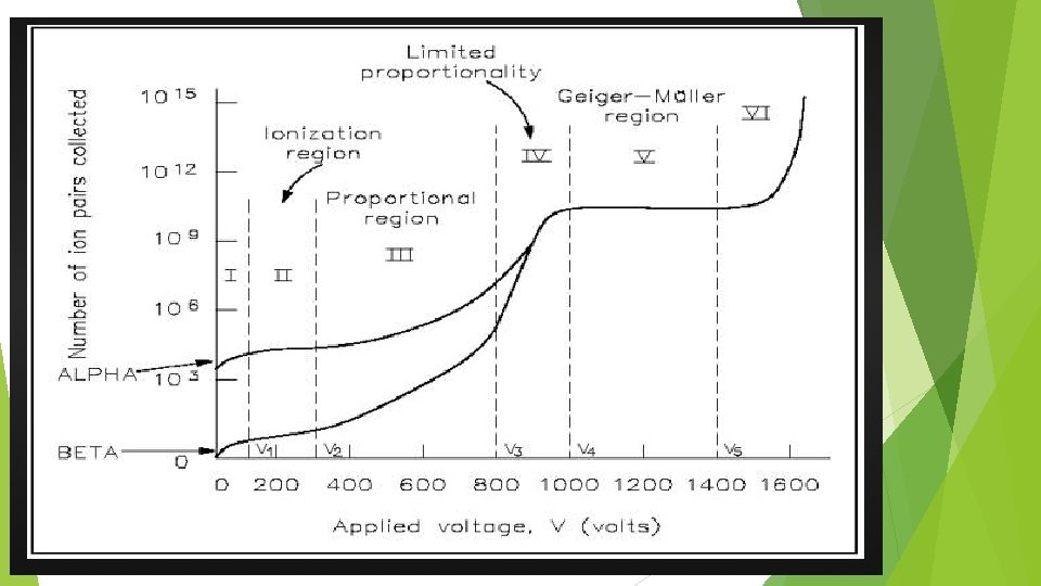
GAS FILLED DETECTORS Most gas-filled detectors belong to a class of detectors called ionization detectors. These detectors respond to radiation by means of ionization-induced electrical currents. The average energy required to produce an ion pair in air is 33 e. V A volume of gas is contained between two electrodes having a voltage difference between them. The negative electrode is called the cathode, the positive electrode the anode. The electrodes are shown as parallel plates, but they may be a pair of wires, concentric cylinders, and so forth.
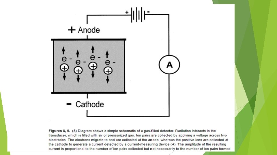

Under normal circumstances, the gas is an insulator and no electrical current flows between the electrodes. Radiation passing through the gas causes ionization, both direct ionization from the incident radiation and secondary ionization from δ rays. The electrons produced by ionization are attracted to the positive electrode and the ionized atoms to the negative electrode, causing a momentary flow of a small amount of electrical current. Gas-filled detectors include ionization chambers, proportional counters, and Geiger Müller (GM) counters.
 The voltage between the electrodes")
IONIZATION CHAMBER Gas between the electrodes. (Most commonly air) The voltage between the electrodes must be sufficient to ensure complete collection of ions and electrons. If the voltage is too low, some of the ions and electrons simply recombine one another without contributing electric current flow. The amount of electrical charge released in an ionization chamber by a single ionizing radiation event is very less. Because of small charge, ionization chamber generally are not used to record or count individual radiation events. Electrometer is used for small amount of current. Two device consisting of electrometer and ionization chamber are survey meter and dose calibrator.

DOSE CALIBRATOR The dose calibrator is the most commonly encountered detector in the nuclear pharmacy because the Nuclear Regulatory Commission requires that all radiopharmaceutical doses be assayed before administration to patients. The dose calibrator has a well ionization chamber configuration to achieve maximum geometric efficiency. The chamber is typically filled with pressurized argon to maximize intrinsic detection efficiency

PROPORTIONAL COUNTER Voltage between the electrodes is sufficient only to collect those charges freed by direction of ionizing radiation. If the voltage is increased to high value, the electron liberated by radiation gains high velocities and accelerated towards positive electrodes causing additional ionization with other atom of the gas - gas amplification. The factor by which ionization is increased called gas ionization factor. Total amount of charge = ionization x gas amplification factor. It is specially designed for gas amplification effect.

USES Used for detecting and counting individual radiation events. Mostly used for non penetrating radiation such as α and β particle

GEIGER-MULLER COUNTER Designed for maximum gas amplification effect. Filled with a special gas mixer – Argon. Gas amplification occurs in the GM counter as a proportion counter. Accelerating electrons also can cause excitation of gas molecules.
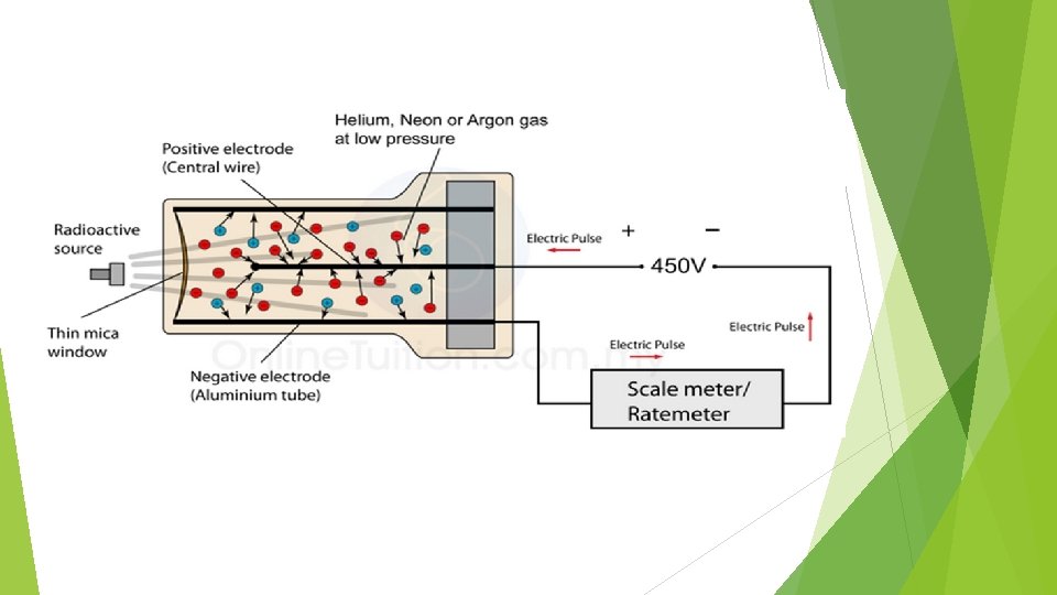

USES Thus GM counter can be used to detect and count the individual ionizing radiation. Used mostly in survey meter for radiation protection purpose. Counting only relatively penetrating radiation.

EXTREMITY AREA MONITOR The extremity and area monitor is a gas-filled detector with a large surface area for maximum efficiency. This device is operated as a proportional counter or Geiger-Müller detector to maximize detection efficiency. The device is frequently wall mounted and can be used as an area monitor or for radionuclide contamination surveying of the hands. When used as an area monitor, the device can be preset to trigger an audible alarm if activity levels exceed an arbitrary level, which is some small multiple of background counting rates. An annual efficiency test should be performed to ensure properation.

GAMMA RAY SPECTROMETER • Gamma-ray spectroscopy is the quantitative study of the energy spectra of gamma-ray sources, in such as the nuclear industry, geochemical investigation, and astrophysics. • Most radioactive sources produce gamma rays, which are of various energies and intensities. When these emissions are detected analysed with a spectroscopy system, a gamma-ray energy spectrum can be produced. • A detailed analysis of this spectrum is typically used to determine the identity and quantity of gamma emitters present in a gamma source, and is a vital tool in radiometric assay

COMPONENTS • The equipment used in gamma spectroscopy includes an energy-sensitive radiation detector, electronics to process detector signals produced by the detector, such as a pulse sorter (i. e. , multichannel analyzer), and associated amplifiers and data readout devices to generate, display, and store the spectrum. • The most common detectors include sodium iodide (Na. I) scintillation counters and highpurity germanium (HPGe) detectors.

SEMICONDUCTOR DETECTORS Semiconductor or solid-state detectors are not as prevalent as the other types of detectors. Semiconductor detectors include personnel dosimeters and intraoperative probes.

PERSONNEL DOSIMETER a pocket solid-state personnel monitoring device that displays cumulative exposure in milliroentgens. These personnel monitoring devices are being used more broadly and have largely replaced the pocket ionization detector for routine real-time estimation of exposure.

INTRAOPERATIVE PROBES Intraoperative probes are a class of semiconductor detectors that is becoming more prevalent in nuclear medicine practice for lymphoscintigraphy applications The probes have a fine tip with a cadmium telluride or cadmium zinc telluride wafer transducer. Most devices are fitted with a removable sleeve aperture, which can improve position discrimination and accuracy with a corresponding decrease in sensitivity. Cadmium telluride has a band gap energy of 1. 47 e. V and an energy resolution of 2. 9% at 122 ke. V.

SCINTILLATION DETECTORS Radioactive materials interacts with matter by causing ionization or excitation of atoms and molecules. Then energy is released. Most of the energy is dissipated as thermal energy, such as molecular vibrations in gases or liquids or lattice vibrations in a crystal. In some materials a portion of the energy is released as visible light. These materials are called scintillators, and radiation detectors made from them are called scintillation detectors.
![MATERIALS USED Sodium iodide activated with thallium [Na. I(Tl)], coupled to PMTs is used](http://slidetodoc.com/presentation_image_h/c040f7f01e32fcf3042880a88745a1dc/image-25.jpg "MATERIALS USED Sodium iodide activated with thallium [Na. I(Tl)], coupled to PMTs is used")
MATERIALS USED Sodium iodide activated with thallium [Na. I(Tl)], coupled to PMTs is used for most nuclear medicine applications. Bismuth germinate (BGO) is coupled to PMT are used in detectors in most PET scanners.

PHOTOMULTIPLIER TUBE The photomultiplier converts light emitted from the crystal into a useful electric signal. The photomultiplier is chosen to have a spectral sensitivity that closely matches the spectral output of the scintillation crystal to which it is coupled. Just inside the entrance window of the photomultiplier is the photocathode, an electrode coated with photosensitive material. On average, for every five scintillation photons striking the photocathode, one electron is emitted

TYPES The scintillator materials used for detectors in nuclear medicine are of two general types: Organic substances in the form of liquid solution. Inorganic substances in solid crystals.

ORGANIC LIQUID SCINTILLATOR The scintillation process in organic scintillator is an inherent molecular property. In these system the scintillator is dissolved in a solvent material in a glass or plastic vial and a radioactive sample is added to the mixture. The vial is then placed in a light tight enclosure between a pair of PM tubes to detect the scintillation events.

INORGANIC SOLID SCINTILLATOR Inorganic scintillators are crystalline solids that scintillate because of characteristics of their crystal structure. They are composed of certain materials which have the property of emitting light when ionizing radiation interact with them Impurity in the atom of the crystal matrix cause disturbance in the normal structure and are sometime called activator centre. Efficient detection of gamma rays requires the material of the scintillator to be with high density and high atomic number
- Slides: 29