
Quantum Optics and Laser Science group Blackett Laboratory
Quantum Optics and Laser Science group Blackett Laboratory Probing molecular structure and dynamics using laser driven electron recollisions Sarah Baker 30 th April 2009 Page 1 © Imperial College London
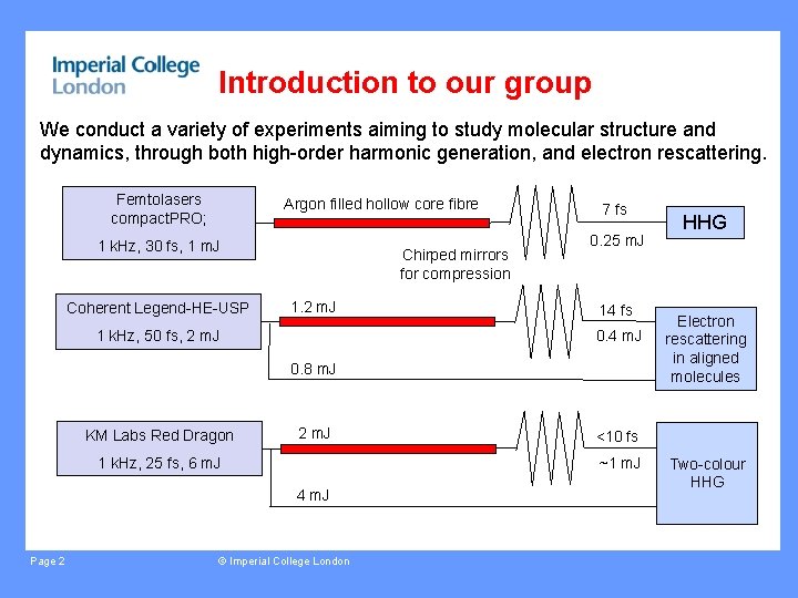
Introduction to our group We conduct a variety of experiments aiming to study molecular structure and dynamics, through both high-order harmonic generation, and electron rescattering. Femtolasers compact. PRO; Argon filled hollow core fibre 1 k. Hz, 30 fs, 1 m. J Coherent Legend-HE-USP Chirped mirrors for compression 1. 2 m. J 1 k. Hz, 50 fs, 2 m. J 7 fs 0. 25 m. J 14 fs 0. 4 m. J 0. 8 m. J KM Labs Red Dragon 2 m. J 1 k. Hz, 25 fs, 6 m. J Page 2 © Imperial College London Electron rescattering in aligned molecules <10 fs ~1 m. J 4 m. J HHG Two-colour HHG

Introduction to our group We conduct a variety of experiments aiming to study molecular structure and dynamics, through both high-order harmonic generation, and electron rescattering. Underlying process is laser driven electron recollision… Returning electron wavepacket can have energy components up to ~ 150 e. V, or wavelength 1 angstrom Recollision lasts ~ 1 fs Page 3 © Imperial College London HHG Electron rescattering in aligned molecules Two-colour HHG
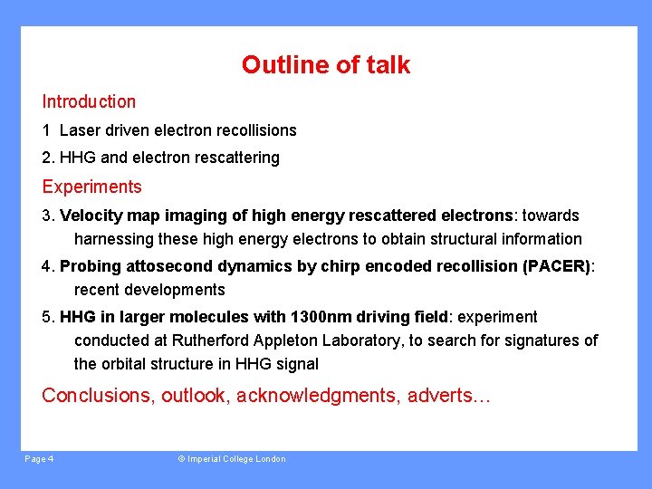
Outline of talk Introduction 1 Laser driven electron recollisions 2. HHG and electron rescattering Experiments 3. Velocity map imaging of high energy rescattered electrons: towards harnessing these high energy electrons to obtain structural information 4. Probing attosecond dynamics by chirp encoded recollision (PACER): recent developments 5. HHG in larger molecules with 1300 nm driving field: experiment conducted at Rutherford Appleton Laboratory, to search for signatures of the orbital structure in HHG signal Conclusions, outlook, acknowledgments, adverts… Page 4 © Imperial College London
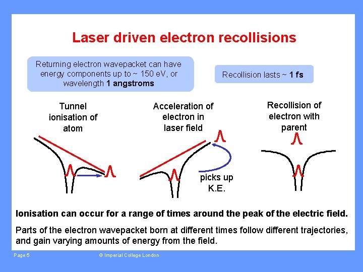
Laser driven electron recollisions Returning electron wavepacket can have energy components up to ~ 150 e. V, or wavelength 1 angstroms Tunnel ionisation of atom Recollision lasts ~ 1 fs Acceleration of electron in laser field Recollision of electron with parent picks up K. E. Ionisation can occur for a range of times around the peak of the electric field. Parts of the electron wavepacket born at different times follow different trajectories, and gain varying amounts of energy from the field. Page 5 © Imperial College London
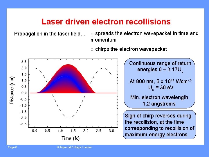
Laser driven electron recollisions Propagation in the laser field… o spreads the electron wavepacket in time and momentum o chirps the electron wavepacket Continuous range of return energies 0 – 3. 17 Up At 800 nm, 5 x 10¹⁴ Wcm-2: Up = 30 e. V Min. electron wavelength 1. 2 angstroms Sign of chirp reverses during the recollision, at the time corresponding to recollision of maximum energy electrons Page 6 © Imperial College London
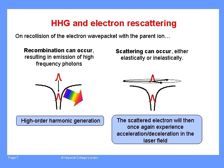
HHG and electron rescattering On recollision of the electron wavepacket with the parent ion… Recombination can occur, resulting in emission of high frequency photons High-order harmonic generation Page 7 © Imperial College London Scattering can occur, either elastically or inelastically. The scattered electron will then once again experience acceleration/deceleration in the laser field
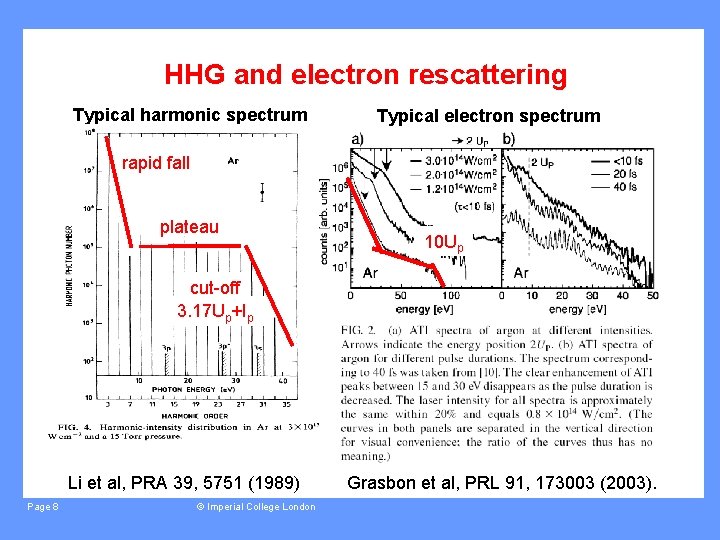
HHG and electron rescattering Typical harmonic spectrum Typical electron spectrum rapid fall plateau 10 Up cut-off 3. 17 Up+Ip Li et al, PRA 39, 5751 (1989) Page 8 © Imperial College London Grasbon et al, PRL 91, 173003 (2003).
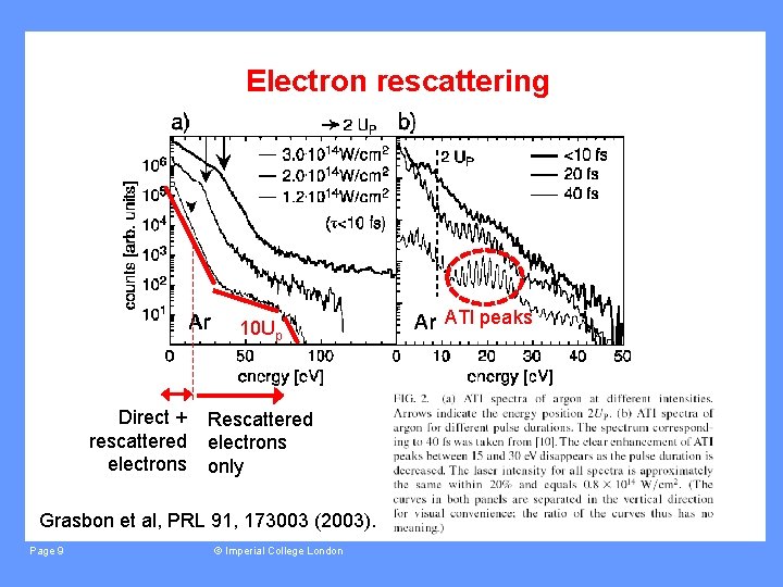
Electron rescattering 10 Up Direct + rescattered electrons Rescattered electrons only Grasbon et al, PRL 91, 173003 (2003). Page 9 © Imperial College London ATI peaks
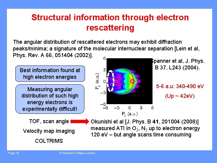
Structural information through electron rescattering The angular distribution of rescattered electrons may exhibit diffraction peaks/minima; a signature of the molecular internuclear separation [Lein et al, Phys. Rev. A 66, 051404 (2002)]. Spanner et al, J. Phys. B 37, L 243 (2004). Best information found at high electron energies 5 -6 a. u: 340 -490 e. V Measuring angular distribution of such high energy electrons is experimentally difficult! TOF, scan angle Velocity map imaging (Up ~ 42 e. V) Okunishi et al [J. Phys. B 41, 201004 (2008)] measured ATI in O 2, N 2 up to electron energy 120 e. V – but angle scans time consuming COLTRIMS Page 10 © Imperial College London
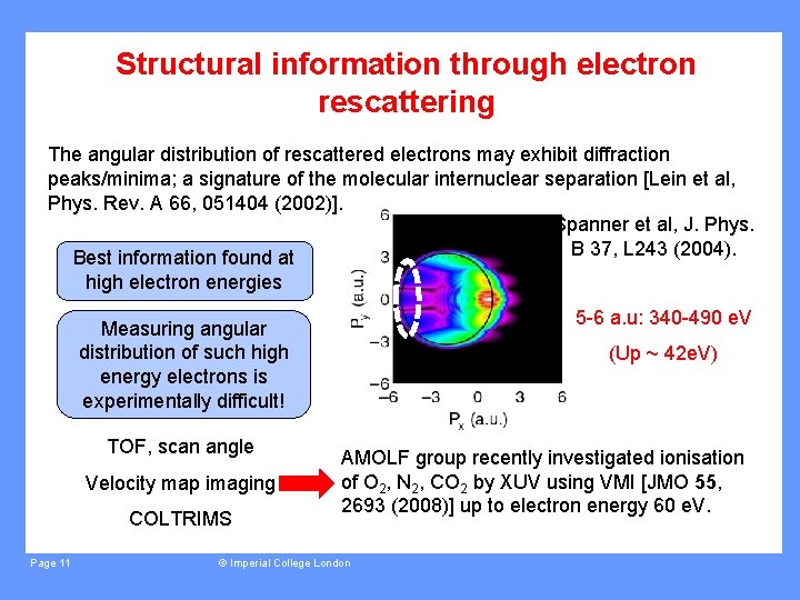
Structural information through electron rescattering The angular distribution of rescattered electrons may exhibit diffraction peaks/minima; a signature of the molecular internuclear separation [Lein et al, Phys. Rev. A 66, 051404 (2002)]. Spanner et al, J. Phys. B 37, L 243 (2004). Best information found at high electron energies 5 -6 a. u: 340 -490 e. V Measuring angular distribution of such high energy electrons is experimentally difficult! TOF, scan angle Velocity map imaging COLTRIMS Page 11 (Up ~ 42 e. V) AMOLF group recently investigated ionisation of O 2, N 2, CO 2 by XUV using VMI [JMO 55, 2693 (2008)] up to electron energy 60 e. V. © Imperial College London
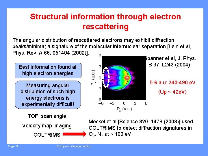
Structural information through electron rescattering The angular distribution of rescattered electrons may exhibit diffraction peaks/minima; a signature of the molecular internuclear separation [Lein et al, Phys. Rev. A 66, 051404 (2002)]. Spanner et al, J. Phys. B 37, L 243 (2004). Best information found at high electron energies 5 -6 a. u: 340 -490 e. V Measuring angular distribution of such high energy electrons is experimentally difficult! TOF, scan angle Velocity map imaging COLTRIMS Page 12 (Up ~ 42 e. V) Meckel et al [Science 320, 1478 (2008)] used COLTRIMS to detect diffraction signatures in O 2, N 2 at ~ 100 e. V © Imperial College London
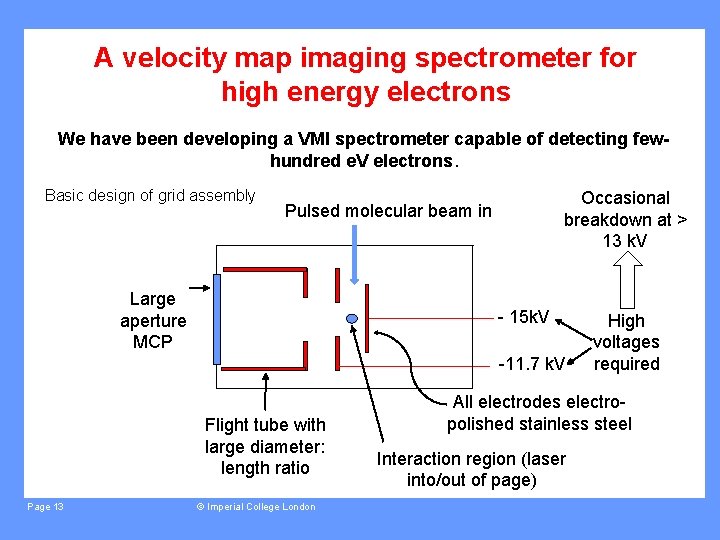
A velocity map imaging spectrometer for high energy electrons We have been developing a VMI spectrometer capable of detecting fewhundred e. V electrons. Basic design of grid assembly Occasional breakdown at > 13 k. V Pulsed molecular beam in Large aperture MCP - 15 k. V -11. 7 k. V Flight tube with large diameter: length ratio Page 13 © Imperial College London High voltages required All electrodes electropolished stainless steel Interaction region (laser into/out of page)
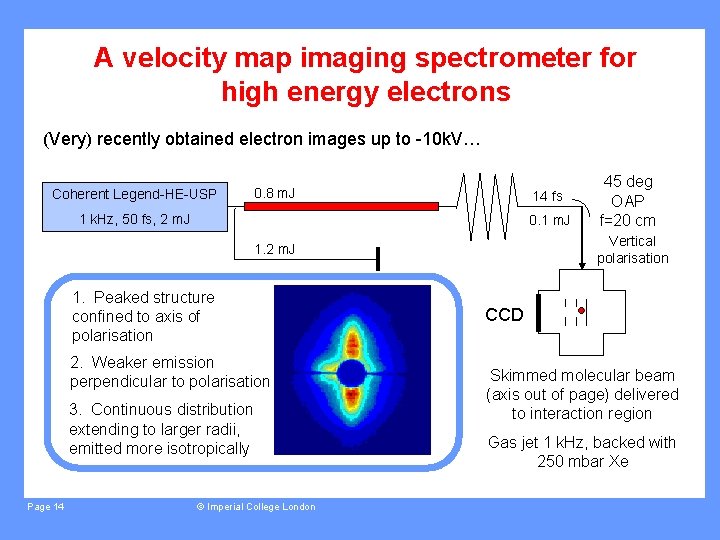
A velocity map imaging spectrometer for high energy electrons (Very) recently obtained electron images up to -10 k. V… Coherent Legend-HE-USP 0. 8 m. J 14 fs 1 k. Hz, 50 fs, 2 m. J 0. 1 m. J Vertical polarisation 1. 2 m. J 1. Peaked structure confined to axis of polarisation 2. Weaker emission perpendicular to polarisation 3. Continuous distribution extending to larger radii, emitted more isotropically Page 14 © Imperial College London 45 deg OAP f=20 cm CCD Skimmed molecular beam (axis out of page) delivered to interaction region Gas jet 1 k. Hz, backed with 250 mbar Xe
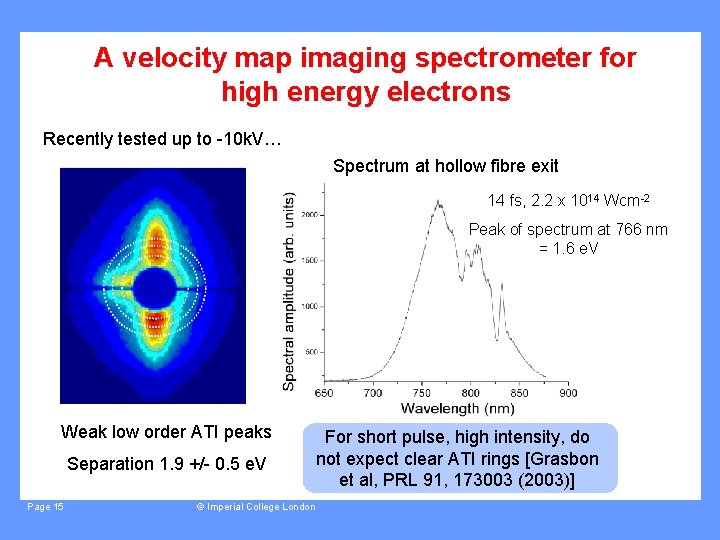
A velocity map imaging spectrometer for high energy electrons Recently tested up to -10 k. V… Spectrum at hollow fibre exit 14 fs, 2. 2 x 10¹⁴ Wcm-2 Peak of spectrum at 766 nm = 1. 6 e. V Weak low order ATI peaks Separation 1. 9 +/- 0. 5 e. V Page 15 © Imperial College London For short pulse, high intensity, do not expect clear ATI rings [Grasbon et al, PRL 91, 173003 (2003)]
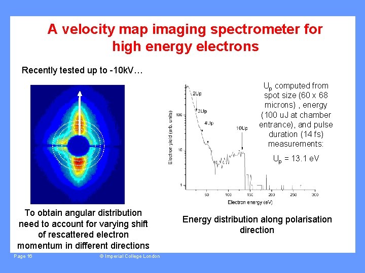
A velocity map imaging spectrometer for high energy electrons Recently tested up to -10 k. V… Up computed from spot size (60 x 68 microns) , energy (100 u. J at chamber entrance), and pulse duration (14 fs) measurements: Up = 13. 1 e. V To obtain angular distribution need to account for varying shift of rescattered electron momentum in different directions Page 16 © Imperial College London Energy distribution along polarisation direction
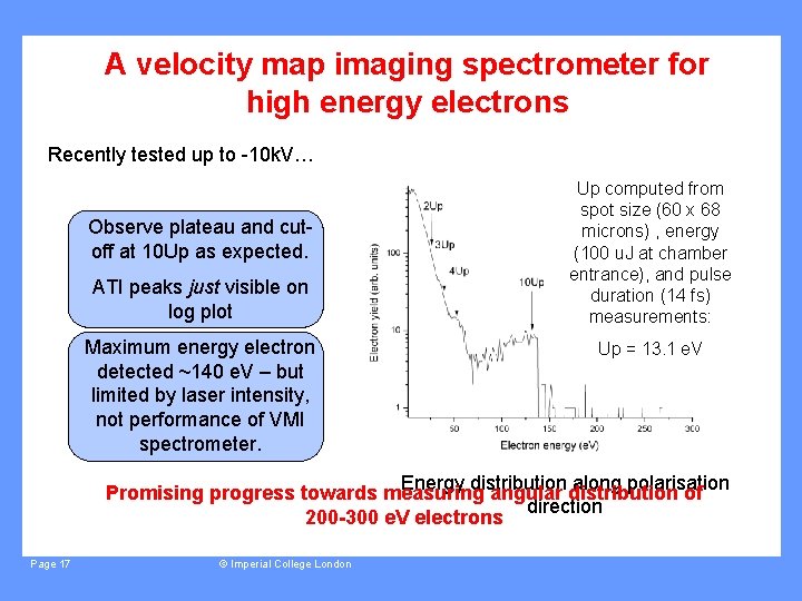
A velocity map imaging spectrometer for high energy electrons Recently tested up to -10 k. V… Observe plateau and cutoff at 10 Up as expected. ATI peaks just visible on log plot Maximum energy electron detected ~140 e. V – but limited by laser intensity, not performance of VMI spectrometer. Up computed from spot size (60 x 68 microns) , energy (100 u. J at chamber entrance), and pulse duration (14 fs) measurements: Up = 13. 1 e. V Energy distribution along polarisation Promising progress towards measuring angular distribution of direction 200 -300 e. V electrons Page 17 © Imperial College London
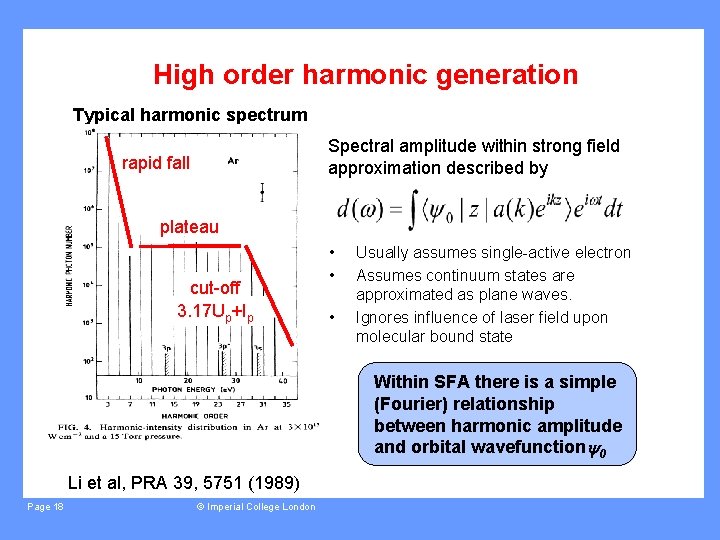
High order harmonic generation Typical harmonic spectrum Spectral amplitude within strong field approximation described by rapid fall plateau cut-off 3. 17 Up+Ip • • • Usually assumes single-active electron Assumes continuum states are approximated as plane waves. Ignores influence of laser field upon molecular bound state Within SFA there is a simple (Fourier) relationship between harmonic amplitude and orbital wavefunction 0 Li et al, PRA 39, 5751 (1989) Page 18 © Imperial College London
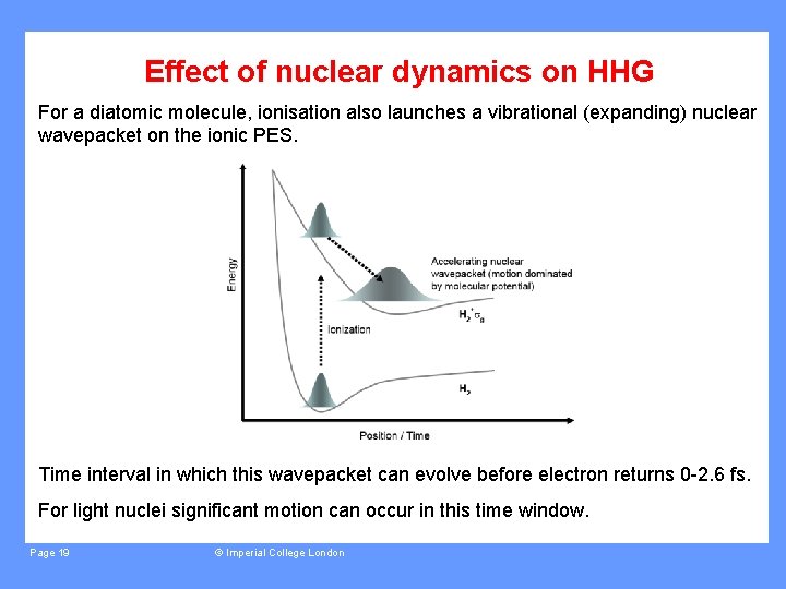
Effect of nuclear dynamics on HHG For a diatomic molecule, ionisation also launches a vibrational (expanding) nuclear wavepacket on the ionic PES. Time interval in which this wavepacket can evolve before electron returns 0 -2. 6 fs. For light nuclei significant motion can occur in this time window. Page 19 © Imperial College London
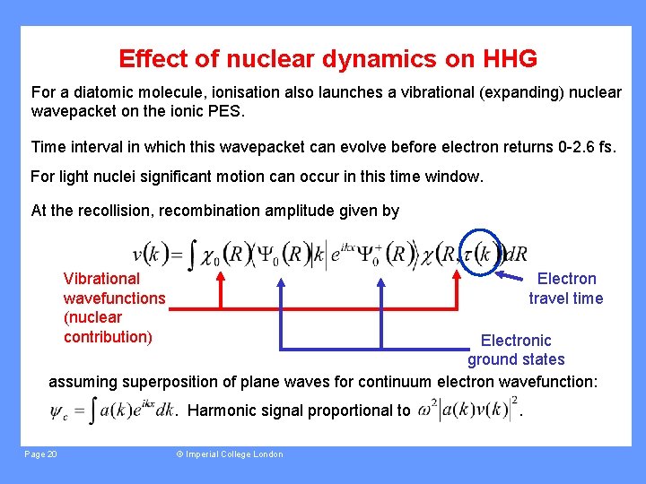
Effect of nuclear dynamics on HHG For a diatomic molecule, ionisation also launches a vibrational (expanding) nuclear wavepacket on the ionic PES. Time interval in which this wavepacket can evolve before electron returns 0 -2. 6 fs. For light nuclei significant motion can occur in this time window. At the recollision, recombination amplitude given by Electron travel time Vibrational wavefunctions (nuclear contribution) Electronic ground states assuming superposition of plane waves for continuum electron wavefunction: . Harmonic signal proportional to Page 20 © Imperial College London .
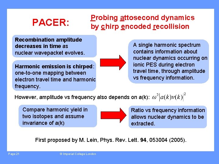
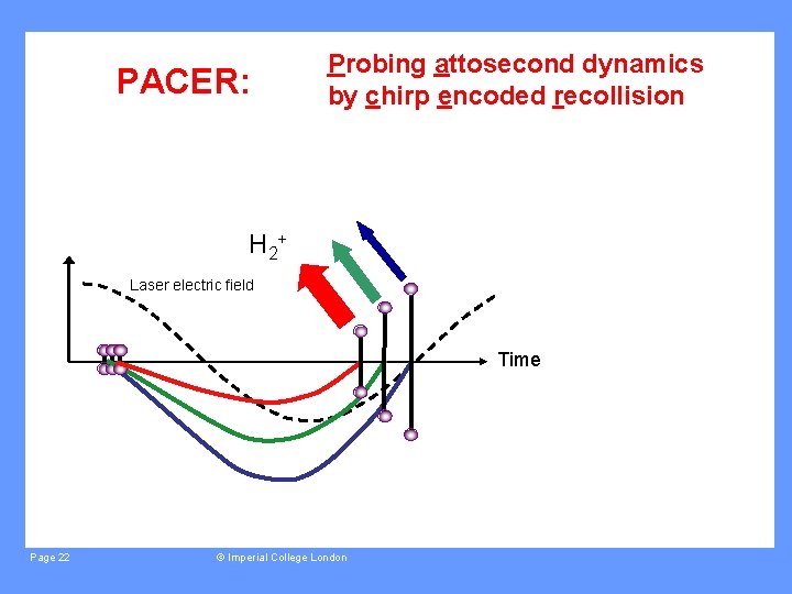
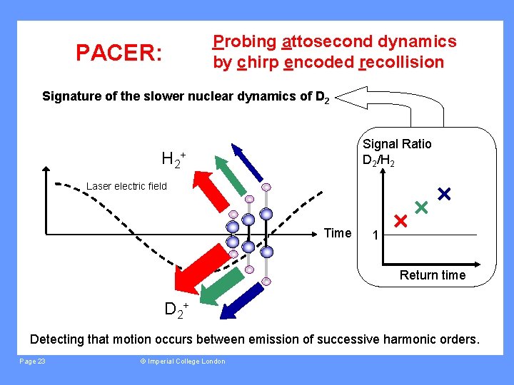
PACER: Probing attosecond dynamics by chirp encoded recollision Recombination amplitude decreases in time as nuclear wavepacket evolves. Harmonic emission is chirped: one-to-one mapping between electron travel time and harmonic frequency. A single harmonic spectrum contains information about nuclear dynamics occurring on ionic PES during electron travel time, through amplitude vs frequency information. However, amplitude vs frequency also depends on a(k): Compare harmonic yield in two isotopes and assume invariance of a(k) Ratio vs frequency information allows nuclear dynamics to be extracted. First proposed by M. Lein, Phys. Rev. Lett. 94, 053004 (2005). Page 21 © Imperial College London
PACER: Probing attosecond dynamics by chirp encoded recollision H 2+ Laser electric field Time Page 22 © Imperial College London
Probing attosecond dynamics by chirp encoded recollision PACER: Signature of the slower nuclear dynamics of D 2 H 2 Signal Ratio D 2/H 2 + Laser electric field Time 1 Return time D 2+ Detecting that motion occurs between emission of successive harmonic orders. Page 23 © Imperial College London
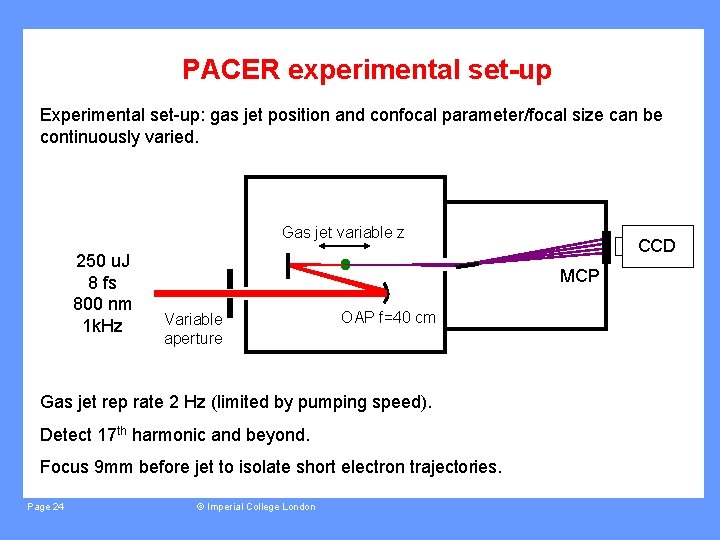
PACER experimental set-up Experimental set-up: gas jet position and confocal parameter/focal size can be continuously varied. Gas jet variable z 250 u. J 8 fs 800 nm 1 k. Hz MCP Variable aperture OAP f=40 cm Gas jet rep rate 2 Hz (limited by pumping speed). Detect 17 th harmonic and beyond. Focus 9 mm before jet to isolate short electron trajectories. Page 24 CCD © Imperial College London
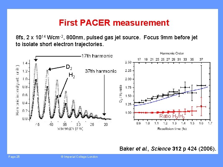
First PACER measurement 8 fs, 2 x 10¹⁴ Wcm-2, 800 nm, pulsed gas jet source. Focus 9 mm before jet to isolate short electron trajectories. D 2 H 2 Ratio H 2/H 2 Baker et al. , Science 312 p 424 (2006). Page 25 © Imperial College London

First PACER measurement 8 fs, 2 x 10¹⁴ Wcm-2, 800 nm, pulsed gas jet source. Focus 9 mm before jet to isolate short electron trajectories. Blue curve: averaged over randomly aligned angle distribution. Use BO potentials for H 2+ and D 2+. Single molecule response, allowing for short trajectories only. Ratio H 2/H 2 Includes two-centre interference effects. Includes effect of reabsorption of generated XUV. Baker et al. , Science 312 p 424 (2006). Page 26 © Imperial College London
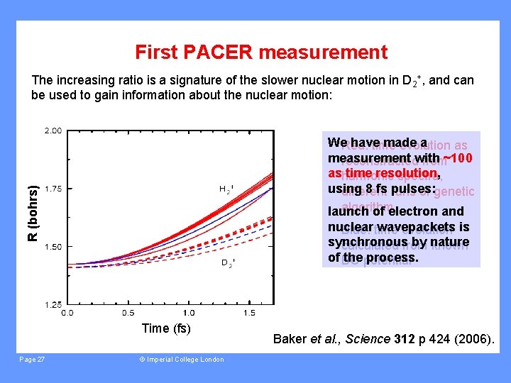
First PACER measurement The increasing ratio is a signature of the slower nuclear motion in D 2+, and can be used to gain information about the nuclear motion: R (bohrs) We. Red: have made a time evolution as measurement reconstructedwith from~100 asharmonic time resolution, spectra, using 8 fs pulses: different runs of genetic algorithm launch of electron and nuclear wavepackets Blue: time evolution is synchronous by nature calculated from known of BO the potential process. Time (fs) Page 27 © Imperial College London Baker et al. , Science 312 p 424 (2006).
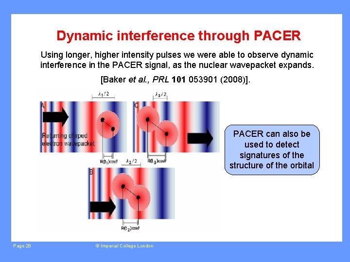
Dynamic interference through PACER Using longer, higher intensity pulses we were able to observe dynamic interference in the PACER signal, as the nuclear wavepacket expands. [Baker et al. , PRL 101 053901 (2008)]. PACER can also be used to detect signatures of the structure of the orbital Page 28 © Imperial College London
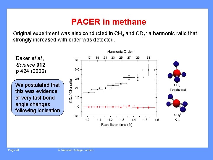
PACER in methane Original experiment was also conducted in CH 4 and CD 4: a harmonic ratio that strongly increased with order was detected. Baker et al. , Science 312 p 424 (2006). We postulated that this was evidence of very fast bond angle changes following ionisation Page 29 © Imperial College London
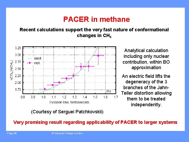
PACER in methane Recent calculations support the very fast nature of conformational changes in CH 4 Analytical calculation including only nuclear contribution, within BO approximation An electric field lifts the degeneracy of the 3 branches of the Jahn. Teller distortion allowing them to be treated independently. (Courtesy of Serguei Patchkovskii) Very promising result regarding applicability of PACER to larger systems Page 30 © Imperial College London
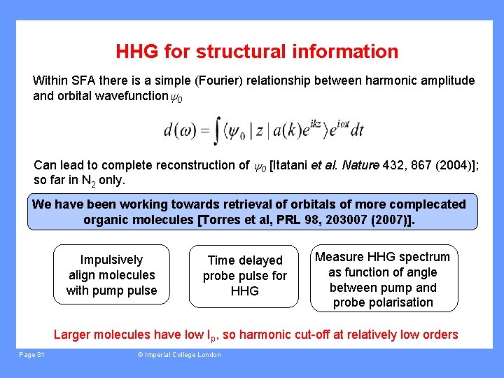
HHG for structural information Within SFA there is a simple (Fourier) relationship between harmonic amplitude and orbital wavefunction 0 Can lead to complete reconstruction of 0 [Itatani et al. Nature 432, 867 (2004)]; so far in N 2 only. We have been working towards retrieval of orbitals of more complecated organic molecules [Torres et al, PRL 98, 203007 (2007)]. Impulsively align molecules with pump pulse Time delayed probe pulse for HHG Measure HHG spectrum as function of angle between pump and probe polarisation Larger molecules have low Ip, so harmonic cut-off at relatively low orders Page 31 © Imperial College London
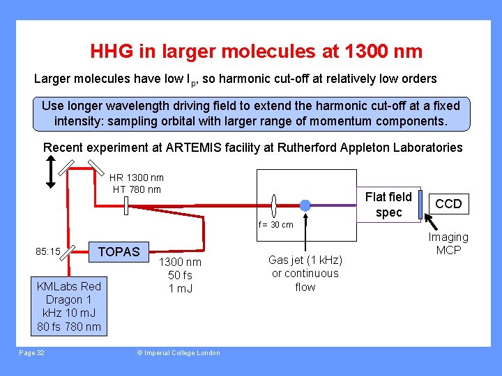
HHG in larger molecules at 1300 nm Larger molecules have low Ip, so harmonic cut-off at relatively low orders Use longer wavelength driving field to extend the harmonic cut-off at a fixed intensity: sampling orbital with larger range of momentum components. Recent experiment at ARTEMIS facility at Rutherford Appleton Laboratories HR 1300 nm HT 780 nm Flat field spec CCD f = 30 cm 85: 15 TOPAS KMLabs Red Dragon 1 k. Hz 10 m. J 80 fs 780 nm Page 32 1300 nm 50 fs 1 m. J © Imperial College London Gas jet (1 k. Hz) or continuous flow Imaging MCP
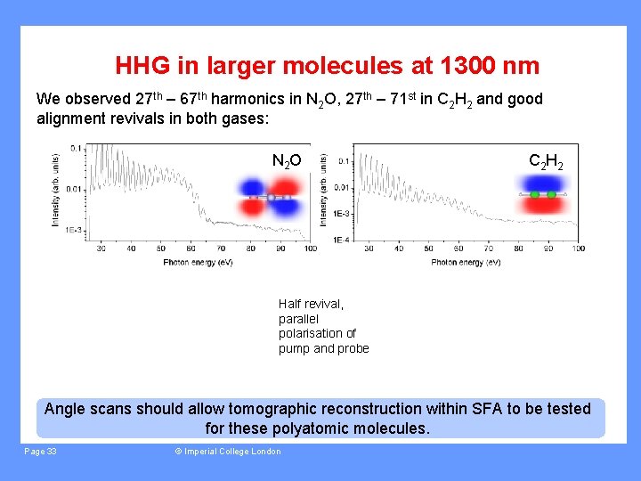
HHG in larger molecules at 1300 nm We observed 27 th – 67 th harmonics in N 2 O, 27 th – 71 st in C 2 H 2 and good alignment revivals in both gases: N 2 O C 2 Half revival, parallel polarisation of pump and probe Angle scans should allow tomographic reconstruction within SFA to be tested for these polyatomic molecules. Page 33 © Imperial College London
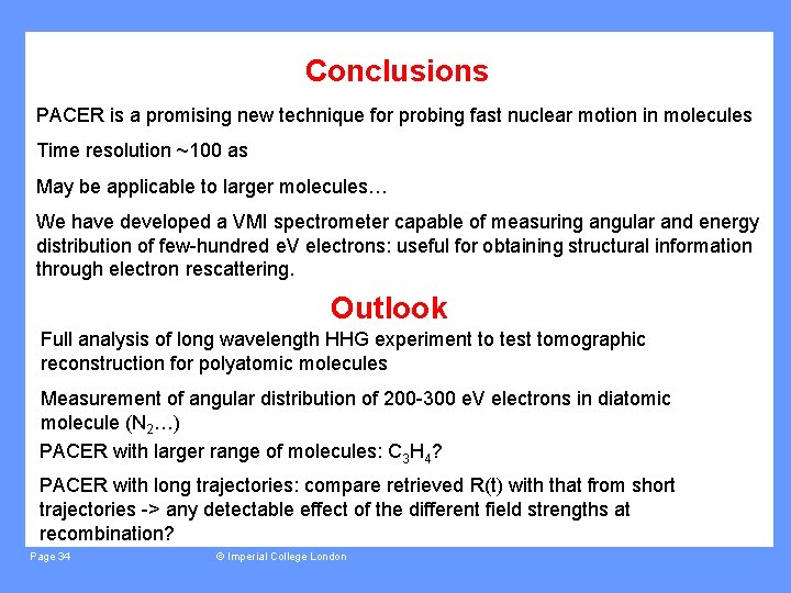
Conclusions PACER is a promising new technique for probing fast nuclear motion in molecules Time resolution ~100 as May be applicable to larger molecules… We have developed a VMI spectrometer capable of measuring angular and energy distribution of few-hundred e. V electrons: useful for obtaining structural information through electron rescattering. Outlook Full analysis of long wavelength HHG experiment to test tomographic reconstruction for polyatomic molecules Measurement of angular distribution of 200 -300 e. V electrons in diatomic molecule (N 2…) PACER with larger range of molecules: C 3 H 4? PACER with long trajectories: compare retrieved R(t) with that from short trajectories -> any detectable effect of the different field strengths at recombination? Page 34 © Imperial College London
People Jon Marangos John Tisch PACER work: Electron VMI: Long wavelength HHG: Joe Robinson Manfred Lein Ciprian Chirila Delphine Darios Marco Siano David Holland (STFC) Tom Siegel Ricardo Torres Leonardo Brugnera Imma Procino Jonathan Underwood Staff at ARTEMIS facility Adverts! E. Springate, I. C. E. Turcu, C. Froud Two posters: Delphine Darios; VMI of high energy electrons Imma Procino; Retrieval of molecular axis alignment from Coulomb explosion imaging experiments without cylindrical symmetry Page 35 © Imperial College London
- Slides: 35