Quantum Dots David S Baskin M D Department

Quantum Dots David S. Baskin, M. D. Department of Neurosurgery The Methodist Hospital Neurological Institute

Medical Treatment Today: A Fundamental Size Mismatch. . . Body Components artificial hips therapeutic radiation scalpel catheters Our watchmaker tool m dm cm mm body organs blood neurons most vessels cells nucleus mm cell organelles supramol. disease starts here proteins nm mm How we water flowers. . assemblies small molecules atoms m dm cm mm nm Drugs

Quantum Dots • Single crystal a few nm in diameter • Size and shape precisely controlled during synthesis • Smaller than the Bohr exciton radius of the compound • Energy is quantized, related to size-quantum confinement • Radiative recombination of exciton and hole yields narrow emission, symmetric energy, long half life • Very different from usual type of florescence

Electrons “Check in but They Don’t Check Out”

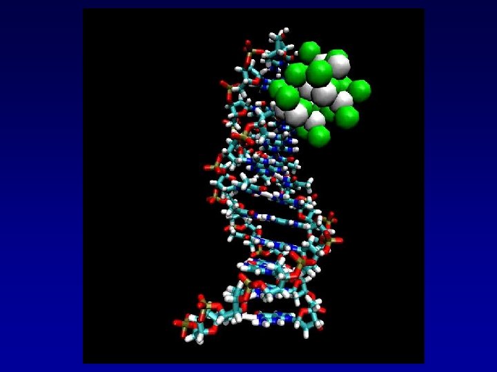
Quantum dot structure
 Polymer Coating Passivation Shell (Zn. S) Semiconductor Nanocrystal (Cd. Se)")
Quantum Dots Biomolecule (SA) Polymer Coating Passivation Shell (Zn. S) Semiconductor Nanocrystal (Cd. Se) • Broad excitation spectrum • Narrow emission band • Brightness • Photostability • Single molecule sensitivity • Bioconjugates (Streptavidin, Protein A, Ig. G. . . ) • Non-toxic • Donors for FRET

Quantum Dot Size

Size of Quantum Dot Determines Color

Quantum Dot Tags

Summary of Properties • • • Broad Absorption Narrow and Tunable Emission Resistant to Photobleaching Strong Luminescence Long Luminescence Time Florescence Resonance Energy Transfer (FRET)

FRET

Nano Protease Sensor

Maltose Nanosensor

Simultaneous labeling of m. Rna and Protein in a single cell

Carbon Allotropes Property of certain substances of existing in forms with different chemical structures

Single-walled Nanotubes Single-walled nanotubes are most beneficial for biological applications because their properties can be easily controlled They travel in and out of cells without difficulty

Fluorescent Nanotube Labeling First enzyme-driven technique for fluorescent labeling of single walled carbon nanotubes.

Chemical pegs to attach Q-dots are biotinylated tyramide radicals created by horseradish peroxidase Peroxidation to attach chemical pegs to nanotube surface Simple Variation of classic biotinylation

Chemical pegs to attach Q-dots are biotinylated tyramide radicals created by horseradish peroxidase

Fluorecent Nanotube Labeling After the reaction, the biotin-tagged SWNTs were combined with streptavidin-FITC or streptavidin-Q-dots (Evident Technologies) by mixing at 1: 1 ratio (v/v).

Fluorecent Nanotube Labeling Linking semiconductor nanocrystals, quantum dots, to the surface of nanotubes resulted in their fluorescent visualization, whereas conventional fluorophores bound to SWNTs directly, or through biotin-streptavidin linkage, were completely quenched by FRET

Fluorescent SWNTs labeled by quantum dots Band on the left shows nanotubes labeled with quantum dots

Electrophoresis test of fluorescent nanotube labeling: fluorophore directly attached to nanotubes • Conventional fluorophores (lanes 2, 3, 4) are quenched when attached to nanotubes • Quantum dots remain brightly fluorescent when attached to nanotubes (upper band in lane 1)

Our approach to visualize nanotubes with Q-dots labeled with streptavidin are attached to a biotinylated nanotube.

Electron microscopy of a nanotube labeled with Q-dots • Nanotubes are covered with streptavidin molecules (white dots) • Beads-on-a-string appearance of nanotube labeled with Q-dots

Q-dot-labeled fluorescent nanotubes Individual quantum dots can be seen fluorescing on a single nanotube molecule

Potassium Ion Channels Lai and Yan, 2006

Voltage Gated Potassium Ion Channel Kv 4. 2

Schematics of surface labeling by Qdots Labeling of molecules on the surface of cells with Qdots The membrane protein, in this case Kv 4. 2 carrying an extracelular myc-epitope, is recognized by a specific primary antibody, which in turn is recognized by a biotinylated Fab fragment of a secondary antibody. A streptavidin-conjugated Qdot then binds to the biotin o the molecule (Kv 4. 2)/ antibody complex

EGFP and Myc-tagged Kv 4. 2 channel cotransfected neuron labeled live with Myc-Qdot Ab trans EGFP QDot FM 4 -64

EGFP and Myc-tagged Kv 4. 2 channel cotransfected neuron labeled live with Myc-Qdot Ab QDot labeling KV 4. 2 pyramidal Neuron in culture-dendrite And Soma FM 4 -64 Synaptic Extrasynaptic FM 4 -64 Synaptic marker QDot

Single Particle Tracking Kv 4. 2 Potassium Ion Channel

Neuroblastoma Cell Labeling Integrin Antibodies

Q-Dots Can Be Used to Target Cancer Cells in vivo Q-dots attached to anti-cancer antibodies recognizes cancer cells

Recent Advances in Quantum Computing Chang-Purdue University ”The Latest in Quantum Dot Switches” • • • 200 nm Two Dot Switches Hundreds of dots now possible with our technology

Quantum Computer • • • Uses quirky world of Quantum mechanics No longer binary-Simultaneous states possible Infinite parallel processing Cryptography-RSA protocol using prime numbers-Seconds vs one billion years Artificial Intelligence barrier could be broken. Nine billion neurons in the brain Teleportation-The “Quantum Internet”

Better Quantum Dots
")
Luciferase (Fireflies)

Quantum Mirages

Quantum Potentials Technology Rules

Many Thanks to my Mentors
- Slides: 42