Purpose To recognize three basic shapes of bacterial

Purpose � To recognize three basic shapes of bacterial cells.

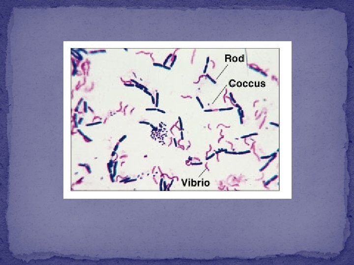
The three common shapes of bacteria : 1 -Coccus 2 - Bacillus 3 - Spiral

1 -Coccus having one of the following arrangements: �Diplococcus: a pair of cocci �Streptococcus: a chain of cocci �Tetrad: a square of 4 cocci �Sarcina: a cube of 8 cocci �Staphylococcus: cocci in irregular, often grape-like clusters

2 - Bacillus �Bacillus: a single bacillus �Streptobacillus: bacilli in chains �Coccobacillus: oval and similar to a coccus

3 - Spiral �Vibrio: an incomplete spiral or comma-shaped �Spirillum: a thick, rigid spiral �Spirochete: a thin, flexible spiral

Simple stain

Simple stain : �The simple stain is a very simple staining procedure involving only one stain. �You may choose from methylene blue, safranin, and crystal violet.

Simple stain : 1. Prepare the smear. - place a small drop of water on a clean slide. Drag the sterile inoculating needle tip through the edge of colony. - Gently spread the mixture into a circle to spread out.

Simple stain : 2. Let the smear air dry completely.

Simple stain : 3. Heat-Fix the smear. � Smears are heat-fixed by quickly passing the slide through a flame two or three times. � This causes the microbes to stick to the slide and not get washed off during the staining process.

Simple stain : 4. Stain the smear. � Place the slide on a rack over the sink. Flood the smear with stain and let it for 60 -90 seconds. Rinse gently and blot dry.

Simple stain : 5. Then, place a drop of oil directly on the stained smear. Turn the oil lens into position and fine focus to observe the cells.

Result
")
Coccus (cocci pl. )
")
Bacillus (Bacilli pl. )
")
Spirillum (Spirilli pl. )
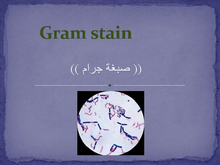

It is used to differentiate between gram-positive and gram-negative bacteria, which have distinct and consistent differences in their cell walls.

The gram stain is called a differential stain because it stain cell differently based on their cell wall structure.

The cell wall structure

Gram-positive bacteria Have a thick peptidoglycan layer surrounds the cell. The stain gets trapped into this layer and the bacteria turned purple. Gram-negative bacteria have a thin peptidoglycan layer that does not retain crystal violet stain. Instead, it has a thick lipid layer which dissolved easily upon decoulorization with Alcohol. Therefore, cells will be counterstained with safranin and turned red.

The material : Cultures of : Staphylococcus aureus, Bacillus subtilis, E. coli 1. Crystal violet. 2. Iodine solution. 3. Alcohol 95%. 4. Safranin. 5. Water.

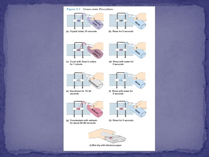
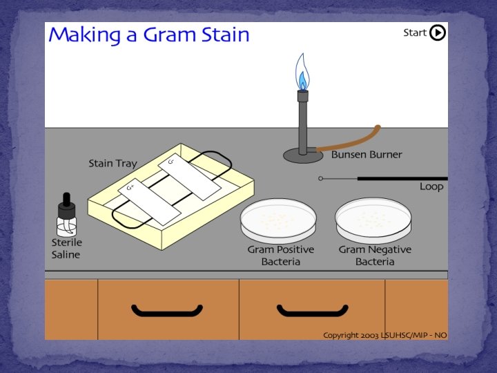
The method

The bacteria under the microscope

–ve Gram +ve

Results: Shape: Cocci Arrangment: irregular clusters Colour: Violet Gram’s reaction: Gram’s +ve Name of microorganism: Staphylococci

Results: Shape: Bacilli Arrangment: Chains Colour: Violet Gram’s reaction: Gram’s +ve Name of microorganism: Bacillus

- Slides: 32