Pupils Optic nerve Done by Khaled Samara Felasten

Pupils & Optic nerve Done by : Khaled Samara Felasten Abed

The pupil • The pupil is a hole located in the centre of the iris of the eye that allows light to strike the retina. It appears black because light rays entering the pupil are either absorbed by the tissues inside the eye directly, or absorbed after diffuse reflections within the eye that mostly miss exiting the narrow pupil.

Movements of the pupil are controlled by the parasympathetic and sympathetic nervous systems. The pupils constrict ( miosis ) when the eye is illuminated (parasympathetic activation, sympathetic relaxation) and dilate ( mydriasis ) in the dark (sympathetic activation, parasympathetic relaxation). When the eyes focus on a near object, they converge and the pupils constrict (the near response ). The pupils are normally equal in size but some 20% of people may have noticeably unequal pupils ( anisocoria ) with no associated disease.

Anatomy & pupilary reflexes • Afferent and efferent arms. • Normal pupillary reflexes • Afferent Pupilary Defect

5 Afferent Pathway of Pupillary Light Reflex ● Light enters the pupil and stimulates the retina. ● Retinal ganglion cells transmit the light signal to the optic nerve ● The optic nerve enters the optic chiasm where the nasal retinal fibers cross to contralateral optic tract. while the temporal retinal fibers stay in the ipsilateral optic tract ● Fibers from the optic tracts project and synapse in the pretectal nuclei in the dorsal midbrain in the collicular region ● The pretectal nuclei project fibers to the ipsilateral Edinger-Westphal nuclei and also to the contralateral Edinger-Westphal nucleus via the posterior commissure

6 Efferent Pathway of Pupillary Light Reflex ● The Edinger-Westphal nucleus projects pre-ganglionic parasympathetic fibers, which exit the midbrain and travel along the oculomotor nerve (CN III) and then synapse on post-ganglionic parasympathetic fibers in the ciliary ganglion ● Ciliary ganglion post-ganglionic parasympathetic fibers (short ciliary nerves) innervate the sphincter muscle of the pupils resulting in pupillary constriction. The physiological result is that light shined in one eye will result in pupillary constriction in both the ipsilateral pupil (direct pupillary light reflex) and the contralateral pupil (consensual pupillary light reflex).

Normal Pupillary Light Response : Row 1: pupils in a dark room without light stimulation. Row 2: intact direct and consensual responses for right eye Row 3: intact direct and consensual responses for left eye
")
Causes of pupil disorders • Ocular causes • Neurological causes. • Other causes (drugs)

Ocular causes of pupillary abnormalities Several diseases of the eye cause pupil irregularity and alter pupil reactions: • anterior uveitis, when posterior synechiae give the pupil an irregular appearance • the sequelae of intraocular surgery • blunt trauma to the eye, which may rupture the sphincter muscle, causing irregularity or fixed dilation ( traumatic mydriasis ) • an acute and severe rise in ocular pressure – as in acute glaucoma.

Traumatic mydriasis Acute Angle-closure Glaucoma POSTERIOR SYNECHIA SECONDARY TO HLA B 27 ANTERIOR UVEITIS

Neurological causes of abnormal pupil • Interruption of the sympathetic or parasympathetic nerve supply. • As seen in : Horner's syndrome , Aide's pupil , Argyll Robertson pupil and pupil defects due to mid brain lesions

Horner's syndrome presentation • Meiosis : small pupil on the affected side which is more prominent in dark due to dilatation of the opposite pupil • Mild ptosis on the affected side • Anhydrosis ( decreases sweating ) on the affected side which depends on the level of the lesion. • Enophthalmos

Causes • Syringomyelia , an expanding cavity within the spinal cord, sometimes extending into the medulla (syringobulbia), which compresses the pathway. • Small - cell carcinoma at the lung apex which catches the cervical sympathetic chain. • Neck injury, disease or surgery. • Cavernous sinus disease – catching the sympathetic carotid plexus in the sinus.

Horner’s syndrome may also be congenital, in which case the iris colour may be altered when compared to the other eye ( heterochromia ).

Relative afferent pupillary defect Row 1: Unstimulated pupils in a dark room Row 2: Stimulation of the right eye produces bilateral pupillary constriction, indicating intact afferent right limb, and intact bilateral efferent limbs. Row 3: When moving the light source from the right to left eye, the left eye paradoxically dilates. This indicates a faulty left eye afferent limb, most likely from left optic nerve dysfunction. Row 4: Demonstrates again that the right afferent pathway is functioning normally and that the problem is with the left eye’s afferent pathway.

Light near dissociation Impaired reaction of the pupils to light, while the near response to accommodation is retained. It is seen with - Adie’s tonic pupil, - Argyll Robertson pupil - peri-aqueductal brainstem lesions such as Parinaud’s syndrome. Other causes include: diabetes and multiple sclerosis.

Adie’s tonic pupil • It is due to a ciliary ganglionitis which denervates the parasympathetic supply to the iris and ciliary body. • Cause anisocoria • More in females 2: 1 • More common in young patients

Presentation • Patient may complain of poor accommodation specially when change from far to near or vice versa • Reading problems

Pupillary finding • • • Large pupil Poor reaction to light Slow and sustained meiosis on accommodation Vermiform movement of the iris Supersensitive pupil to small concentration of Pilocarpine muscarenic

Argyll Robertson pupil • The pupils are bilaterally small and irregular. They do not react to light but respond to accommodation. • Seen mainly in neurosyphilis • Loss of light response and intact accommodation. • Feathery appearance of the iris stroma • Loss of iris architecture details

Mid –brain pupil • Lesion in the region of the pretectal nuclear complex disturbing the retinocortical fibers but preserves the accommodative fibers • Seen in preiaqueductal (Parinaud”s ) syndrome

Other causes of abnormal pupil • Coma • Drugs • CN III palsy
. •")
Drugs • Topical drugs as muscarinic blockade (cyclopentolate , tropicamide & atropine ). • Alpha receptor agonists as ( phenylephrine & adrenaline ) • Muscarinic agonist causing constriction as Pilocarpine • Systemic drugs as ( Atropine , adrenaline & morphine )

Thank

Optic Nerve Diseases Felasten Abed

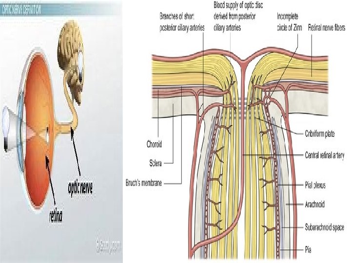
Optic nerve • The optic nerve is formed by the axons arising from the retinal ganglion cell layer , which form the nerve fibre layer of the retina (papillae). • The optic disc or nerve head is the point where the axons from the retinal ganglion cells leave the eye. • The nerve head appears as a white circular structure in the back of the eye. There are no photoreceptors on this structure. physiological blind spot. 1� • it passes out of the eye through the cribriform plate of the sclera. That allow the nerve fibers to pass through many holes and into the extraocular (outside of the eyeball) space. As the fibers pass through, they become covered by a sheath formed by dura , archnoid and pia , continuous with that surrounding the brain. It is bathed in CSF. • The central retinal vein and artery enter the eye in the center of the optic nerve

Optic nerve head swelling: it is a swelling of the optic disc. In response to many ocular or systemic disorders

• Normally we have clear and distinct neuro-retinal RIM , small telangiectated vesseels on the surface , and we can differentiate the retina from papillae( nerve fiber) , And the optic cup is easily appreciated • In optic nerve swelling there is Swollen retina , prominent capillaries , not clear neuro-retinal RIM

swelling of the optic disc.

Causes of ON head swelling : • Different causes but almost comparable appearance causes: • Increased ICP –> (papilledema). • Space occuping leasions • Papillitis • Malignant hypertension • Central retinal vein occlusion ( CRVO) • Sub dural or sub arachnoid hemorrhage • Ischemic optic neuropathy

DDx of optic nerve head swelling : Pseudopapilledema : Some normal optic nerve heads may appear to be swallen due to crowding of the nerve fibres entering the disc • 1) small hypermetropic disc • 2) ON drusen • 3) Myelinated nerve fibers • 4) Dysplasia ( MC congenital optic nerve anomaly)

Symptoms of increase icp • Obscuration of vision : transient visual loss due to change position ( from lying to standing ) • No acute or prolonged loss of vision in the beginning • Headache worse on awaking & cough • Nausea & retching • Diplopia due to CN VI palsy ( it has the longest pathway and its usually fense intracranially so involved in increas icp) • Focal neurological symptoms ( in SOL ) • History of trauma suggesting subdural hemorrhage

Signs of high ICP: • Vision may be affected • Swollen optic disc with loss of margin details , prominent capillaries & absent venous pulsation which may occur normally in 5 -20% of normal population ( so if there is venous pulsation its rule out high ICP ) • Large blind spot on VF • VF constriction if chronic papilledema • Focal neurological signs if space occupying lesion suspected

Investigations: • Usually history is suggestive but we need brain imaging to rule out secondary causes: • 1) CT • 2)MRI • 3) MRV • 4) Lumber puncture to measure ICP (8 -12 mmgh) • 5) multiteam care

Treatment : • Raised ICP with no other space occupying lesion or ventricle dilatation previously called benign raised ICP. Need to decrease ICP to prevent permanent visual field losses : By decrease production of csf or increase drainge of csf • Medication ( acetazolamide ) ( used also in glaucoma ) • VP ( ventriculoperitoneal) shunts. • Optic nerve decompression ( small hole around the optic nerve sheath ) to make CSF leak (fenestration) • Neurosurgery consults for tumours and SOL

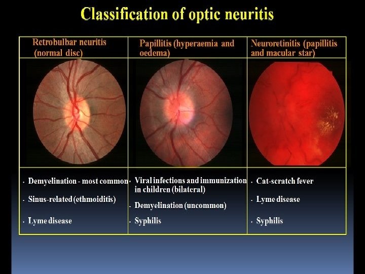
Optic neuritis : Optic neuritis”: is an inflammation or demyelination of the optic nerve. • It may affect the part of the nerve and disc within the eyeball , anterior and visible part of ON (papillitis) • the portion behind the eyeball , posterior to sclera (retrobulbar optic neuritis).

Causes: • Certain autoimmune diseases such as multiple sclerosis commonest cause 25% of MS patients come firstly with symptoms of ON • Infections such as viruses (especially in children), measles, meningitis, syphilis, sinusitis, tuberculosis, and human immunodeficiency virus (HIV) • Malnutrition. E. g. vit B complex deficiency. • Intraocular inflammation (uveitis)

Optic Neuritis presentation • Acute loss of vision that may progress over few days then slowly improves. • Pain upon eye movement due to the contact between the recti ( medial rectus ms) and the inflammed ON • History of viral illness • Focal neurological symptoms as in Multiple sclerosis (MS )

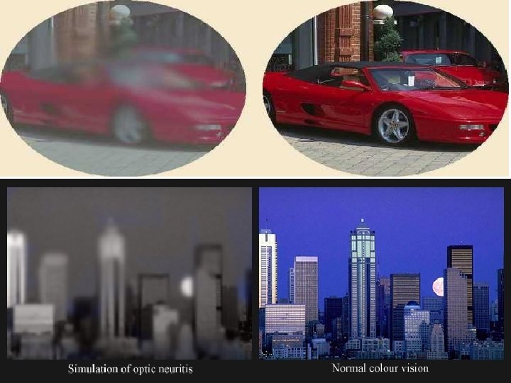
Examination: • • Reduced VA , VF Reduced color vision Central scotoma and large blind spot RAPD –relative afferent pupillary defect- ( swinging flash light sign ) very important sign to diagnose Optic neuritis • Swollen OD in Papillitis • Normal OD in retrobulbar neuritis • Focal neurological signs in MS

Papillitis

Management • MRI to rule out or diagnose MS • Treatment of the primary cause if known • Steroid rule to speed up the resolution phase but not affect final prognosis

on MRI hyperintense changes in on multiple lesions on more than one level peri-ventricular , infratentorrial and spinal cord lesions >5 mm more likely the diagnosis is MS

Prognosis • Variable • Mostly vision will recover slowly but some of the ON functions as color contrast and color vision will be affected permanently • Visual loss may be permanent • Recurrent attacks will lead to cumulative damage of on functions

Ischemic optic neuropathy • There is closure in bv • May be artetitic or non-artetitic • Symptoms mostly sudden loss of vision or vf defect
 • Autoimmune disease occurring in elderly patients")
Aretritic type: • Giant Cell Arteritis (GCA) • Autoimmune disease occurring in elderly patients over 60 years • Affect medium sized arteries with internal elastic lamina • Presentation : Sudden loss of vision Scalp tenderness Jaw claudication ( due to affection of masseter muscle) Shoulder pain Malaise
 • Swollen")
GCA signs • Decreased VA • VF defect typically lower half (altitudinal) • Swollen and hemorrhagic disc • Normal retina and blood vessels • Some discs are small in size ( the fellow eye ). it’s common in hypermetropic patient • Tender temporal artery
 giant cell granulomas infiltration • 2) small lumen • 3)")
Histological findings: • 1) giant cell granulomas infiltration • 2) small lumen • 3) wall infiltrated by chronic type-inflammatory cell ( T-cell)

Investigations • ESR & CRP high in most cases • Temporal artery biopsy ( very important to do it because this disease may affect other mediam sized artery such as coronary artery and you must give steroid to prevent systemic complication ) • In non-arteritic ischemic optic neuropathy do : CBC to R/O anemia Blood pressure ( hyper and hypo ) - - HTN Blood sugar - - D. M Autoimmune workup

Treatment • In GCA immediate treatment with steroid even before confirming the diagnosis is mandatory to prevent systemic complication and protect the other eye. • Long term steroid treatment ( tapering dose ) • Steroid precautions as ( DM , TB &HTN ) • No treatment available for the non-arteritic ( management of underlying conditions ) • Medical consult and follow up

Prognosis • Visual loss is usually permanent • Fellow eye may be affected in GCA rapidly • In the non-artetitic the rate of fellow eye involvement is 40 -50 %

Optic atrophy Causes: • Optic nerve compression : Hx of orbital or chiasmal disease. • Ischemic optic neuropathy : Hx of sudden uni. Visual loss in the past • Glaucoma : the optic disc is pathologically cupped • ON : Hx of previous loss of vision / signs of MS • Inherited diseases : In the 1 st few years of life • Drugs and toxins : may follow chemical toxins • Tobacco , Alcohol & vitamine deficiency

• Signs & symptoms: • • • Loss of vision Loss of brightness & color discrimination Diminished or absent Pupillary reflex V. F. defect Blindness ( end result) • Treatment: • Optic nerve can’t regenerate, so visual loss is irreversible.

Observed clinically as : disc pale & white, margins are distinct
- Slides: 56