
Pulse Oximetry Capnography Ray Taylor Valencia Community College



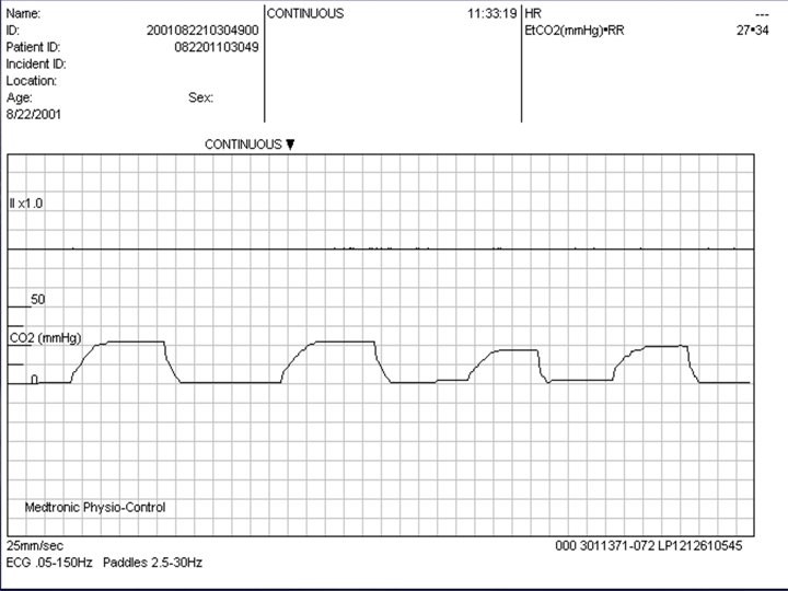
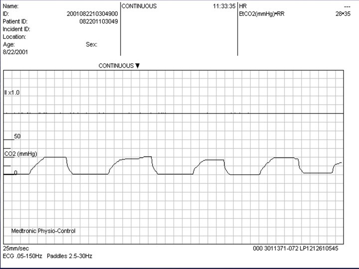




- Slides: 123
Pulse Oximetry & Capnography Ray Taylor Valencia Community College Department of Emergency Medical Services
Notice § § § All rights reserved. Slide show used with permission only for the purposes of educating emergency medical providers (EMTs and Paramedics) No portion of this presentation may be reproduced, stored in a retrieval system in any form or by any means (including but not limited to electronic, mechanical, photocopying etc. ) without prior written permission from the author
CAPNOGRAPHY IS THE VITAL SIGN FOR VENTILATION (what we should evaluate) OXIMETRY IS THE VITAL SIGN FOR OXYGENTATION (what we have historically used)
Capnography § Definition: – Noninvasive measurement of the partial pressure of C 02 in exhaled air – Provides instantaneous information about § Ventilation – How effectively C 02 is being eliminated by the pulmonary system § Perfusion – How effectively C 02 is being transported through the vascular system § Metabolism – How effectively C 02 is being produced by cellular metabolism
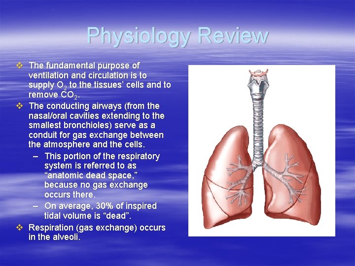
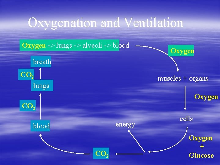
Physiology Review v The fundamental purpose of ventilation and circulation is to supply O 2 to the tissues’ cells and to remove CO 2. v The conducting airways (from the nasal/oral cavities extending to the smallest bronchioles) serve as a conduit for gas exchange between the atmosphere and the cells. – This portion of the respiratory system is referred to as “anatomic dead space, ” because no gas exchange occurs there. – On average, 30% of inspired tidal volume is “dead”. v Respiration (gas exchange) occurs in the alveoli.
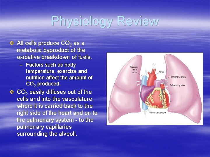
Physiology Review v All cells produce CO 2 as a metabolic byproduct of the oxidative breakdown of fuels. – Factors such as body temperature, exercise and nutrition affect the amount of CO 2 produced. v CO 2 easily diffuses out of the cells and into the vasculature, where it is carried back to the right side of the heart and on to the pulmonary system - to the pulmonary capillaries surrounding the alveoli.
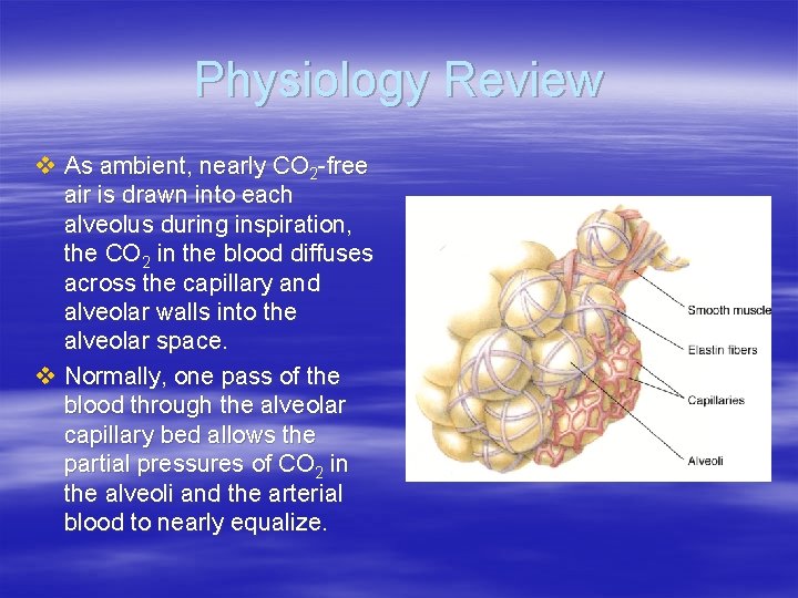
Physiology Review v As ambient, nearly CO 2 -free air is drawn into each alveolus during inspiration, the CO 2 in the blood diffuses across the capillary and alveolar walls into the alveolar space. v Normally, one pass of the blood through the alveolar capillary bed allows the partial pressures of CO 2 in the alveoli and the arterial blood to nearly equalize.
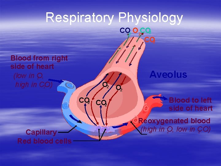
Respiratory Physiology CO 2 O 2 CO 2 Blood from right side of heart (low in O, 2 high in CO) 2 Aveolus O 2 CO 2 . Capillary Red blood cells Blood to left side of heart Reoxygenated blood (high in O, low in CO) 2 2
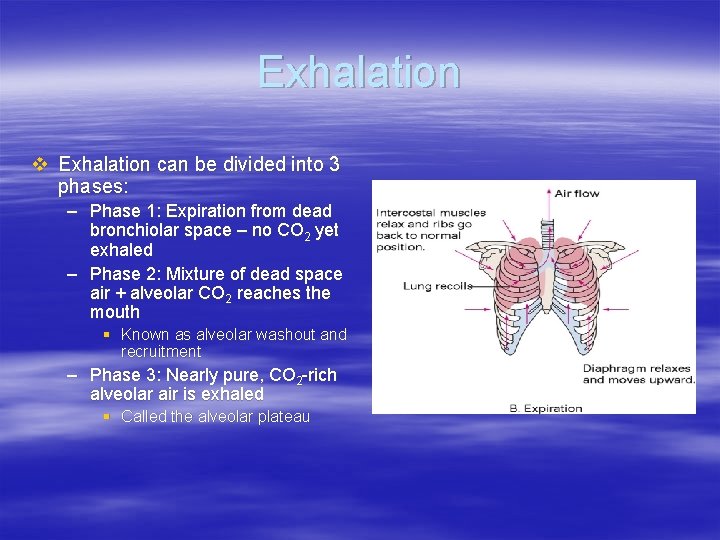
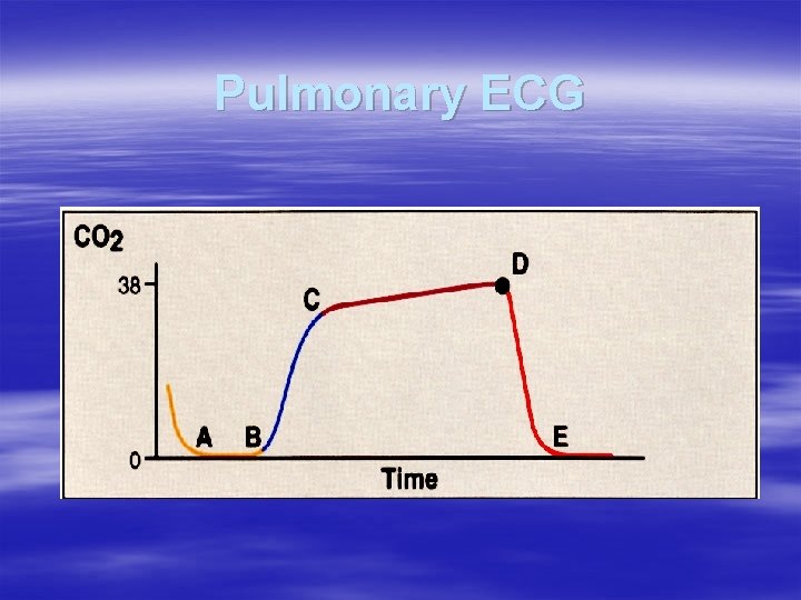
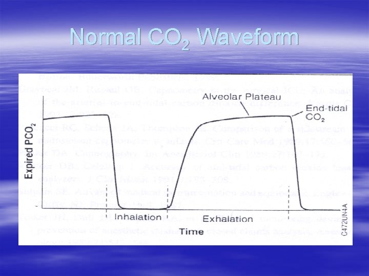
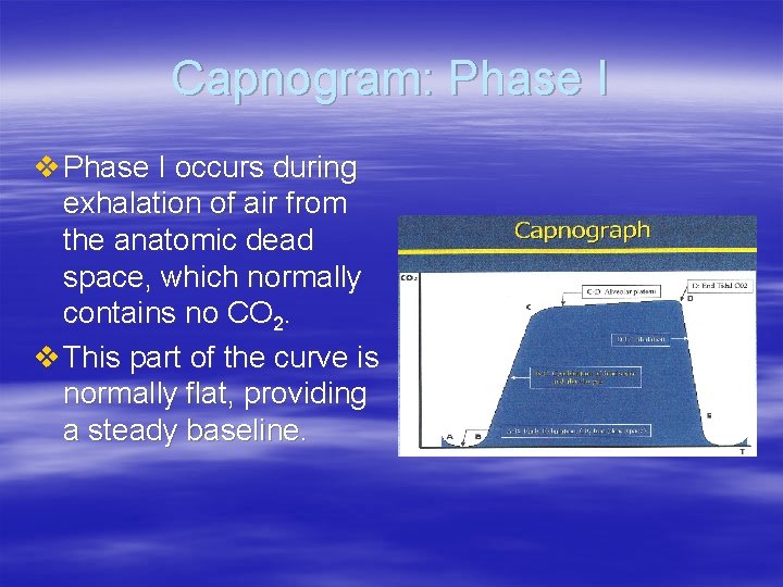
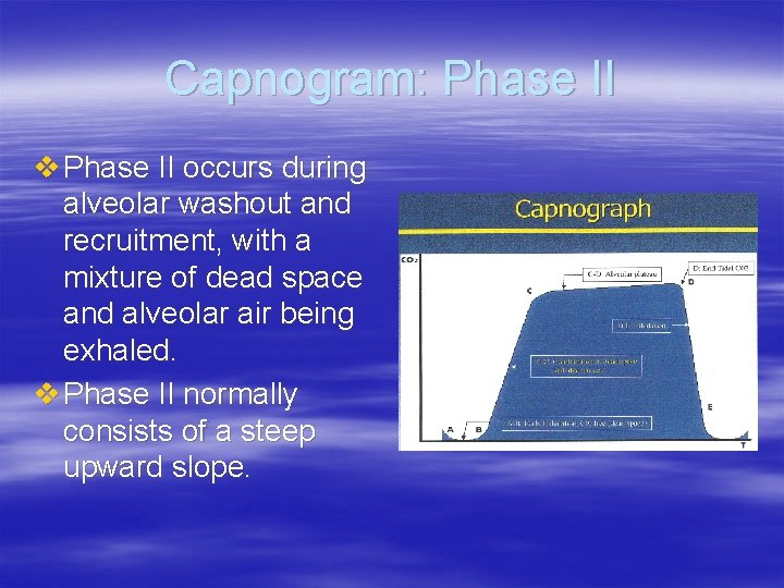
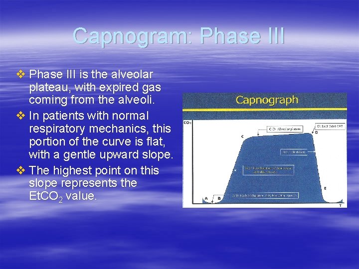
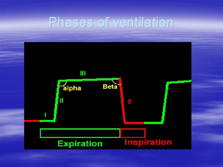
Exhalation v Exhalation can be divided into 3 phases: – Phase 1: Expiration from dead bronchiolar space – no CO 2 yet exhaled – Phase 2: Mixture of dead space air + alveolar CO 2 reaches the mouth § Known as alveolar washout and recruitment – Phase 3: Nearly pure, CO 2 -rich alveolar air is exhaled § Called the alveolar plateau
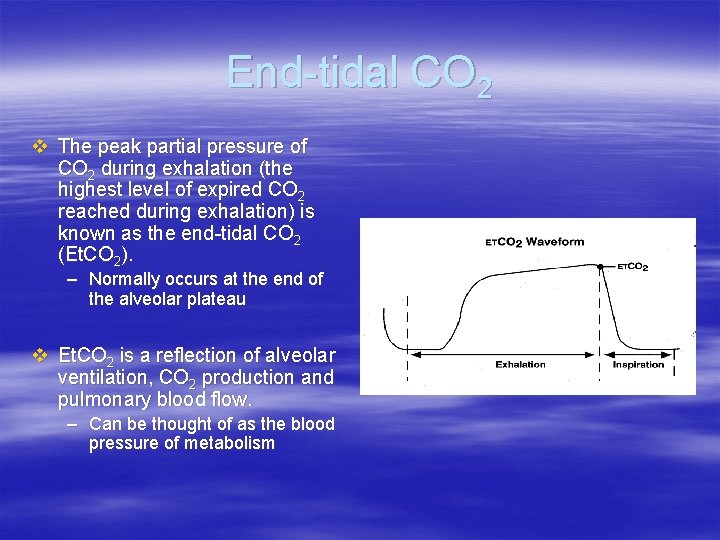
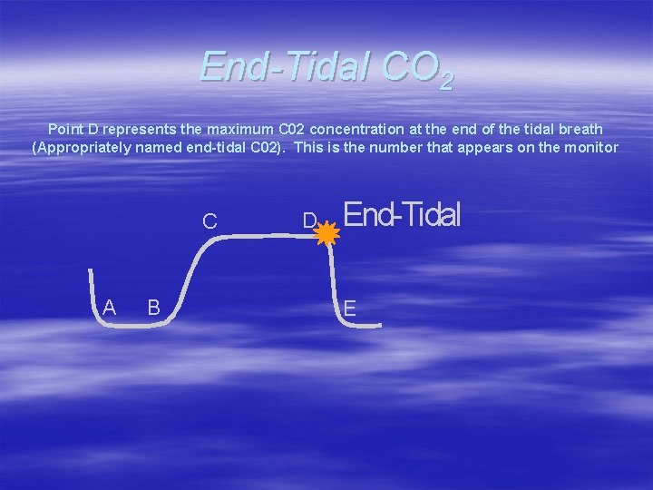
End-tidal CO 2 v The peak partial pressure of CO 2 during exhalation (the highest level of expired CO 2 reached during exhalation) is known as the end-tidal CO 2 (Et. CO 2). – Normally occurs at the end of the alveolar plateau v Et. CO 2 is a reflection of alveolar ventilation, CO 2 production and pulmonary blood flow. – Can be thought of as the blood pressure of metabolism
End-tidal CO 2 v. In healthy people, the Et. CO 2 is within 5 mm. Hg of the partial pressure of CO 2 in arterial blood (Pa. CO 2). – Normal values of both are between 35 -45 mm. Hg. § 4. 5% – 6 %
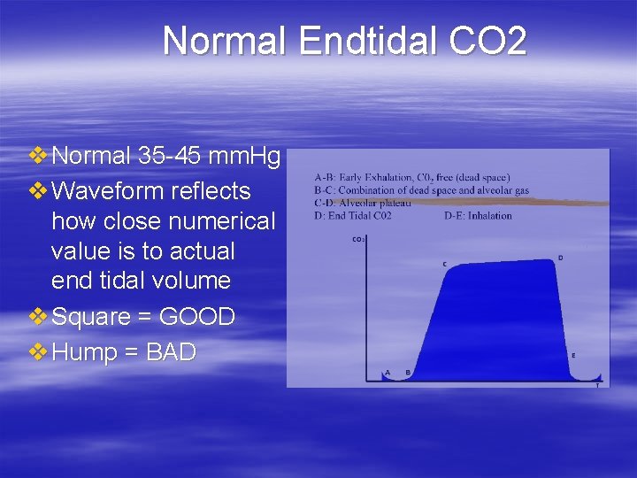
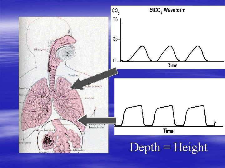
Normal Endtidal CO 2 v Normal 35 -45 mm. Hg v Waveform reflects how close numerical value is to actual end tidal volume v Square = GOOD v Hump = BAD
Depth = Height
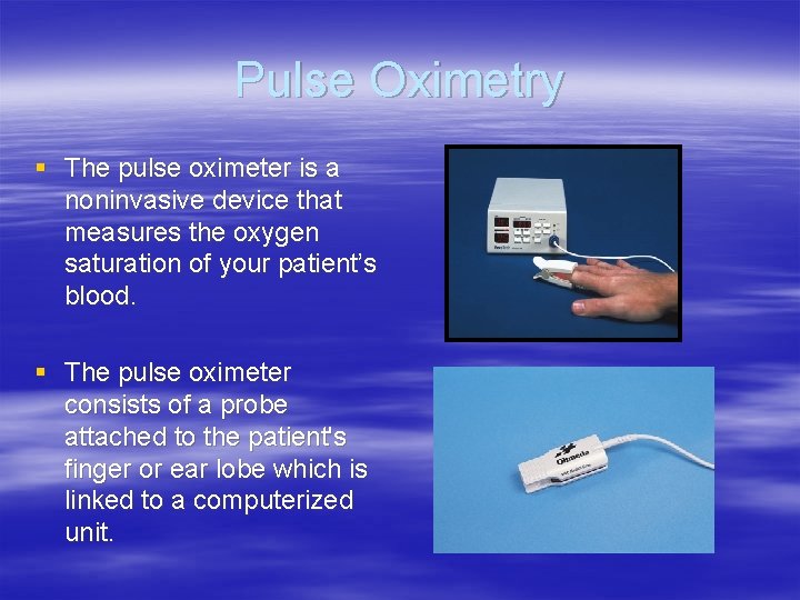
Pulse Oximetry § The pulse oximeter is a noninvasive device that measures the oxygen saturation of your patient’s blood. § The pulse oximeter consists of a probe attached to the patient's finger or ear lobe which is linked to a computerized unit.
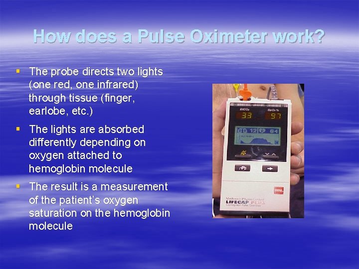
How does a Pulse Oximeter work? § The probe directs two lights (one red, one infrared) through tissue (finger, earlobe, etc. ) § The lights are absorbed differently depending on oxygen attached to hemoglobin molecule § The result is a measurement of the patient’s oxygen saturation on the hemoglobin molecule
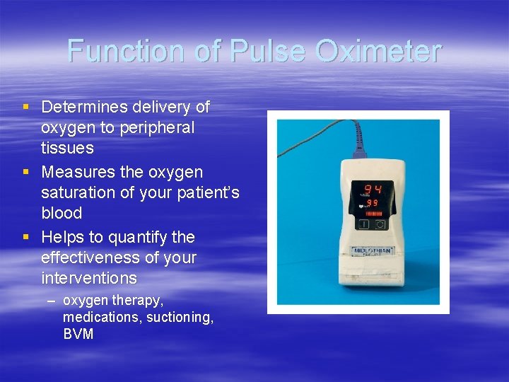
Function of Pulse Oximeter § Determines delivery of oxygen to peripheral tissues § Measures the oxygen saturation of your patient’s blood § Helps to quantify the effectiveness of your interventions – oxygen therapy, medications, suctioning, BVM
Indications § A depressed respiratory drive (e. g. , narcotic overdose) § An increased resistance in the respiratory airways (e. g. , asthma) § A reduced capacity of the blood to transport oxygen (e. g. , shock, anemia) § All patients (additional vital sign)
Sa. O 2 v. Sp. O 2 § Pa. O 2 is actual amount of oxygen dissolved in arterial blood – measured by blood gases – expressed in mm. Hg § Sa. O 2 is percentage of hemoglobin saturated by oxygen – measured by blood gases – expressed in percentage § Sp. O 2 is percentage of hemoglobin saturated by oxygen – measured by pulse oximeter – expressed in percentage
Normal Pulse Oximetry Readings §Normal lab values range between 95 -100% §Readings between 93% and 97% may be normal for some patients (COPD) §Oxygen (at minimum) should be applied for readings below 90%
Pitfalls of the Pulse Oximeter § Certain medical conditions can alter the machines interpretation, and give false readings § Certain environmental conditions can also produce false reading
In the following situations the pulse oximeter readings may not be accurate: § A reduction in peripheral pulsatile blood flow produced by peripheral vasoconstriction (hypovolemia, severe hypotension, cold, cardiac failure, some cardiac arrhythmias) or peripheral vascular disease. § The presence of methemoglobin will prevent the oximeter from working accurately and the readings will tend towards 85%, regardless of the true saturation.
In the following situations the pulse oximeter readings may not be accurate: § Carboxyhemoglobin( hemoglobin combined with carbon monoxide [COHb]) is lumped in with oxyhemoglobin (O 2 Hb), thus producing incorrect readings. § A person could have 15% COHb on board (or more), plus 80% O 2 Hb, but the pulse. Ox reading would still be 95% saturation.
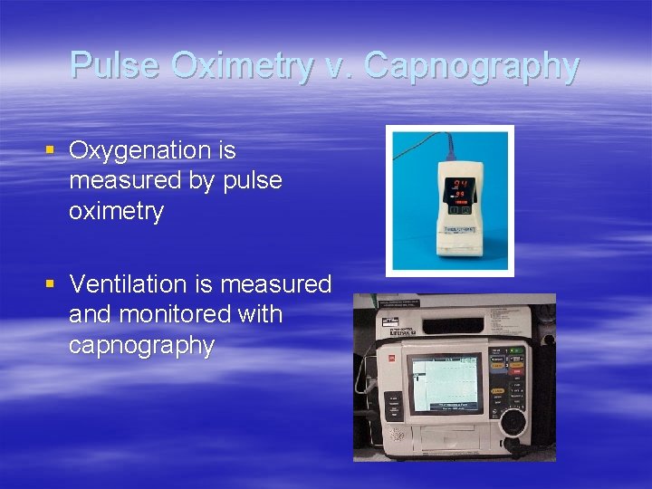
Pulse Oximetry v. Capnography § Oxygenation is measured by pulse oximetry § Ventilation is measured and monitored with capnography
Comparison of Capnograhy and Oximetry v Capnographs and pulse oximeters present different views of the same cardiopulmonary processes. Oximeters measure saturated hemoglobin in peripheral blood and provide additional information about the adequacy of lung perfusion and oxygen delivery to the tissues. – Many sources recommend monitoring both Sp. O 2 and Et. CO 2 on intubated and non-intubated patients. v However, pulse oximetry is a late indicator of O 2 supply, and is less sensitive than capnography. It does not afford a complete picture of ventilatory status.
Comparison v Accurate pulse oximetry measurement is dependent upon adequate peripheral perfusion and may be unreliable in patients who have compromised peripheral circulation. v Capnography continuously and nearly instantaneously measures pulmonary ventilation and is able to rapidly detect small changes in cardio-respiratory function before oximeter readings change. v Healthy patients can maintain Sa. O 2 > 90% for minutes even with inadequate ventilation.
Terminology § Capnos = smoke § Capnometer = number (Et. CO 2) § Capnograph = number + digitized signal
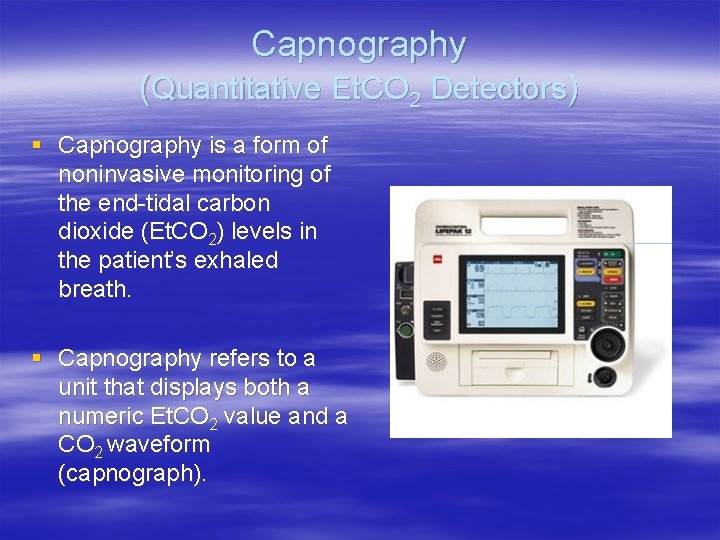
Capnography (Quantitative Et. CO 2 Detectors) § Capnography is a form of noninvasive monitoring of the end-tidal carbon dioxide (Et. CO 2) levels in the patient’s exhaled breath. § Capnography refers to a unit that displays both a numeric Et. CO 2 value and a CO 2 waveform (capnograph).
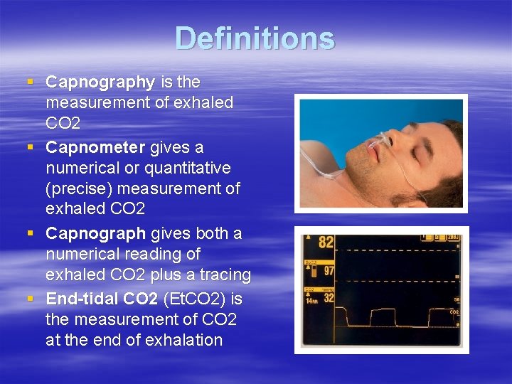
Definitions § Capnography is the measurement of exhaled CO 2 § Capnometer gives a numerical or quantitative (precise) measurement of exhaled CO 2 § Capnograph gives both a numerical reading of exhaled CO 2 plus a tracing § End-tidal CO 2 (Et. CO 2) is the measurement of CO 2 at the end of exhalation
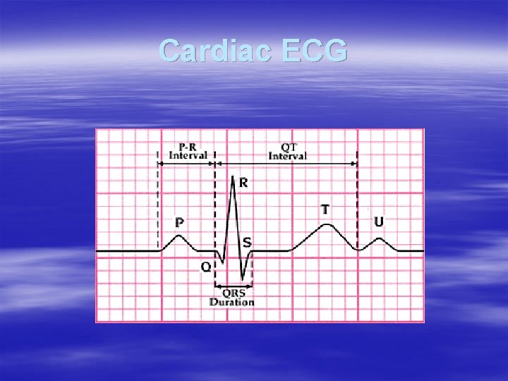
Cardiac ECG
Pulmonary ECG
Oxygenation and Ventilation Oxygen -> lungs -> alveoli -> blood Oxygen breath CO 2 lungs muscles + organs Oxygen CO 2 energy blood CO 2 cells Oxygen + Glucose
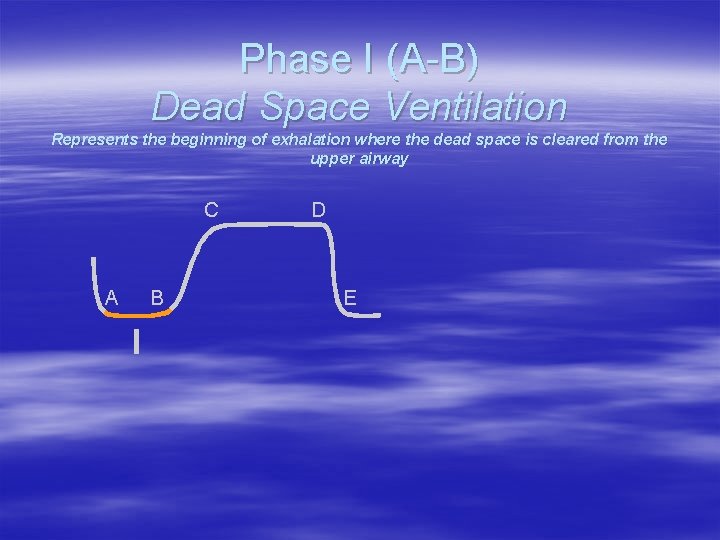
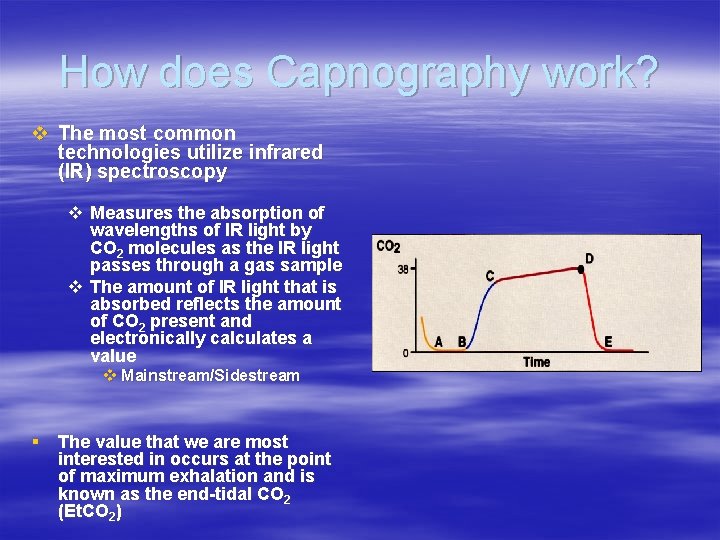
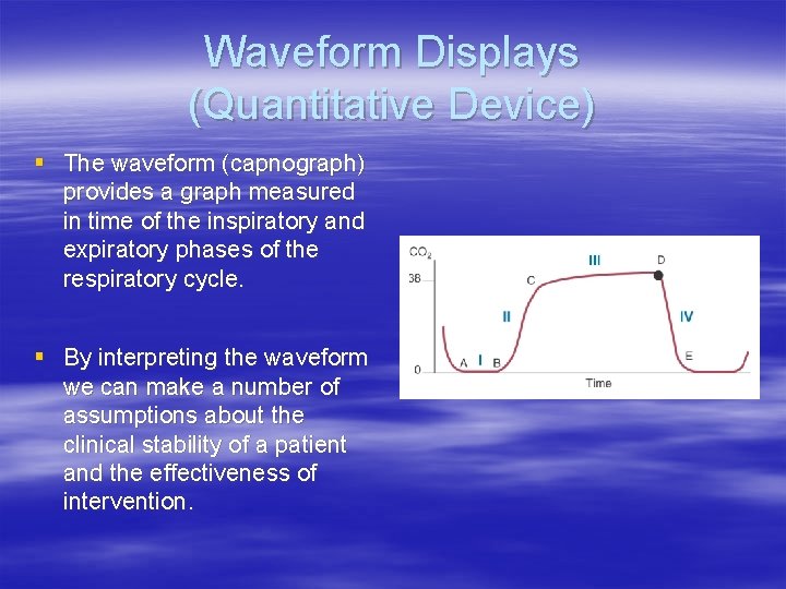
Phase I (A-B) Dead Space Ventilation Represents the beginning of exhalation where the dead space is cleared from the upper airway C A B I D E
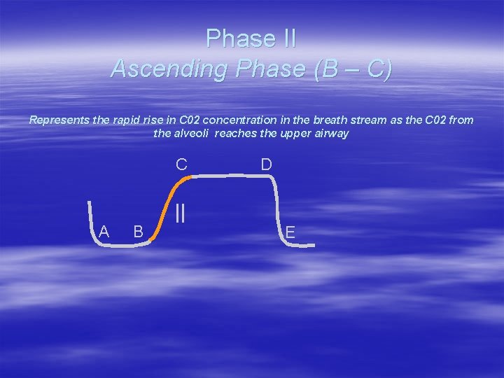
Phase II Ascending Phase (B – C) Represents the rapid rise in C 02 concentration in the breath stream as the C 02 from the alveoli reaches the upper airway C A B II D E
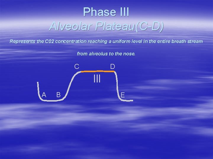
Phase III Alveolar Plateau(C-D) Represents the C 02 concentration reaching a uniform level in the entire breath stream from alveolus to the nose. C D III A B E
End-Tidal CO 2 Point D represents the maximum C 02 concentration at the end of the tidal breath (Appropriately named end-tidal C 02). This is the number that appears on the monitor C A B D End-Tidal E
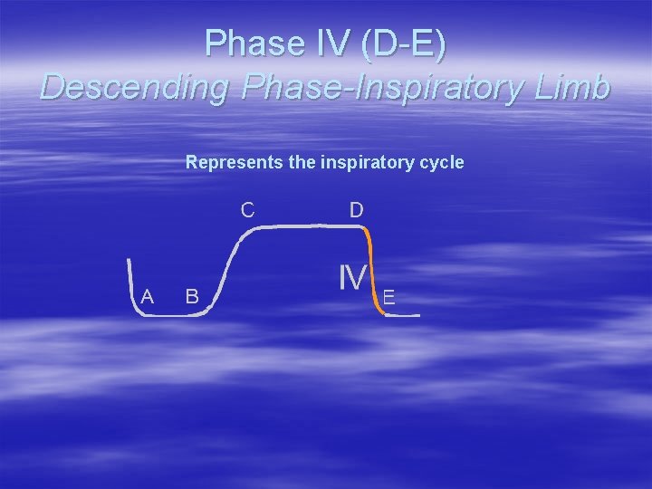
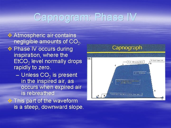
Phase IV (D-E) Descending Phase-Inspiratory Limb Represents the inspiratory cycle
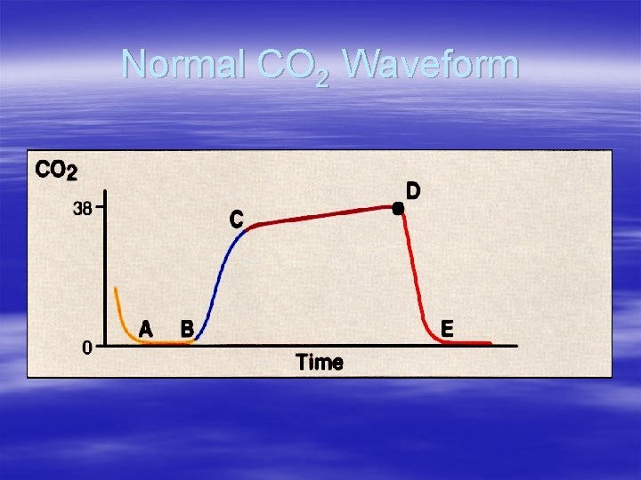
Normal CO 2 Waveform
Normal CO 2 Waveform
How does Capnography work? v The most common technologies utilize infrared (IR) spectroscopy v Measures the absorption of wavelengths of IR light by CO 2 molecules as the IR light passes through a gas sample v The amount of IR light that is absorbed reflects the amount of CO 2 present and electronically calculates a value v Mainstream/Sidestream § The value that we are most interested in occurs at the point of maximum exhalation and is known as the end-tidal CO 2 (Et. CO 2)
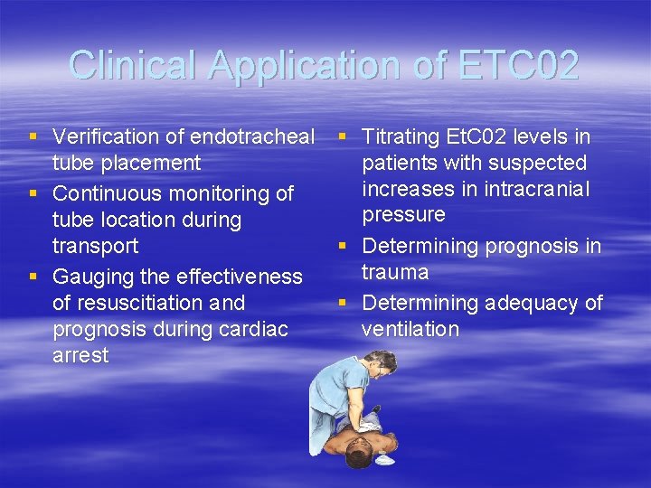
Clinical Application of ETC 02 § Verification of endotracheal § Titrating Et. C 02 levels in tube placement patients with suspected increases in intracranial § Continuous monitoring of pressure tube location during transport § Determining prognosis in trauma § Gauging the effectiveness of resuscitiation and § Determining adequacy of prognosis during cardiac ventilation arrest
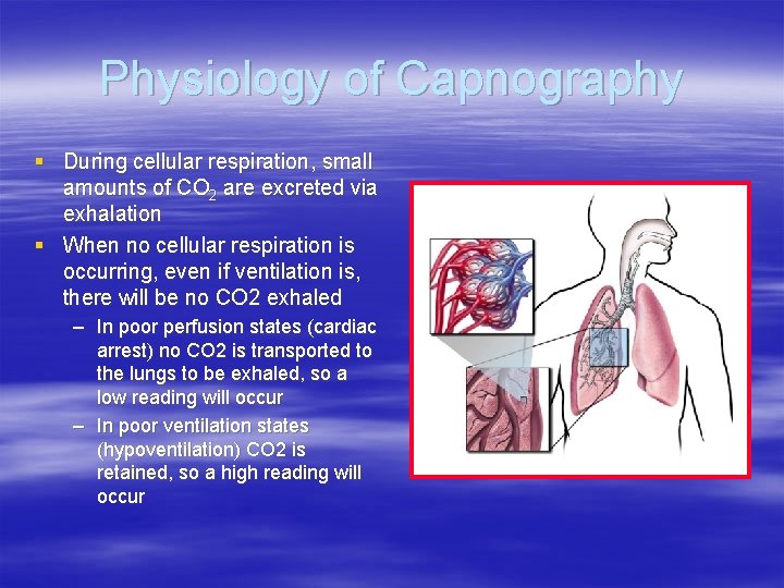
Physiology of Capnography § During cellular respiration, small amounts of CO 2 are excreted via exhalation § When no cellular respiration is occurring, even if ventilation is, there will be no CO 2 exhaled – In poor perfusion states (cardiac arrest) no CO 2 is transported to the lungs to be exhaled, so a low reading will occur – In poor ventilation states (hypoventilation) CO 2 is retained, so a high reading will occur
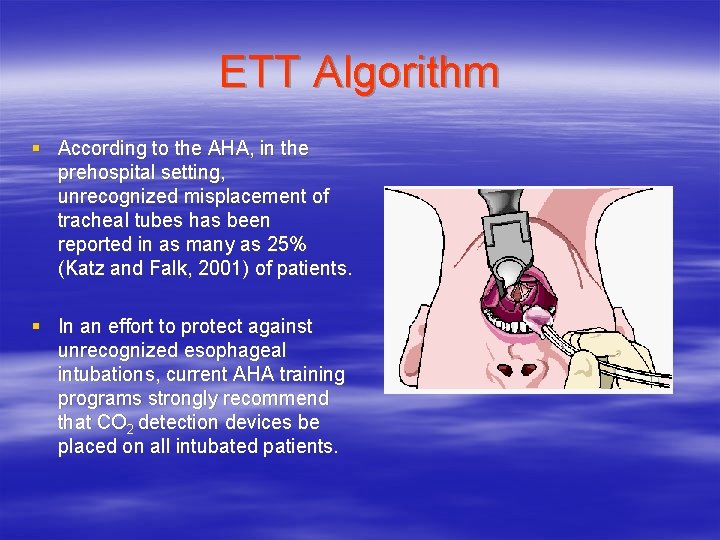
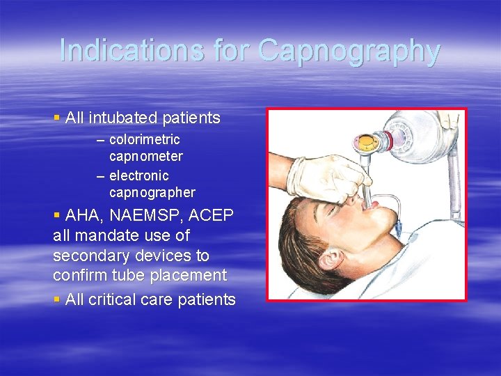
ETT Algorithm § According to the AHA, in the prehospital setting, unrecognized misplacement of tracheal tubes has been reported in as many as 25% (Katz and Falk, 2001) of patients. § In an effort to protect against unrecognized esophageal intubations, current AHA training programs strongly recommend that CO 2 detection devices be placed on all intubated patients.
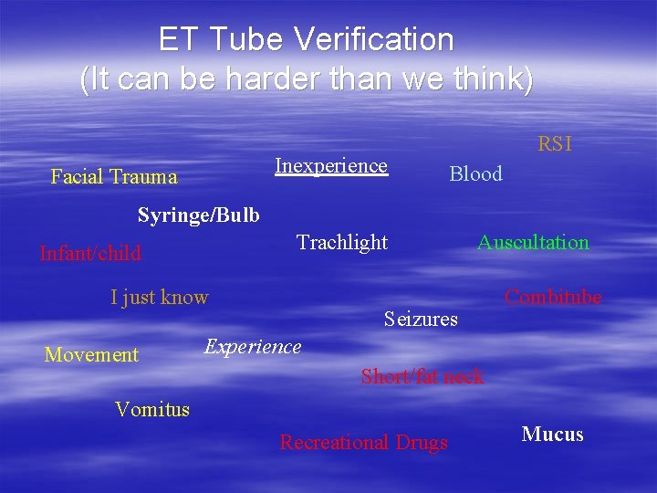
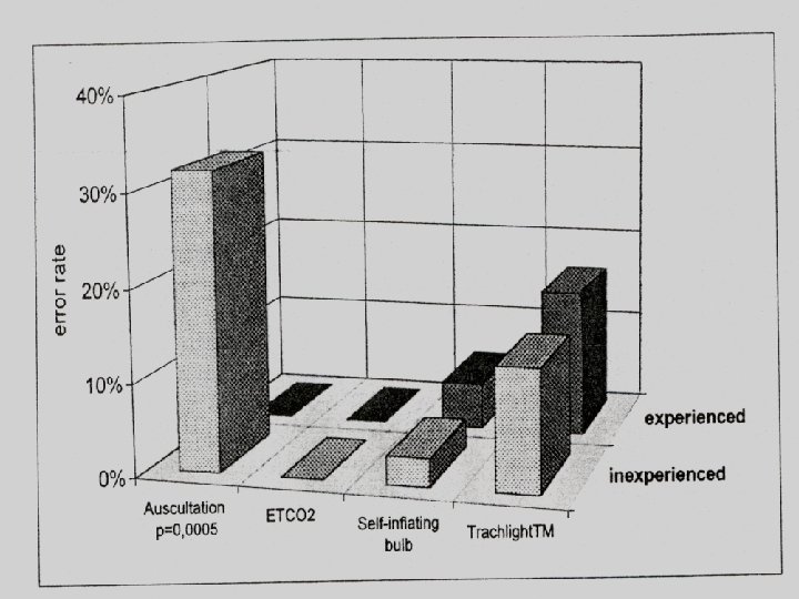
ET Tube Verification (It can be harder than we think) Inexperience Facial Trauma RSI Blood Syringe/Bulb Trachlight Infant/child I just know Movement Auscultation Seizures Combitube Experience Short/fat neck Vomitus Recreational Drugs Mucus
Confirmation of ET Tube Placement v Data confirm that physical assessment procedures to confirm ET tube placement can be misleading. – Movement of air through the esophagus may be difficult to differentiate breath sounds, and may create chest rise. – Breath sounds and/or normal chest wall expansion may be difficult to confirm in victims with traumatic thoracic injury. – Lung sounds can be transmitted to the epigastrium in peds – Misting appears in a high % of esophageally-placed ET tubes. – Difficult to confidently confirm ET tube placement in patients with severe bronchoconstriction. – Unvisualized nasal ET tubes may be difficult to confirm.
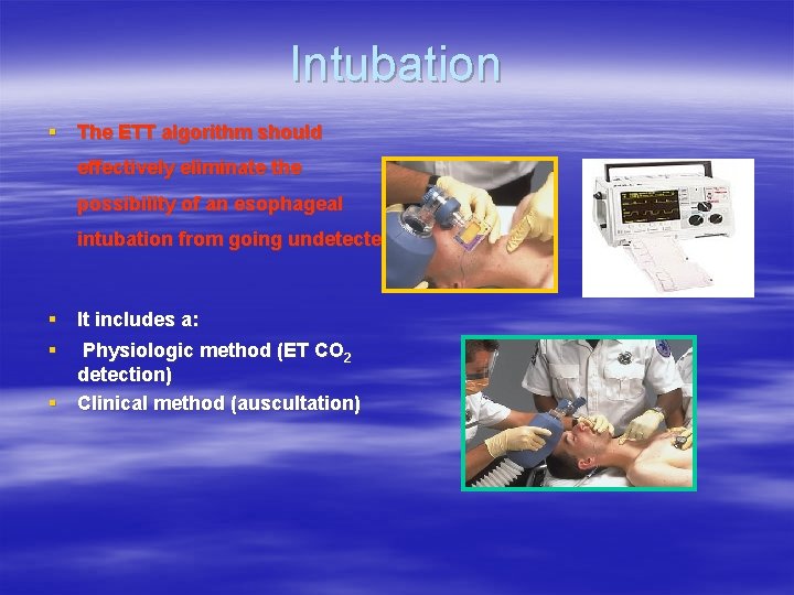
Intubation § The ETT algorithm should effectively eliminate the possibility of an esophageal intubation from going undetected. § It includes a: § Physiologic method (ET CO 2 detection) § Clinical method (auscultation)
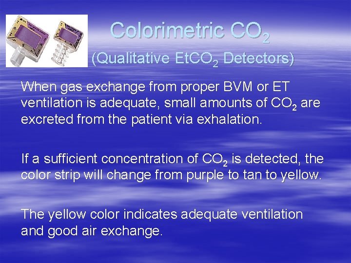
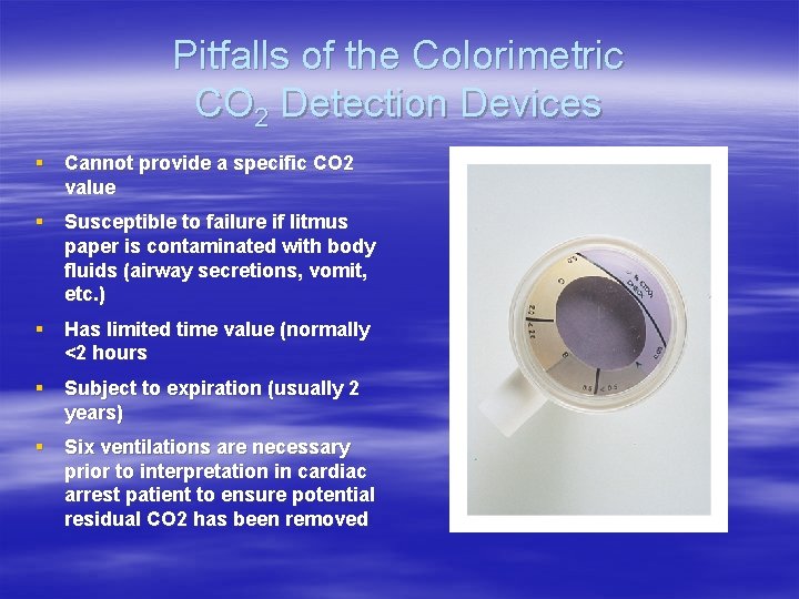
Colorimetric CO 2 (Qualitative Et. CO 2 Detectors) When gas exchange from proper BVM or ET ventilation is adequate, small amounts of CO 2 are excreted from the patient via exhalation. If a sufficient concentration of CO 2 is detected, the color strip will change from purple to tan to yellow. The yellow color indicates adequate ventilation and good air exchange.
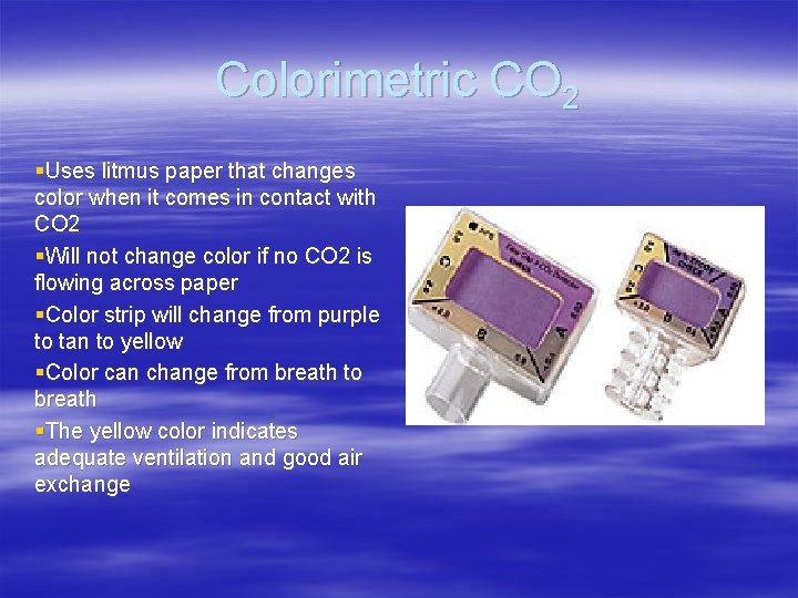
Colorimetric CO 2 §Uses litmus paper that changes color when it comes in contact with CO 2 §Will not change color if no CO 2 is flowing across paper §Color strip will change from purple to tan to yellow §Color can change from breath to breath §The yellow color indicates adequate ventilation and good air exchange
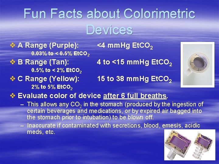
Fun Facts about Colorimetric Devices v A Range (Purple): 0. 03% to < 0. 5% Et. CO 2 v B Range (Tan): 0. 5% to < 2% Et. CO 2 v C Range (Yellow): 2% to 5% Et. CO 2 <4 mm. Hg Et. CO 2 4 to <15 mm. Hg Et. CO 2 15 to 38 mm. Hg Et. CO 2 v Evaluate color of device after 6 full breaths. – This allows any CO 2 in the stomach (produced by the ingestion of certain beverages and medications, or by expired air bagged into the stomach prior to intubation) to be blown off. – Inaccurate if contaminated with secretions, blood, emesis, acidic meds, etc.
Pitfalls of the Colorimetric CO 2 Detection Devices § Cannot provide a specific CO 2 value § Susceptible to failure if litmus paper is contaminated with body fluids (airway secretions, vomit, etc. ) § Has limited time value (normally <2 hours § Subject to expiration (usually 2 years) § Six ventilations are necessary prior to interpretation in cardiac arrest patient to ensure potential residual CO 2 has been removed
Indications for Capnography § All intubated patients – colorimetric capnometer – electronic capnographer § AHA, NAEMSP, ACEP all mandate use of secondary devices to confirm tube placement § All critical care patients
Waveform Displays (Quantitative Device) § The waveform (capnograph) provides a graph measured in time of the inspiratory and expiratory phases of the respiratory cycle. § By interpreting the waveform we can make a number of assumptions about the clinical stability of a patient and the effectiveness of intervention.
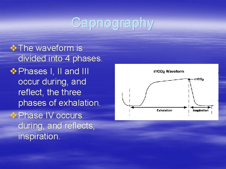
Capnography v The waveform is divided into 4 phases. v Phases I, II and III occur during, and reflect, the three phases of exhalation. v Phase IV occurs during, and reflects, inspiration.
Capnogram: Phase I v Phase I occurs during exhalation of air from the anatomic dead space, which normally contains no CO 2. v This part of the curve is normally flat, providing a steady baseline.
Capnogram: Phase II v Phase II occurs during alveolar washout and recruitment, with a mixture of dead space and alveolar air being exhaled. v Phase II normally consists of a steep upward slope.
Capnogram: Phase III v Phase III is the alveolar plateau, with expired gas coming from the alveoli. v In patients with normal respiratory mechanics, this portion of the curve is flat, with a gentle upward slope. v The highest point on this slope represents the Et. CO 2 value.
Capnogram: Phase IV v Atmospheric air contains negligible amounts of CO 2. v Phase IV occurs during inspiration, where the Et. CO 2 level normally drops rapidly to zero. – Unless CO 2 is present in the inspired air, as occurs when expired air is rebreathed v This part of the waveform is a steep, downward slope.
Phases of ventilation
Normal capnography
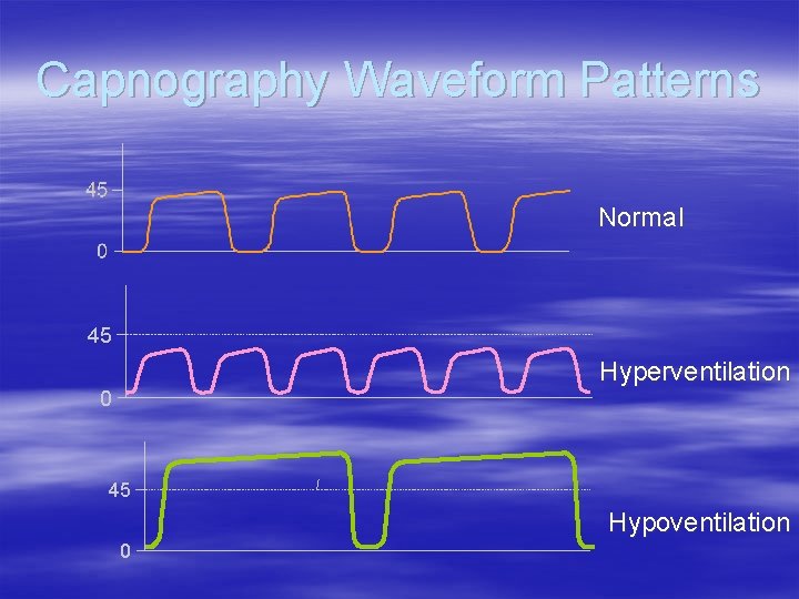
Capnography Waveform Patterns Normal 45 Hyperventilation 0 45 Hypoventilation 0
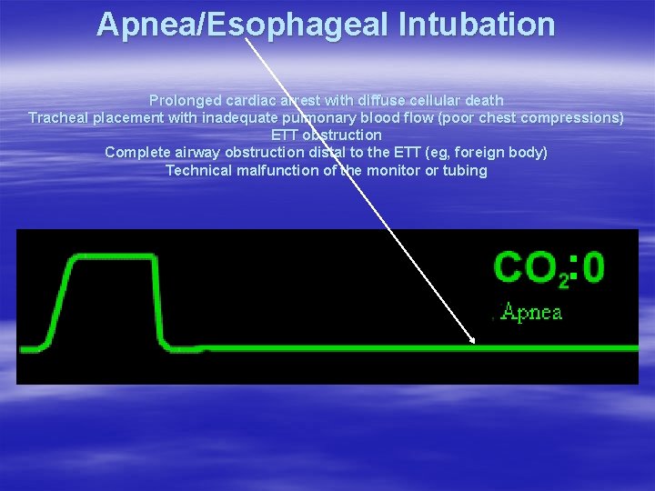
Apnea/Esophageal Intubation Prolonged cardiac arrest with diffuse cellular death Tracheal placement with inadequate pulmonary blood flow (poor chest compressions) ETT obstruction Complete airway obstruction distal to the ETT (eg, foreign body) Technical malfunction of the monitor or tubing
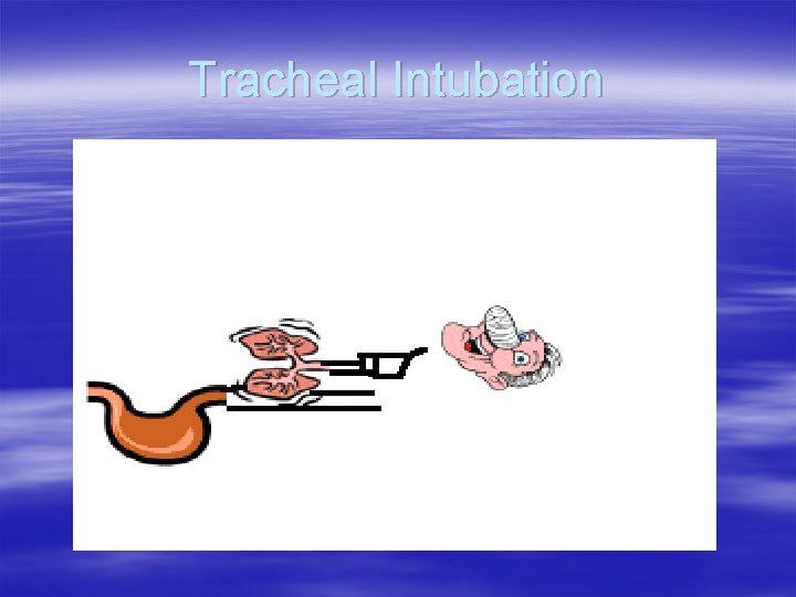
Tracheal Intubation
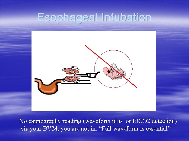
Esophageal Intubation No capnography reading (waveform plus or Et. CO 2 detection) via your BVM, you are not in. “Full waveform is essential”
Unsuccessful Intubation
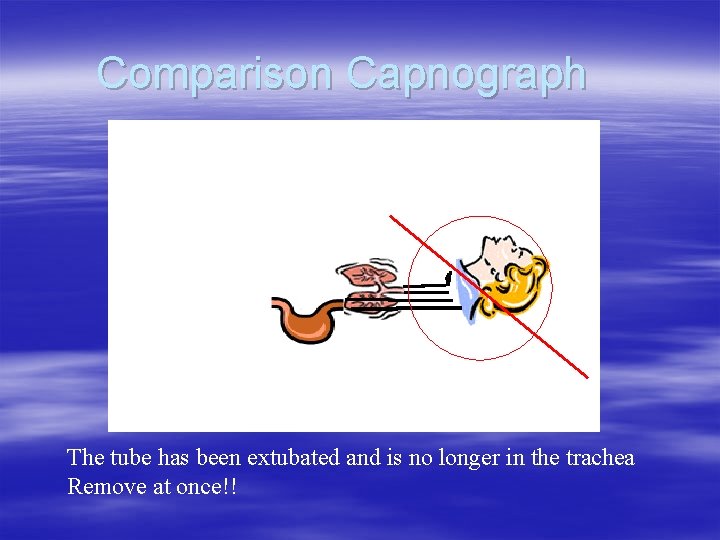
Comparison Capnograph The tube has been extubated and is no longer in the trachea Remove at once!!
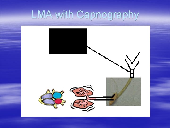
LMA with Capnography
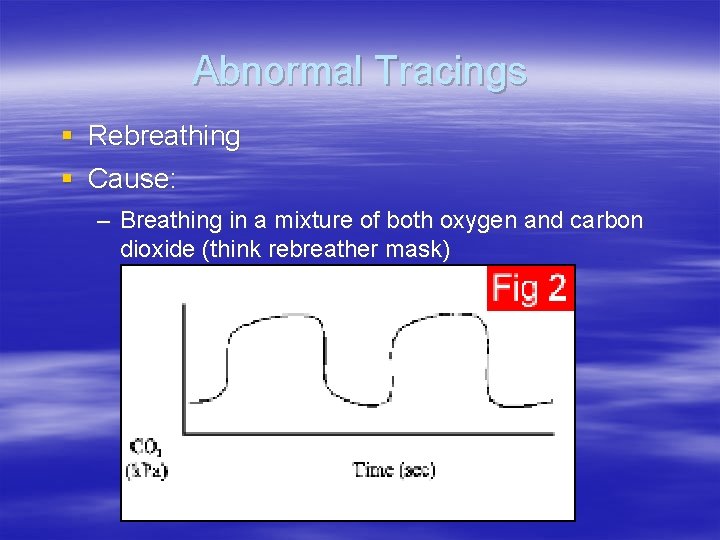
Abnormal Tracings § Rebreathing § Cause: – Breathing in a mixture of both oxygen and carbon dioxide (think rebreather mask)
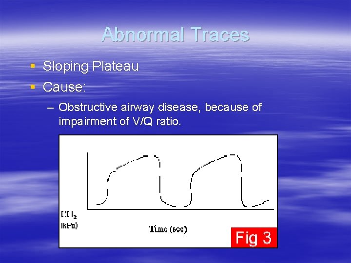
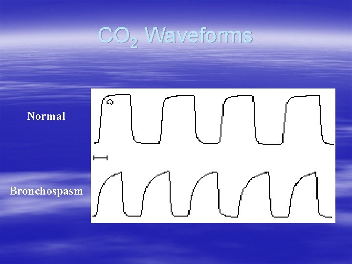
Abnormal Traces § Sloping Plateau § Cause: – Obstructive airway disease, because of impairment of V/Q ratio.
CO 2 Waveforms Normal Bronchospasm
Trending is Key to monitoring Respiratory Failure v Acute exacerbation of COPD v Asthma v Pneumonia v CHF v Respiratory Muscle Fatigue v Hypoventilation Syndromes v IS THE PATIENT RESPONDING TO THERAPY OR NOT?
Trending v Trending of capnography provides a continuous view of the patient’s ventilatory status v Early detection allows for early intervention v Trending is a simple tool that does not stress an already failing system Video versus Snapshot
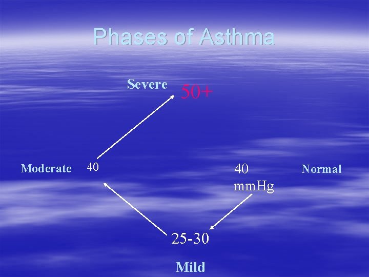
Phases of Asthma § Hyperventilation – mild § Tiring – moderate § Tired – severe
Phases of Asthma Severe Moderate 50+ 40 40 mm. Hg 25 -30 Mild Normal
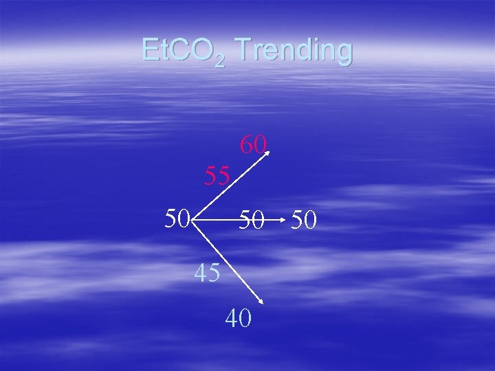
Et. CO 2 Trending 60 55 50 50 50 45 40
Using Capnography in Asthma § Diagnoses presence of bronchospasm – Waveform § Assesses severity of Asthma – Et. CO 2 trends § Gauges response to treatment – Et. CO 2 trends
COPD § Baseline CO 2 is higher – > 50 mm Hg § Follow CO 2 trends to: – Establish baseline – Track response to treatment
COPD § Determine who is a CO 2 retainer – Et. CO 2 trends § COPD vs. CHF – Waveform
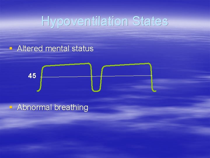
Hypoventilation States § Altered mental status 45 § Abnormal breathing
Hypoventilation
Hypoventilation States § Sedation § Analgesia § ETOH intoxication § Drug Ingestion § Postictal states § Head trauma § Meningitis § Encephalitis
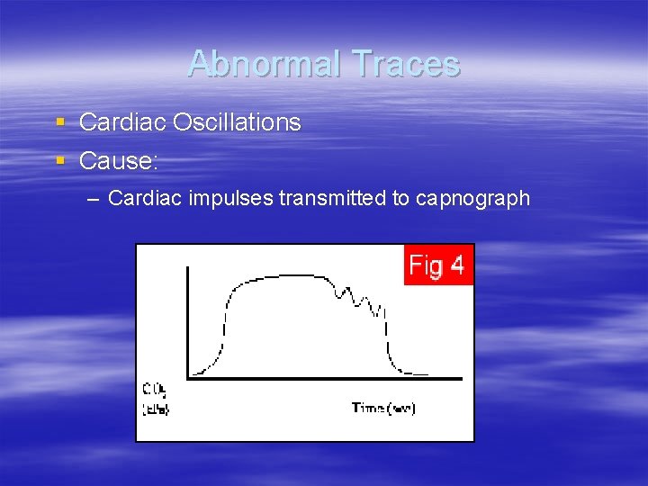
Abnormal Traces § Cardiac Oscillations § Cause: – Cardiac impulses transmitted to capnograph
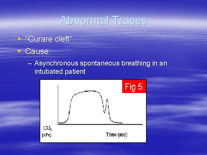
Abnormal Traces § “Curare cleft” § Cause: – Asynchronous spontaneous breathing in an intubated patient
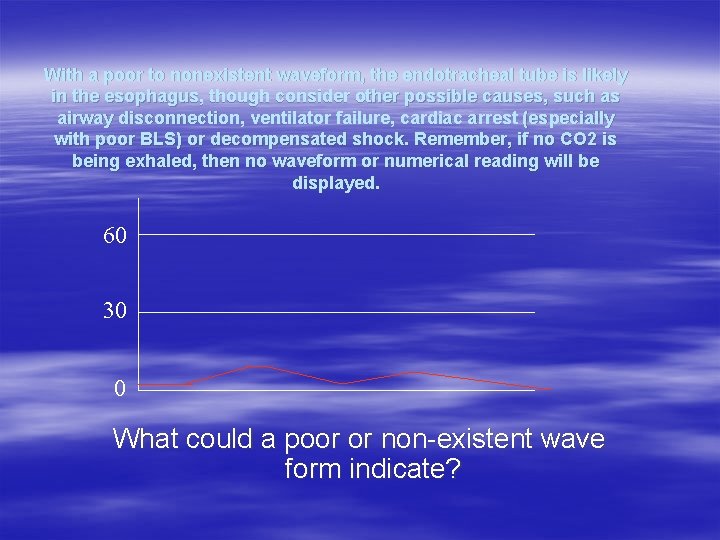
With a poor to nonexistent waveform, the endotracheal tube is likely in the esophagus, though consider other possible causes, such as airway disconnection, ventilator failure, cardiac arrest (especially with poor BLS) or decompensated shock. Remember, if no CO 2 is being exhaled, then no waveform or numerical reading will be displayed. 60 30 0 What could a poor or non-existent wave form indicate?
Case Study #1 v 32 yo 90 kg female presents in acute respiratory distress v Cyanotic, short word sentencing v Respiratory rate is shallow and labored at 24 v Expiratory phase is prolonged due to gas trapping v Heart rate of 140, strong and bounding at the radial v BP 170/88
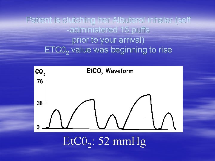
Patient is clutching her Albuterol inhaler (self -administered 15 puffs prior to your arrival) ETC 02 value was beginning to rise Et. C 02: 52 mm. Hg
Treatment v BLS assisted ventilations via Bag Valve Mask with 100% O 2 v Epinephrine 1: 1000 SQ admin v Paramedic prepares for nasal intubation
Patient Arrests v. As the nasal tube is being advanced, patient arrests v. Paramedic pushes the tube in anyway, but it is found to be in the esophagus v. Tube immediately withdrawn
Tube Confirmation v Cords visualized during intubation v Patient is successfully intubated orally v Auscultation reveals no discernible breath sounds anterior chest wall v Abdominal auscultation is equally silent v Minimal chest rise v Positive misting and condensation in the tube v Bulb aspirated syringe flows free-air
What is the next step? v. Auscultation doesn't really help v. Sp. O 2 is not reading (before or after) v. Remains difficult to ventilate – What other tool can you use to confirm the placement of the tube? – What else could be going on with this patient? – Are we 100% sure we are in the trachea?
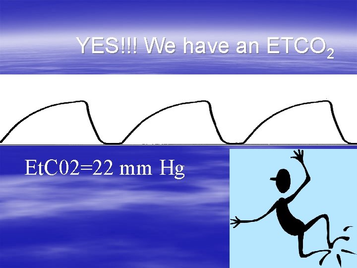
YES!!! We have an ETCO 2 Et. C 02=22 mm Hg
Case Study #2: Cardiac Arrest 42 year old 90 kg, male, involved in motorcycle vs. truck MVA. The patient is pinned under vehicle on highway approx. 8 min from local hospital • Rapid extrication performed from underneath truck • Pt. is unresponsive with weak, irregular carotid at 150 bpm, and agonal respirations weak carotid • Patient becomes pulseless and apneic, BLS/CPR is initiated
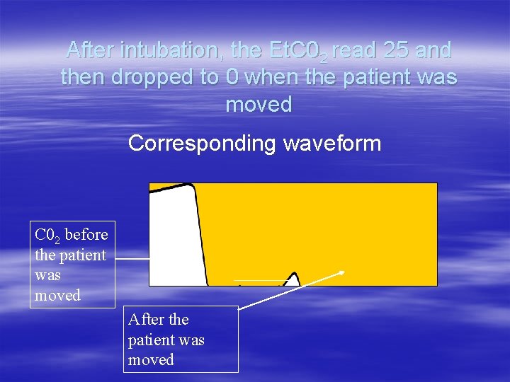
After intubation, the Et. C 02 read 25 and then dropped to 0 when the patient was moved Corresponding waveform C 02 before the patient was moved After the patient was moved
Where is the tube? ? v. Provider checked the number at the lip – It had not changed v. Listened to Breath Sounds – They were less audible v. The Et. C 02 was 25, then dropped to 0 – Normal Et. C 02 is between 35 to 45 mm Hg
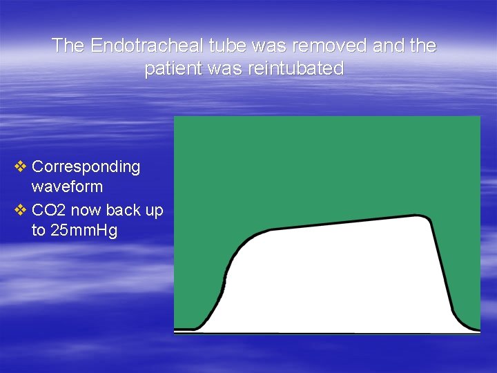
The Endotracheal tube was removed and the patient was reintubated v Corresponding waveform v CO 2 now back up to 25 mm. Hg
Case Study #3 v. A 12 year old boy presented in acute respiratory failure with copious secretions and was successfully orally intubated and placed on a ventilator. v. The patient remained obtunded, cyanotic and had little airway movement on auscultation.
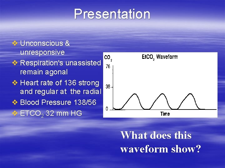
Presentation v Unconscious & unresponsive v Respiration's unassisted remain agonal v Heart rate of 136 strong and regular at the radial v Blood Pressure 138/56 v ETCO 2 32 mm HG What does this waveform show?
A slow upstroke and incomplete emptying. What could you do therapeutically? Answer:
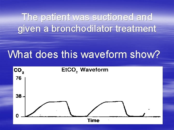
The patient was suctioned and given a bronchodilator treatment What does this waveform show?
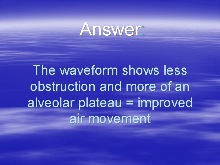
Answer: The waveform shows less obstruction and more of an alveolar plateau = improved air movement
Case Study # 4 70 year old 60 kg female, status post cardiac arrest resuscitation by EMS She was resuscitated in the field and now being transported to hospital. Enroute Pt. is unconscious & unresponsive (orally intubated and on a vent) Vital Signs No spontaneous respirations BP 76/palpated Heart rate is palpable only at the carotid at 136 weak and irregular
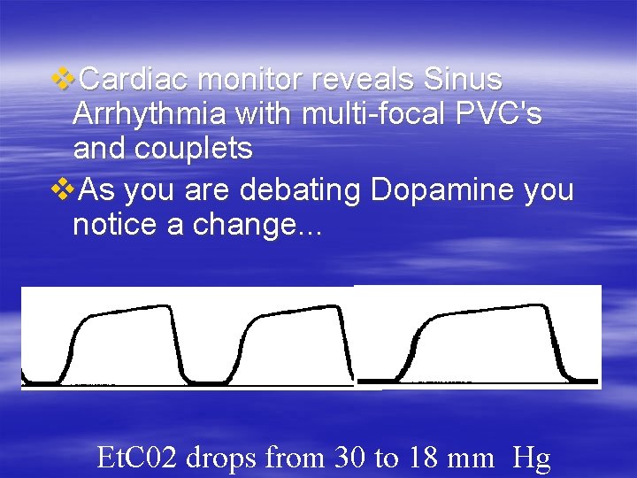
v. Cardiac monitor reveals Sinus Arrhythmia with multi-focal PVC's and couplets v. As you are debating Dopamine you notice a change. . . Et. C 02 drops from 30 to 18 mm Hg
Patient’s perfusion decreases more…………. CO 2 drops further Now unable to palpate a carotid pulse CPR is Initiated
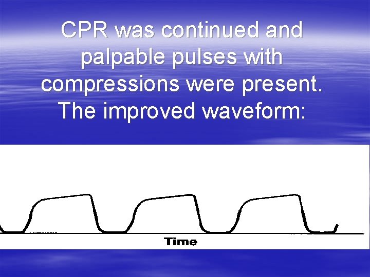
CPR was continued and palpable pulses with compressions were present. The improved waveform:
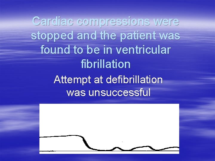
Cardiac compressions were stopped and the patient was found to be in ventricular fibrillation Attempt at defibrillation was unsuccessful
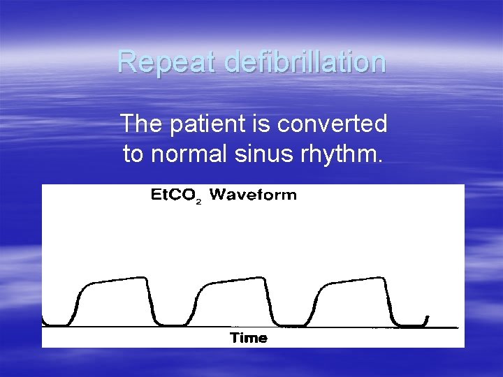
Repeat defibrillation The patient is converted to normal sinus rhythm.
Simple use of Et. C 02: Capnography can be a useful tool in determining the effectiveness of pulmonary perfusion during a cardiac arrest.
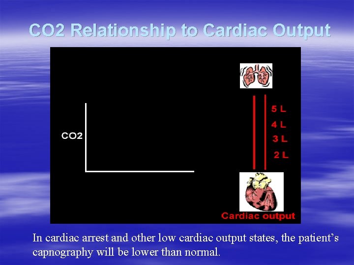
CO 2 Relationship to Cardiac Output In cardiac arrest and other low cardiac output states, the patient’s capnography will be lower than normal.
Evaluation of Efficacy of CPR v Patients in cardiopulmonary arrest produce no Et. CO 2. v During CPR (given a normal blood volume), effective chest compressions circulate enough blood to return CO 2 from the tissue cells to the pulmonary circuit. Combined with effective ventilations, providers will be see improved Et. CO 2 values – most likely lowerthan-normal levels. – “Et. CO 2 concentration varies directly with pulmonary and systemic blood flows under conditions of constant minute ventilation. This relationship holds true even during extremely low blood flow rates. ” Ann Emer Med, 3/1994, 23: 3, p. 571
ETCO 2 is also an indication of successful resuscitation v. Sanders et al, 1989 JAMA noted a threshold for survival of ETCO 2 >10 mmhg v. Successful = 15 (+/-) 4 v. Unsuccessful = 7 (=/-) 5
Cardiac Arrest/Successful Resuscitation
Predicting ROSC in Arrest v Studies have shown a correlation with Et. CO 2 levels during a code and ROSC. Annals of Emerg Med, June, 1995, 25: 6, pages 756 -761 v Et. CO 2 values can predict non-resuscitatable patients. v We may see the development of protocols utilizing Et. CO 2 values in conjunction with the terminating rhythm to determine calling codes in the field. – Most sources cite an Et. CO 2 value of 10 mm. Hg or less as an appropriate and predictive threshold. § This is a terminating – not initial – value, measured after 20 minutes of standard ACLS interventions.
Early Detection of ROSC v If the patient in cardiac arrest has a return of spontaneous circulation, Et. CO 2 levels will rise quickly to a higher level. v Et. CO 2 detection may be the earliest indicator of this improvement in perfusion, and should be confirmed by palpating for central and peripheral pulses. – One study showed a marked rise in Et. CO 2 levels just before conversion of PEA to a perfusing rhythm. § Before any return of measurable BP or palpable pulses! Annals of Emergency Medicine, June, 1995, 25: 6, page 762767
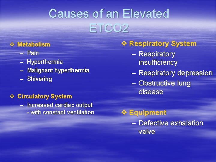
Causes of an Elevated ETCO 2 v Metabolism – Pain – Hyperthermia – Malignant hyperthermia – Shivering v Circulatory System – Increased cardiac output - with constant ventilation v Respiratory System – Respiratory insufficiency – Respiratory depression – Obstructive lung disease v Equipment – Defective exhalation valve
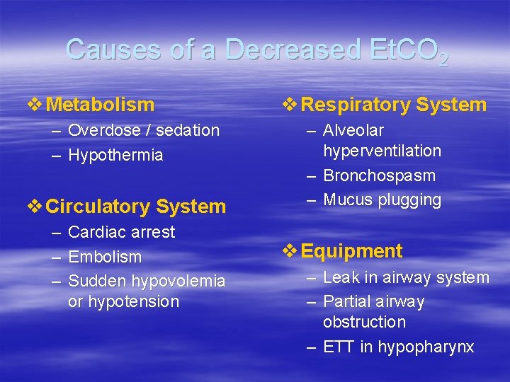
Causes of a Decreased Et. CO 2 v Metabolism – Overdose / sedation – Hypothermia v Circulatory System – Cardiac arrest – Embolism – Sudden hypovolemia or hypotension v Respiratory System – Alveolar hyperventilation – Bronchospasm – Mucus plugging v Equipment – Leak in airway system – Partial airway obstruction – ETT in hypopharynx
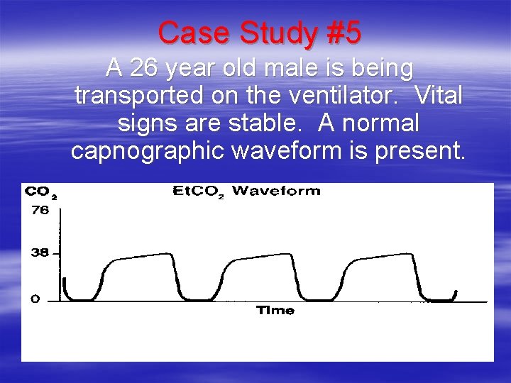
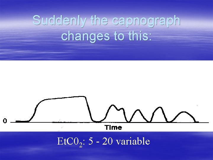
Case Study #5 A 26 year old male is being transported on the ventilator. Vital signs are stable. A normal capnographic waveform is present.
Suddenly the capnograph changes to this: Et. C 02: 5 - 20 variable
What does this mean? What should you check?
Check the patient first Then check the connections between the ETT and breathing circuit And what was found……?
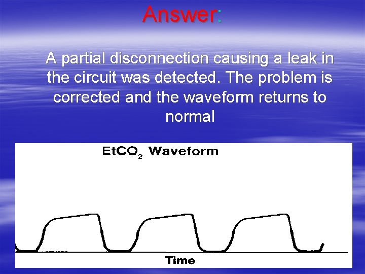
Answer: A partial disconnection causing a leak in the circuit was detected. The problem is corrected and the waveform returns to normal
Summary Points § Detection device -------> Diagnostic monitoring § Static -------------> Dynamic monitoring § Advanced warning of ventilatory status – Don’t be caught off guard – Avoid backing into a critical situation – Crash---------->elective § Objective confirmation of clinical assessment
Thank you!