Pulp Zones of the pulp Odontoblastic zone Cellfree

Pulp

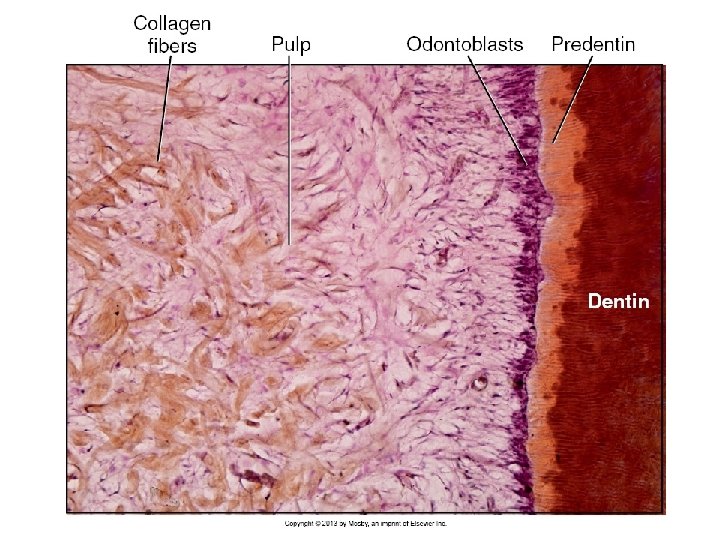
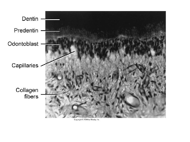
Zones of the pulp • Odontoblastic zone • Cell-free zone of Weil – More prominent in the coronal pulp • Cell-rich zone – More prominent in the coronal pulp • Pulp core

Cells • Odontoblasts • Fibroblasts • Undifferentiated ectomesenchymal cells – Cell-rich zone and core • Inflammatory cells – Macrophages – Lymphocytes • T>B – Dendritic cells • Immuno surveillance

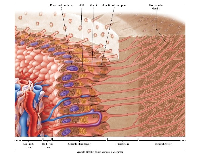
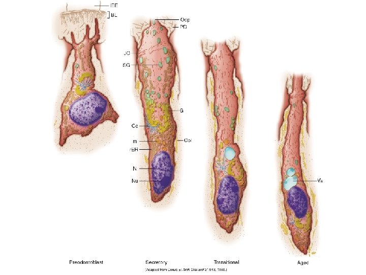
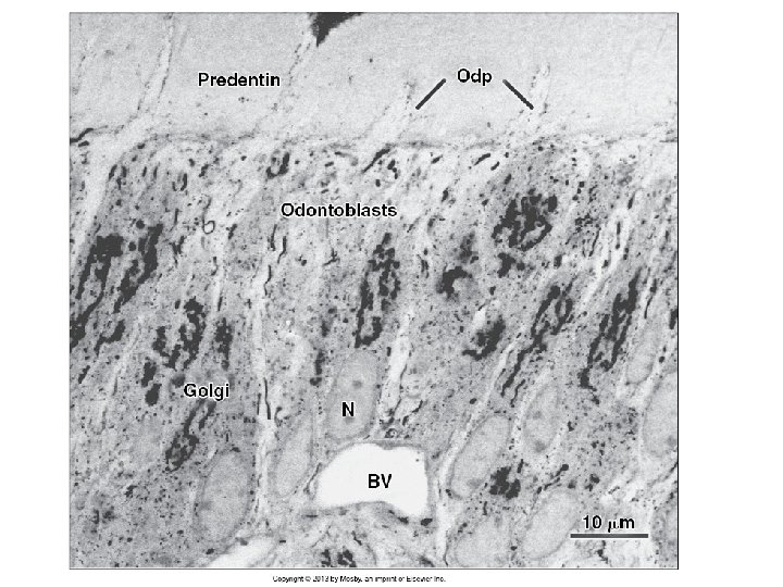
Odontoblasts • Most distinctive cells • End cells: The life span of an odontoblast is generally believed to equal that of the viable tooth • Periphery of the pulp • Palisading pattern • Pseudostratified arrangement – Centripetal arrangement; tangential sectioning (2 D) • # of odontoblasts = # of dentinal tubules • #Coronal cells > #Root cells • Coronal columnar; mid root more cuboidal; apical flat • Active cells - Resting cells - Transitional cells
 and desmosomes • Form collagen similar to fibroblasts")
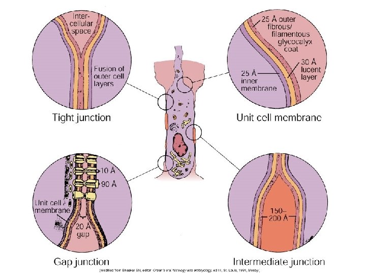
Odontoblasts • Junctions (gap, tight, adherent) and desmosomes • Form collagen similar to fibroblasts • Specialized secretory granules for collagen • Non-collagenous proteins – Different cytoplasmic secretory granules • The odontoblastic process starts as it enters predentin – Devoid of major organelles; has microtubules and filaments – Pinocytotic (specialized fluid endocytosis) activity

Question • The odontoblast needs the ameloblast to differentiate and function. • When the ameloblast is gone how do new odontoblasts develop? What about odontoblasts in the developing root of the tooth as cementum is building?
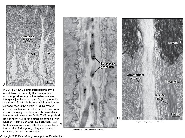

The odontoblastic process • They probably run throughout the length of the dentinal tubule

Tubules • So-called dentinal fluid? Does it really exist? – Proteoglycans – Albumin (water-soluble, carrier) – Tenascin (formative glycoprotein) – Fibronectin (binding protein) – Transferrin (iron delivery)

Fibroblasts • • Most numerous Cell rich zone and core Activity corresponds to shape Producing and absorbing capabilities

Undifferentiated ectomesenchymal cells Represent pool from which connective tissue cells of the pulp are derived Depending on the stimulus, these cells may give rise either odontoblasts or fibroblasts Older pulps: less # of undifferentiated mesenchymal cells thus reducing regenerative potential of the pulp Dental pulp stem cells Mesenchymal stem cells isolated from both deciduous and permanent teeth Self-renewal capacity Can differentiate into odontoblasts, chondrocytes, adipocytes, neurons and osteoblasts

Matrix and ground substance • Type I and III • Ground substance – Glycosaminoglycans • Polysaccharides with repeating disaccharide units – Glycoproteins • Oligosaccharides – Water

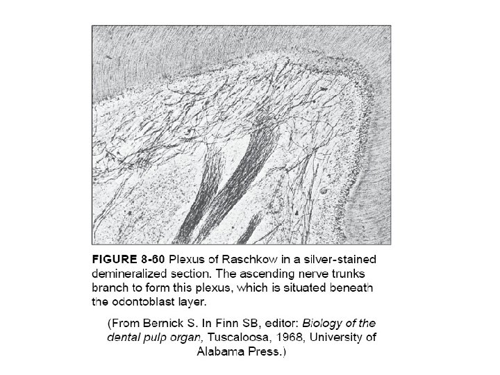
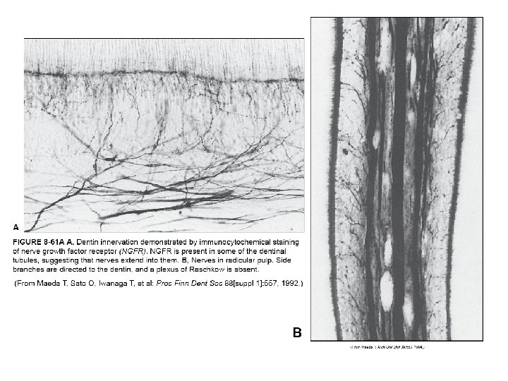
Vascular and nerve supply • Arterioles, venules – – – Extensive supply in the subodontoblastic layer Pericytes Arteriovenous anastomoses Sympathetic nerve supply to the smooth muscle wall of arterioles Afferent free nerve endings • Lymphatics • Nerves – – Cell-free zone (Subodontoblastic plexus of Raschkow) Sensory afferent (V) and sympathetic (sup. cervical ganglion) Unmyelineated Some nerve endingd go into dentinal tubules (minority of tubules containg nerves that occasionally form little branches) – No synapsis between process and nerves
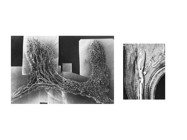

Vascular injection to illustrate blood vessel organization in pulp and periodontium. Larger vessels centrally, and smaller capillaries are in the peripheral pulp.
 • Numerous")
Pulp sensation • Dentin Sensitivity = pain (but also thermal, mechanical, tactile) • Numerous stimuli related to clinical dentistry • Histamine and bradykinine DO NOT produce pain in dentin • 3 mechanisms

Theories • First: no frank evidence • Second: Favored, abandoned, considered – Odontoblast is a neural crest derivative and it can transduce or propagate an impulse • Third: Movement of fluid affecting nerve endings in the plexus of Raschkow

Dentin Sensitivity Nerve response when stimulated Coupled to nerves in pulp

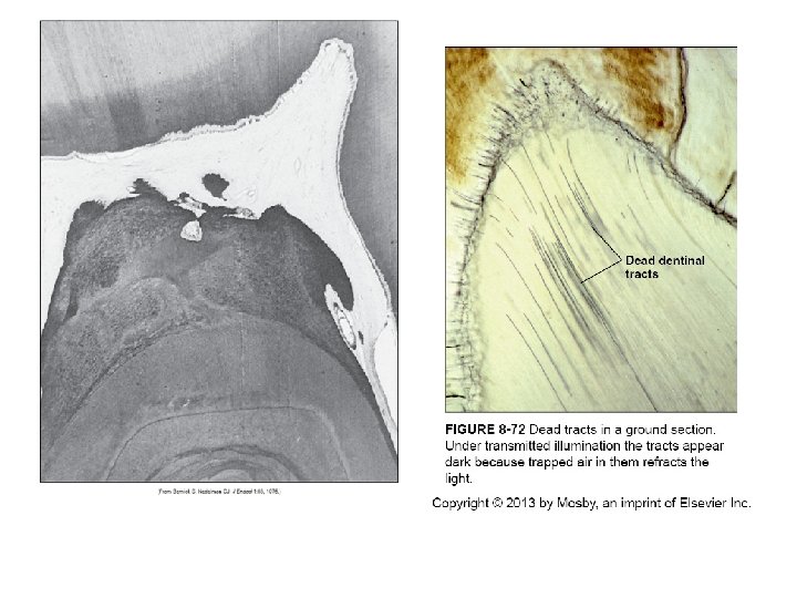
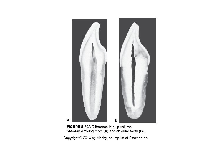
Age changes • • • Decrease in volume Decrease in cell number Dystrophic calcifications Sclerotic dentin Dead tracts of dentin

Pulp stones • Pulp stones or denticles – True or false • Free or attached
- Slides: 30