Pulp cavities of permanent teeth Tooth structure Tooth


- Slides: 43
Pulp cavities of permanent teeth
Tooth structure Tooth consist of 2 parts: • crown • root Crown: is the visible part Root: the part that is embedded in the jaw (not visible)
Tooth structure • Enamel: The hardest, white outer part of the tooth
dentin • Dentin: A layer underlying the enamel. It is a hard tissue • Forms most of tooth structure
Tooth structure • Cementum: A layer of connective tissue that binds the roots of the teeth firmly to the gums and jawbone • pulp
Tooth structure
What is mean dental pulp?
• Dental pulp is a loose connective tissue has a soft, gelatinous consistency • It is like heart of tooth
Function of pulp • Nutritive: transportation of nutrient into dentin • Sensory: mediation of pain sensation • Defensive/reparative: formation of reparative dentin in response to irritation • Formative: formation of dentin
Where is pulp found • Pulp cavity: It is the central space in the dentin contain the dental pulp and housed it • Enclosed entirely by dentin except at apical foramen
Pulp cavity • Divided into: q Coronal • pulp chamber • Pulp horn q Radicular (pulp canal)
Pulp chamber • Is that portion of pulp cavity located in crown
Pulp chamber • Projections extend from the corners of pulp chamber into cusps
Pulp chamber
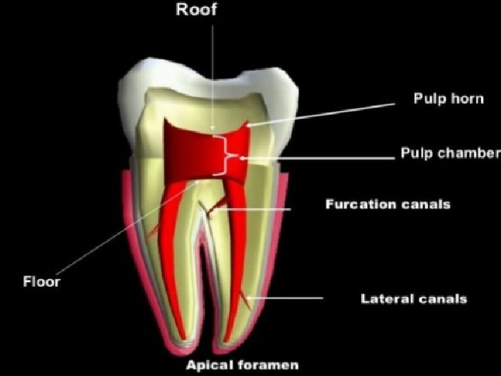
Pulp chamber • Roof: consist of dentin covering the chamber occlusaly • Pulp horn: projection of the roof under a cusp or developmental groove • Floor: consist of dentin parallel to roof • Canal orifice
Pulp cavity • Outline of pulp chamber correspond to shape of crown which it is housed • Outline of pulp canal correspond to shape of roots
Radicular pulp • Pulp space: Portion of pulp cavity from canal orifice to apical foramen
Classification of pulp canals
: Type I One canal extending from pulp chamber to the apex
: Type II Two canals arise from pulp chamber and joining into one short of the apex
Type III • Two separate canals from orifice to apex , exit the root as separate foramina
Type IV • One canal leaving the pulp chamber and divides into two separate canals at apical third with separate apical foramina
Apical foramen • It is the constricted opening near the apex of the root through which the blood supply and nerves pass
Lateral canal • Is a lateral branch of root canal
Delta system • Complex system formed by breaking up of the root canal into multiple tiny canals
Pulp cavities of individual teeth
Maxillary incisors • Pulp chamber wider mesiodistally than buccolingually • Central incisor has three pulp horns corresponding three mamelones • has wider pulp cavity than lateral • Pulp chamber cross section is triangular
Mamelons • protuberances which are present on the cutting edge of an incisor tooth when it first erupts through the gum.
Maxillary lateral incisor • Pulp chamber smaller than central • Has 2 pulp horns conform mamelones
Mandibular incisors • Outline of pulp cavity conform to the crown • 2 pulp horns • Cross section: oval • Pulp cavity of lateral larger than central • Usually one canal but 2 canals also found
Maxillary canine • Widest pulp chamber in mouth labiolingually • One pulp canal, one horn • Cross section oval or triangular • Labioligually: chamber pointed incisally, cervically wide till middle then narrow to the apex
Mandibular canine • Similar to maxillary canine but less dimension
Maxillary first premolar • Labiolingual: • Wide pulp chamber • Two root canals • Buccal horn higher than lingual • Cross section: kidney shape • Mesiodistal: • Canal narrow to apex
Maxillary first premolar • Two pulp horns one under each cusp
Maxillary second premolar • One root and one canal • 2 roots are possible • 2 canals in single root possible • 2 pulp horns • Cross section: oval
Mandibular first premolar • The pulp cavity of this tooth consists of two pulp horns each pulp horn is located within a cusp • The buccal pulp horn is higher • Majority one canal but 2 possible • Cross section: oval, rectangular, round or triangular
Mandibular • Similar to 1 st premolar with increased dimension • 2 -3 pulp horns depend on number of cusps • One root • One canal nd 2 premolar
Maxillary 1 st molar • Cervical cross section is rhomboidal • 3 roots, each root one canal • Mesiobuccal root may have 2 canals • Cross section: canals triangular
Maxillary 1 st molar • Has 4 pulp horns, mesiolingual is the highest
Maxillary 2 nd molar • Similar to first molar • 2 canals in mesiobuccal not common • Orifices of canals much closer
Mandibular 1 st molar • Cross section rectangular • 5 pulp horns • 3 canals, sometimes 4 canals, mesial root has 2 canals
Mandibular 1 st molar • • Ø • • Mesial root has 2 canals one buccal and one lingual Distal root one large canal Mandibular 2 nd molar Similar to 1 st molar but has 4 pulp horns Cross section: triangular