Pulmonary Hypertension What you need to know for


 • Mean")


 V/Q High-Probability Scans CTPA")


 Route FC Goal of Therapy (PI) Bosentan (Tracleer) ERA")

- Slides: 33
Pulmonary Hypertension: What you need to know for boards Jean M. Elwing, MD, FCCP Associate Prof of Medicine Director, Pulmonary Hypertension Program Pulmonary and Critical Care Medicine
Disclosures • Consulting • United Therapeutics, Gilead, Actelion • Research • Actelion, United Therapeutics, Bayer, Gilead, Eiger, Bellerophon, Reata, Arena
Learning Objectives: • Know the hemodynamic definitions of pulmonary hypertension • Be familiar with the clinical evaluation of elevated pulmonary pressures • Review clinical classification • Be aware of optimal treatment strategies for pulmonary hypertension
Ms. M is a 62 -year-old female with scleroderma who presents with progressive shortness of breath. She has notable dyspnea with any routine activity. She was recently seen in the office. She underwent testing for shortness of breath with a chest x-ray that did not reveal significant abnormalities. Pulmonary function tests revealed a low DLCO at 42% predicted. Echocardiogram revealed grade II diastolic dysfunction, elevated pulmonary pressures at 66 mm. Hg with RV dilation/dysfunction. The patient sends a message to the office asking what is the cause of her pulmonary hypertension. How would you respond? A. Pulmonary arterial hypertension. B. WHO group I disease related to scleroderma. C. WHO group II disease related to diastolic dysfunction. D. Unable to classify at this time as patient requires further workup.
Pulmonary hypertension evaluation Screening and evaluation Confirmatory testing • Echocardiogram reveals changes that will prompt further evaluation of pulmonary hypertension. • Echocardiography cannot confirm the diagnosis of pulmonary hypertension. • Right heart catheterization confirms the presence of elevated pulmonary pressures. • Right heart catheterization assesses the origin of elevated pulmonary pressures. • Additional workup required to determine etiology.
Apical Four-Chamber Echocardiogram Ventricular septum diastolic and systolic flattening RA dilation RV severely dilated with reduced function TAPSE 1. 5 Pericardial effusion but no chamber collapse Parasternal Short Axis Estimated PASP PAP estimated by TR jet velocity
Right Heart Cath
Pressure Waveforms in the Right Heart and Pulmonary Artery
Ms. P is 42 -year-old Caucasian female with a past medical history significant for diabetes, hypertension, obesity who was found to have elevated pulmonary pressures on her recent right heart catheterization. Her pulmonary pressures were noted to be 80/30. She is calling the office and asking what is the next step in her management. A. B. C. D. What is the best response? Tell her she has pulmonary hypertension. Recommend she start on targeted therapy for pulmonary hypertension with sildenafil. Let her know she has pulmonary hypertension related to left heart disease as she has multiple risk factors. Obtain additional data to help better understand the origin of the pulmonary hypertension to determine next step in evaluation. Advise her she does have pulmonary hypertension. Recommend diet and exercise as first steps in management.
Pulmonary Hypertension Differentiation by Origin of Increase Pulmonary Pressures m. PAP >25 VC RA RV PA PC PV LA LV PAH Hypoxia CTEPH CETPH Left Heart Ao
Diagnosis of Pulmonary Hypertension Hemodynamic Classification • Definition of Pulmonary Hypertension (PH) • Mean pulmonary artery pressure (m. PAP) >25 mm. Hg at rest • Hemodynamic Characteristics of Pulmonary Arterial Hypertension (PAH) • PH associated with pulmonary artery wedge pressure (PWP) <15 mm. Hg • Pulmonary vascular resistance (PVR) >3 mm. Hg/L/min (Wood units) or 240 dynes/sec/cm-5
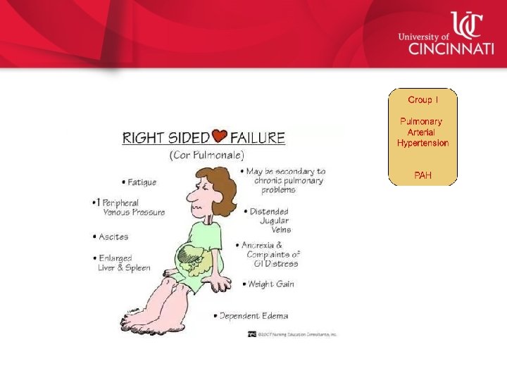
Pulmonary Hypertension PH Group III Group IV Group V Pulmonary Arterial Hypertension Pulmonary Hypertension due to Left Heart Disease Pulmonary Hypertension due to Lung Disease Chronic Thromboembolic Pulmonary Hypertension Multifactorial Mechanisms PAH CTEPH
5 th World Symposium on Pulmonary Hypertension Classification Scheme 3. PH-Lung Disease/Hypoxia – COPD – Interstitial lung disease – Sleep disorder – Alveolar hypoventilation 1. Pulmonary Arterial – – – – Hypertension Idiopathic/Heritable Drugs/toxins Connective tissue disease HIV Portal hypertension Congenital heart disease Schistosomiasis 1 – Pulmonary veno-occlusive disease ’ – Pulmonary capillary hemagiomatosis 4. Chronic Thromboembolic Pulmonary Hypertension – Operable – Inoperable Adapted from: Condliffe R, et al. F 1000 Prime Rep. 2015; 7: 06. 2. PH-Left Heart – Systolic dysfunction – Diastolic dysfunction – Valvular disease 5. Multifactorial/unclear – Hematological • Chronic hemolytic anemia • Myeloproliferative disease • Splenectomy – Systemic Disorders • Sarcoidosis • Langerhans cell histiocytosis • Lymphangioleiomyomatosis • Neurofibromatosis • Vasculitis – Metabolic Disorders • Glycogen storage disease • Gaucher’s disease • Thyroid disorder – Others • Tumour obstruction • Fibrosing mediastinitis • Chronic renal failure
Elevated pulmonary pressures are common and have many causes Most Common Left heart Hypoxia PAH disease CTEPH OSA HFp. EF Obstructive HFr. EF Restrictive Elevated PAP Rare Circulation. 2012; 126: 975 -990. Valvular
Pulmonary Arterial Hypertension WHO Group I Pulmonary Vascular Histopathology Normal Pulmonary Artery Pulmonary Arterial Hypertension
Pulmonary Arterial Hypertension WHO Group I Schematic Progression of PAH Presymptomatic/ Compensated Symptomatic/ Decompensating Usual time of diagnosis Declining/ Decompensated Cardiac output tery r ry a ona m l Pu sure pres r scula a nary v Pulmo ce n a resist Right Heart Dysfunction Right atrial pressure Time Adapted from: Hill NS. Pulmonary Hypertension Therapy. Summit Communications, LLC; 2006: 9.
Pulmonary Arterial Hypertension WHO Group I Demographics and Survival • Reveal Registry – – – March 2006 – Sept 2007 2716 consecutively enrolled Traditional Hemodynamics PAHREVEAL: • – – Historical Data: 1, 3 -, 5 -, and 7 year survival rates 68%, 48%, 35%, and 31% Wedge < 15 1, 3 -, 5 -, and 7 year survival rates 85%, 68%, 57%, and 49% Mean age was 53 ± 14 years 79. 5% female 2. 8 years - symptoms to diagnosis 41. 3% combination PAH therapy Badesch D, Raskob G, Elliott G, et al. Pulmonary Arterial Hypertension: Baseline Characteristics From the REVEAL. Chest. 2010; 137(2): 376 -387.
Risk factors for PAH • Family members with pulmonary hypertension • Drugs and toxin exposures • Connective tissue disease • HIV infection • Liver disease with portal hypertension • Congenital heart disease • Schistosomiasis CHEST. 2010; 137(2): 376 -387. IPAH REVEAL Registry APA H Demographics PPH N PVO D Drugs/To xins HIV Other PCH HPA H
Examination findings in PAH Skin: Changes c/w CTD Lungs: CTA w/o wheeze / crackles Neck: HJR, JVD Liver: Hepatomegaly Pulsatile liver Heart: Heave, RRR, Increased P 2, TR SM Abdomen: Ascites Joint: Changes c/w CTD Extremities: Edema Digits: Cool, Cyanotic
Pulmonary testing findings in PAH • Pulmonary function testing • Preserved pulmonary mechanics • Isolated low DLCO • Example: FEV 1 88%, FVC 86%, DLCO 42% • Lack of significant parenchymal disease on CT chest • Enlarged PA/RA/RV • EKG with RAD, RVH, RV strain • No or mild hypoxemia at overnight pulse oximetry • No or controlled OSA on polysomnogram
Echocardiogram changes in PAH • Echocardiogram in PAH • RA/RV • RAE, RV dilation, RV dysfunction, decreased TAPSE, elevated PAP • Echocardiogram in left heart disease • LAE, LVH, LV dilation, LV dysfunction, grade II/III diastolic dysfunction, mitral/aortic valvular disease http: //commons. wikimedia. org
Pulmonary Hypertension due to Left Heart Disease WHO Group II • Pulmonary venous hypertension – Left-sided valvular heart disease – Diastolic or systolic heart failure – Elevation of pulmonary artery wedge pressure – Respond to treatment for left sided heart disease – Negative response to treatments for PAH
Pulmonary Hypertension due to Lung Disease and/or Hypoxia WHO Group III • Chronic obstructive pulmonary disease • • – Majority with mild PH (m. PAP < 35 mm. Hg) – Leads to moderate PH < 10%, severe < 5% Interstitial lung disease Sleep disordered breathing Alveolar hypoventilation disorders Chronic exposure to high altitude – Resolves with descent Weitzenblum E, Chaouat A. Severe pulmonary hypertension in COPD: is it a distinct disease? Chest 2005; 127: 1480 -2. Group III Pulmonary Hypertension due to Lung Disease
Mr. W is a 44 -year-old male with history of multiple back surgeries who presents with progressive shortness of breath. He has notable dyspnea with walking on a flat surface more than 15 feet. He has had recurrent exertional syncope. A recently performed echocardiogram revealed severe RV dilation and dysfunction with estimated PA pressures at 110 mm. Hg. A CTPA performed during recent ER visit did not reveal acute pulmonary embolism. Serologies are negative. HIV is negative. He has no known connective tissue disease, anorexigen exposure, family history of PAH or history of DVT/PE. What additional testing is recommended prior to right heart catheterization to better determine etiology of pulmonary hypertension? A. Ig. E level to assess for asthma as a contributor. B. CT abdomen and pelvis to assess for malignancy. C. V/Q to evaluate for chronic thromboembolic disease. D. Cardiac stress test to evaluate for CAD. E. None of the above. Workup is complete. Patient is ready for right heart catheterization.
V/Q Scan More Sensitive Than Multidetector CT Pulmonary Angiography (CTPA) V/Q High-Probability Scans CTPA Sensitivity 96. 2% 51. 3% Specificity 94. 6% 99. 3% Accuracy 95. 2% 82. 8% Negative Predictive Value 97. 9% 79. 7% Positive Predictive Value 90. 3% 97. 6% N=227 undergoing both V/Q and CTPA at a single center. Tunariu N, et al. J Nucl Med. 2007; 48: 680 -684.
Chronic Thromboembolic Pulmonary Hypertension WHO Group IV • Thromboembolic disease – 4% of patients followed for 2 years after PE – 10% with antiphospholipid antibodies • Thromboembolic obstruction of proximal pulmonary arteries – 0. 1 to 0. 5% pulmonary emboli survivors – Up to 2500 patients/year in US are candidates PTE • Thromboembolic obstruction of distal pulmonary arteries – 30% 5 year survival without intervention AU - Pengo V; Lensing AW; Prins MH, et al. Incidence of chronic thromboembolic pulmonary hypertension after pulmonary embolism. NEJM 2004; 350: 2257 -64. Fedullo PF, Auger WR, Kerr K, et al. Chronic Thromboembolic Pulmonary Hypertension. NEJM 2001; 345: 1465 -1472.
Pulmonary Hypertension due to Chronic Thrombotic and/or Embolic Disease WHO Group IV Fibrous bands across a pulmonary artery Fibrous band in peripheral pulmonary artery due to remote pulmonary embolus www-medlib. med. utah. edu
What is the optimal treatment? ? ? Pulmonary Hypertension due to Chronic Thrombotic and/or Embolic Disease WHO Group IV Surgery with pulmonary thromboendarterectomy (PTE) www. uptodate. com
Mrs. C. presents to the ER with progressive shortness of breath. She reports that she is no longer able to walk up a flight of stairs. She’s becoming breathless with vacuuming. She became presyncopal when caring for her grandchildren today and presented to the ER because she was concerned about her heart. She underwent labs which were unremarkable and a chest x-ray that revealed prominent pulmonary arteries. EKG revealed right ventricular hypertrophy. She underwent a bedside echocardiogram that revealed enlarged right ventricle. There was concern about PE and she underwent a CTPA which was negative. She was admitted and underwent further evaluation with serologies that were notable for positive SCL 70. Skin findings were notable for sclerodactyly and Raynaud’s. Further workup of her pulmonary hypertension revealed no desaturations at rest or with sleep. She did undergo a V/Q to assess for chronic thromboembolic disease which was negative. She proceeded to right heart catheterization and findings are noted below. What is the best next step in her treatment? A. B. C. D. E. Initiation of supplemental oxygen. Initiation of anticoagulation. Exercise rehabilitation program. Initiation of targeted pulmonary vasodilator therapy. Calcium channel blocker therapy.
Pulmonary vasodilator therapy for PAH Humbert M, Sitbon O, Simonneau G, Treatment of Pulmonary Arterial Hypertension. NEJM. 2004; 351: 1425 -39.
Drug Name Class Indication (PI) Route FC Goal of Therapy (PI) Bosentan (Tracleer) ERA (non-select) WHO Group 1 PO II-IV *EC and decrease rate of clinical worsening Macitentan (Opsumit) ERS WHO Group 1 PO II-III Improve morbidity Ambrisentan (Letairis) ERA (selective) WHO Group 1 PO II-III *EC and delay clinical worsening Sildenafil (Revatio) PDE-I 5 WHO Group 1 PO / IV II-IV *EC Tadalafil (Adcirca) PDE-I 5 WHO Group 1 PO II-IV *EC, delay clinical worsening Riociquat (Adempas) GC WHO Group 1, 4 PO II-IV EC Selexipag (Uptravi) IP receptor Ag WHO Group 1 PO II-IV Morbidly and Mortality Oral treprostinil (Orenitram) Oral prostacyclin WHO Group 1 PO II-IV EC as monotherapy Epoprostenol (Flolan, Veletri) Prostacyclin IPAH and PAH w/ Scleroderma IV III-IV *EC and Survival IPAH *EC Scleroderma Treprostinil (Remodulin) Prostacyclin WHO Group 1 IV, SQ, PO II-IV Decrease PAH symptoms related Exercise Iloprost (Ventavis) Prostacyclin WHO Group 1 Inhaled III-IV *EC, Improve *FC, delay deterioration Inhaled Treprostinil (Tyvaso) Prostacyclin WHO Group 1 Inhaled III *EC EC = Exercise Capacity FC =Functional Class
Summary: • Pulmonary hypertension is the condition with multiple etiologies • Elevated PASP on echocardiogram is frequently likely related to left heart disease or hypoxic lung disease than PAH • Full evaluation is necessary to determine optimal treatment strategies • Surgery is intervention of choice for CTEPH • Three major pathways are targeted in medical therapy for PAH.