PULMONARY HYPERTENSION ETIOPATHOGENESIS CLASSIFICATION PART I Presented by

PULMONARY HYPERTENSION ETIOPATHOGENESIS & CLASSIFICATION PART- I Presented by: Dr RAKESH JAIN Senior Resident, Dept of cardiology Medical College, CALICUT July 15 TH, 2013

DEFINITION OF PAH Current hemodynamic definition is a Ø m. PAP >25 mm Hg and Ø PCWP, LA pressure, or LVEDP ≤ 15 mm Hg, and Ø PVR>3 Wood units m. PAP ≥ 30 mm. Hg on exercise: No supportive evidence Circulation. 2009; 119: 2250 -2294 J Am Coll Cardiol. 2009; 53: 1573 -1619

CLASSIFICATION’S OF PH 1. Clinical Classification 2. Histopathological classification

WHO Geneva, Switzerland 1973 • In 1973 WHO was first to attempted the classification of pulmonary hypertension into two categories. 1. Primary PH (Histopathological pattern) I. Arterial plexiform II. Veno-occlusive and III. Thromboembolic 2. Secondary PH

Evian Classification 1998 • Expanded prior 1973 classification from 2 groups to 5 major groups. • Based on defining categories of PH that shared similar histopathology, clinical characteristics & therapeutic options.

Retaining “PPH” Avoided term “secondary PH” Rich S, Evian, France , WHO September 6– 10, 1998
 1.")
Venice 2003 classification; revised from Evian 1998 Group 1. Pulmonary artery hypertension (PAH) 1. Modest change (Mechanism based ) 2. Abandon term PPH 3. Moved pulmonary venoocclussive disease & pulmonary hemangiomatosis to under PAH. 4. Miscellaneous group added 1. 1 Idiopathic (IPAH) 2 1. 2 Familial (FPAH) 1. 3 Associated with (APAH) 1. 3. 1 Collagen vascular disease 1. 3. 2 Congenital systemic-to-pulmonary shunts 1. 3. 3 Portal hypertension 1. 3. 4 HIV infection 1. 3. 5 Drugs and toxins 3 1. 3. 6 Other (thyroid disorders, glycogen storage disease, Gaucher disease, splenectomy, hereditary haemorrhagic telangiectasia, haemoglobinopathy) 1. 4 Associated with significant venous or capillary involvement 1. 4. 1 Pulmonary veno-occlusive disease 1. 4. 2 Pulmonary capillary haemangiomatosis 1. 5 Persistent pulmonary hypertension of the newborn Group 2. Pulmonary hypertension with left heart disease Group 3. Pulmonary hypertension associated with lung disease and/or hypoxaemia Group 4. Pulmonary hypertension due to chronic thrombotic and/or embolic disease Group 5. Miscellaneous (sarcoidosis, histiocytosis X, 4 lymphangiomyomatosis, compression of pulmonary vessels)

(J Am Coll Cardiol 2004; 43: 5 S– 12 S

PAH Non PAH Dana Point Classification of. PH PH, 2008 1. Histologic 1. Mediatinal 2. Clinicaladenopathy presentation 2. Crackles risk 3. Ground factors glass 4. Familial opacities occurrence 4. Low DLco Galiè N et 3. Common al. Eur Heart J 2009; 30: 2493 -537 5. Hemosiderin 5. BMPR 2 association laden macrophages in BAL Galiè N et al. Eur Resp J 2009; 34: 1219 -63

Fallacies of Dana Point, 2008 • Pulmonary arterial hypertension is not limited to Group I (this is inappropriately suggested by its designation as ‘pulmonary arterial hypertension) Ø It may be associated with pulmonary venous hypertension Ø It may be posttrombotic (Group IV) or hypoxic (Group III). • The group ‘miscellaneous’ includes: compression of pulmonary veins, which should be in Group II (pulmonary venous hypertension). • Application to pediatric PAH sometimes difficult. Facts not addressed Ø Fetal origin of PVD. Ø Developmental mechanism. Ø Inconsistent approach of neonatal PVD & importance of perinatal maladaptation, mal-development & pulmonary hypoplasia. Ø Heterogeneity of risk factors compared to adult.
 Panama Classification (2011) for pediatric pulmonary hypertensive vascular disease")
Pulmonary Vascular Research Institute (PVRI) Panama Classification (2011) for pediatric pulmonary hypertensive vascular disease • Recognition that application of Dana Point Classification to pediatric PAH sometimes difficult. • Aim: to improve diagnostic strategies promote appropriate clinical investigations and improve understanding of disease pathogenesis, physiology and epidemiology.
 continue…. • “Pediatric Pulmonary Vascular Hypertensive Disease” – term used &")
Panama Classification (2011) continue…. • “Pediatric Pulmonary Vascular Hypertensive Disease” – term used & not pulmonary HT. • Excludes patients with pulmonary hypertension but without elevated pulmonary vascular resistance – ie patients with large systemic to pulmonary connections. • Such patients do not require drug Rx for PHT but rather closure of defect.

Definition: PVRI Panama Classification 2011 • Definition of pediatric pulmonary hypertensive vascular disease: § m. PAP > 25 mm. HG § PVR index >3. 0 Wood units m 2

Panama Classification 2011
 • Grade I:")
Heath-Edwards classification of pulmonary vascular changes in congenital heart disease (1958) • Grade I: Medial hypertrophy. • Grade II: Cellular intimal proliferation. • Grade III: Occlusive changes.

Heath-Edwards classification of pulmonary vascular changes • Grade IV: Dilation: Vessel is dilated, and media is abnormally thin. • Grade V: Plexiform lesion: There is cellular intimal proliferation, clustered around are numerous thin-walled vessels that terminate as capillaries in alveolar wall. • Grade VI: Acute necrotizing arteritis: A severe reactive inflammatory exudate is seen through all layers of the vessel.

ETIOPATHOGENESIS

Normal pulmonary circulation • High flow, low pressure and low resistance circulation • Unique double arterial blood supply • Pulmonary arteries: § Elastic: conducting vessel, ≥ 500 μm, highly distensible § Muscular: 100 -500 μm, no elastin, non distensible § Arterioles: ≤ 100 μm, thin intima and single elastic lamina • Bronchial arteries: nutrition to the airways

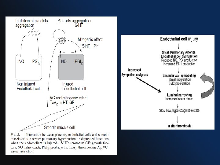
PATHOPHYSIOLOGY • Panvasculopathy predominantly affecting small PA • Exact mechanism is unknown, abnormalities in pulmonary artery endothelial & smooth muscle cells (PASMCs) with varying degrees of I. Vasoconstriction, II. Vascular proliferation, III. Thrombosis, and IV. Inflammation contribute to the development of pulmonary hypertension
 25, 2243– 2278")
Postulated pathobiology in PAH European Heart Journal (2004) 25, 2243– 2278

VASOCONSTRICTION • Genetic predisposition for increased pulmonary vascular reactivity and vasoconstriction. • Voltage-dependent and calcium-dependent potassium channels (PASMCs) modulate pulmonary vascular tone. • Abnormal functions PASMCs. are involved in the initiation or progression of pulmonary hypertension.

Molecular mechanisms of vasoconstriction-mediated remodeling Ca dependa nt ca release Circulation 98: 1400, 1998
. •")
VASCULAR PROLIFERATION • Striking feature is intimal proliferation (May cause complete vascular occlusion). • Enhanced growth factor release and intracellular signaling lead to Ø PASMC proliferation and migration. Ø ↑ extracellular matrix synthesis (elastin, collagen, and fibronectin)

• PASMCs favor ↓ apoptosis and ↑ proliferation. • Impaired apoptosis: multifactorial Ø ↑ expression of antiapoptotic protein survivin Ø activation of transcription factors such as HIF-1α Ø mitochondrial and ion channel dysregulation. • Enhanced proliferation: Ø ↑ serotonin Ø ↑ Transforming growth factor-β (TGF-β)
 having key role in PAH acting through serotonin transporter")
• Serotonin (platelet-dense granules) having key role in PAH acting through serotonin transporter (SERT) • SERT is abundantly expressed in the lung and appears specific to PASMCs. • It causes Ø vasoconstrictor Ø ↑ SMC hypertrophy and hyperplasia
, rho")
Molecular mechanisms of cellular proliferation–mediated remodeling 2 1 3 reactive oxygen species (ROS), rho kinase (ROCK), and mitogen-activated protein kinases (MAPK) receptor-mediated Smads (R-Smads) inhibitors of DNA binding 3 (Ids) 5 HT : 5 hydroxy tryptamine Curr Opin Pharmacol 9: 281, 2009

Inflammation CCL 2 = chemokine ligand 2 CCL 5 = chemokine ligand 5 CX 3 CL 1 chemokine ligand 1 (fractalkine) CX 3 CR 1 = chemokine receptor 1 ROK = rho kinase RANTES regulated upon activation, normal T cell expressed and secreted J Am Coll Cardiol 54: S 10, 2009

THROMBOSIS • Widespread occlusion of arteries/arterioles and thrombosis in situ. • Studies of pulmonary vascular histopathology in IPAH showed the prevalence rates of thrombotic lesions in > 50%. • Chronic warfarin anticoagulation has been associated with a marked survival advantage in several longitudinal studies.

Role of Genetics in Pulmonary Arterial Hypertension • Reported in approximately 6% to 10% of patients with PAH. • Mutations in 3 receptors of the TGF-ß family identified in heritable PAH I. Bone morphogenetic protein receptor 2 (BMPR 2) II. Activin receptor-like kinase type 1, and III. Endoglin • 50% to 90% of mutations in BMPR 2.
 • Chromosome 2 q 33,")
BMPR 2 (bone morphogenetic protein receptor type II gene) • Chromosome 2 q 33, codes for BMPR-II receptor • Genetic anticipation and incomplete penetrance (20%). • BMPR 2 mutations Ø 70% with familial PAH Ø 25% with IPAH Ø 15% of PAH related to fenfluramine use • Normally, it modulate vascular cell growth & is critical for the maintenance and/or normal response to injury of the pulmonary vasculature. • Haploinsufficiency for BMPR-II leads to Ø EC proliferation Ø PASMC hypertrophy, and Ø fibroblast deposition.
 Smads: cytoplasmic signaling proteins Smads p")
DAN : differential screening-selected gene aberrative in neuroblastoma) Smads: cytoplasmic signaling proteins Smads p 38 MAPK: p 38 mitogen-activated protein kinase PKA: protein kinase A LIMK 1: LIM motif-containing protein kinase 1
 & endoglin • Rare mutations • Also members")
ALK 1 gene (activin-like kinase 1) & endoglin • Rare mutations • Also members of TGF-β superfamily • Associated with PAH in Ø Hereditary hemorrhagic telangiectasia and Ø IPAH
 polymorphisms • Encoded by single gene on chromosome 17 q 11.")
Serotonin transporter (SERT) polymorphisms • Encoded by single gene on chromosome 17 q 11. 2 • L allele induces greater rate of SERT gene transcription than S allele. • Overexpression is associated PASMC hyperplasia. • One study has shown that the L-allelic variant is found to be present in homozygous form in 65% of IPAH patients but in only 27% of control subjects.
 • Elevated levels are seen in PAH patients. •")
The role of Endothelin-1 (ET-1) • Elevated levels are seen in PAH patients. • Levels correlate with disease severity & prognosis. • Deleterious effects mediated through ETA and ETB receptors Ø Fibrosis Ø Hypertrophy and cell proliferation Ø Inflammation Ø Vasoconstriction

The role of Prostacyclin and Thromboxane A 2 • Arachidonic acid metabolites • Prostacyclin: low levels in patients with PAH Ø potent vasodilator Ø inhibits platelet activation Ø Antiproliferative properties • Thromboxane A 2: high levels in patients with PAH Ø potent vasoconstrictor Ø promotes proliferation Ø platelet activation In PAH, the balance between these 2 molecules is shifted toward thromboxane A 2, favoring thrombosis, proliferation, and vasoconstriction.

The role of nitric oxide • • • Potent vasodilator Inhibitor of platelet activation Possesses anti-proliferative properties Vasodilatory effect is mediated by c. GMP Rapidly degraded by phosphodiesterases (PDEs-5) Decreased endothelial NOS (NOS 3) has been observed in PAH patients
 • A member of glucagon-growth hormone-releasing superfamily • Pharmacologic profile")
Vasoactive intestinal peptide (VIP) • A member of glucagon-growth hormone-releasing superfamily • Pharmacologic profile similar to prostacyclins. • Serum and lung tissue VIP levels decreases in PAH results in Ø platelet activation, and Ø PASMC proliferation.

Figure. Schematic depicting the potential “hits” involved in the development of PAH. A rise in [Ca 2+]cyt in PASMCs (due to decreased Kv channel activity and membrane depolarization, which opens VDCCs; upregulated TRPC channels) Circulation 2009; 119: 2250 -2294

Distribution of PH types PAH 4. 2% No Dx 6. 8% Lung disease 9. 7% Left heart disease 78. 7% Gabbay E, PAH an uncommon cause of pulmonary Hhypertension : the Armadale echocardiography study. Am J Resp Crit Care Med 2007; 175: A 713.
")
Mean PAP in patients with different causes of pulmonary hypertension (PH)

Survival in PAH 1. 0 Congenital heart disease 0. 9 0. 8 0. 7 0. 6 Percent survival 0. 5 IPAH 0. 4 CTD 0. 3 0. 2 HIV 0. 1 0 0 1 2 3 Years Mc. Laughlin VV et al. Chest. 2004; 126: 78 S-92 S. 4 5
 • Reserved for patients with neither a family history")
Idiopathic Pulmonary Arterial Hypertension (IPAH) • Reserved for patients with neither a family history nor an identifiable risk factor. • Rare disease • prevalence ~ 6 per million • Female/male ratio of 1. 7: 1 • Mean age 37 years.

Heritable Pulmonary Arterial Hypertension • • Approximately 6% to 10% of patients with PAH 1 50% to 90% mutations is in BMPR 22 Family history may or may not be present. PAH associated with BMPR 2 mutations have more severe disease with less vasoreactivity than those with IPAH without BMPR 2 mutations. 3 1. Genet Med. 2005; 7: 169 – 74 2. J Med Genet. 2000; 37: 741– 5 3. J Am Coll. Cardiol. 2009; 54

Familial/Heritable Pulmonary Arterial Hypertension: The ‘Two-Hit’ Hypothesis According to the hypothesis, vascular abnormalities characteristic of PAH are triggered by accumulation of genetic and/or environmental insults in a susceptible individual. A combination of germline BMPR 2 mutation (‘first hit’) and the ingestion of appetite suppressants (‘second hit’) were used to generate the clinical disease.

Pulmonary Arterial Hypertension Associated With Congenital Heart Disease Friedman WF, ed. Proceedings of the National Heart, Lung, and Blood Institute Pediatric Cardiology Workshop. Pulmonary hypertension. Pediatric Res. 1986; 20: 816 -817.

PAH associated with heart Defects with Increased Pulmonary Blood Flow • more frequently when PBF is extremely high. • Especially true for L→R shunt entering RV or PA directly (i. e. post-tricuspid shunt, such as VSD or PDA), experiencing higher incidence of severe & irreversible pulmonary vascular damage than pre-tricuspid shunt, as in ASD. • Important feature is RV well adaptive, sustaining an increased afterload for many years or decades.

PAH PATHOPHYSIOLOGY chronic high flow and high pressure Stretching of pulmonary arteries Endothelial dysfunction smooth muscle cell dysfunction (↓NO , ↓PGI 2, ↑ET , Tx. A 2) Vasoconstriction vascular SMC proliferation and migration (↑ S 100 A 4/Mrs 1 calcium binding protein) Peripheral pulmonary arterial development through morphometric changes: extension of muscle into peripheral arteries, percent wall thickness, and artery number (alveolararterial [ALV/Art] ratio) as they relate to age. PAH Platelet dysfunction ↑Serotonin

EISENMENGER SYNDROME • “Eisenmenger syndrome” was coined by Paul Wood. • Defined as CHD with initial large systemic-to-pulmonary shunt that induces progressive pulmonary vascular disease and PAH, with resultant reversal of the shunt and central cyanosis. • Represent most advanced form of PAH associated with CHD • Histopathologic and pathobiologic changes are similar to idiopathic.

PAH associated with heart Defects with Decreased Pulmonary Blood Flow • Condition like ü PA with intact IVS ü TOF • Associated because of § Hypoplasia of pulmonary arteries. § Intra-acinar pulmonary arteries are small and few in number. § Alveolar development is impaired (mostly reduction in alveolar number) § ↑ Hematocrit resulting in in-situ thrombus.

Persistent Pulmonary Hypertension of the Newborn • Normal: arterial dilates during transition from fetal to neonatal circulation (NO dependent) 3 types of PPHN • Hypoplastic type: Lungs underdeveloped, vascular bed is hypoplastic & abnormally muscular. Ex: congenital diaphragmatic hernia or Oligohydraminos • Hypertrophic type: Lung is maldeveloped, vascular bed is abnormally muscular. Ex: chronic fetal distress • Reactive type: Lung is maladapted, vessels not dilated appropriately at birth. Ex ↑ vasoconstrictive [Tx. A 2, NE, leukotrienes] may be responsible and may result from streptococcal infection or acute asphyxia at birth

Pulmonary Hypertension Associated left heart disease • Most common cause of PH • As a consequence of § § § Left ventricular dysfunction (MC) 1 Mitral and aortic valve disease Cardiomyopathy Cor-triatum and Pericardial disease 1. Clin Chest Med. 28 2007: 233 -241.

Increase in LA pressure Pathophysiology Backward transmission of the pressure to PV/PC Initially, PVR & pressure gradient across the lungs falls (reflecting distention of compliant small vessels, recruitment of additional vascular channels, or both) Further increases in LA pressure ↑ PAP & PVP, constant PBF, PG between PA & PV and PVR remains constant When PVP ≥ 25 mm. Hg chronically Abnormal formation and thickening of a neointima, medial hypertrophy, thickening and rupture of the basement membranes , Pulmonary hemosiderosis extensive fibrosis, Pulmonary lymphatics may become markedly distended ↑PVR Disproportionate elevation in PAP, PG between PA & PV ↑, PBF ~ or ↓

Pulmonary Hypertension Associated with Hypoxic Lung Diseases • Common cause of mild pulmonary hypertension • Conditions associated are ü ü Chronic Obstructive Pulmonary Disease Interstitial Lung Diseases Sleep-Disordered Breathing Alveolar Hypoventilation Disorders • Mechanism: Hypoxia induces Ø Ø vasoconstriction & muscularization of distal vessels medial hypertrophy of more proximal arteries loss of vessels & lung parenchyma Intimal thickening (appears to be an early event). The development of plexiform lesions is not observed.

Pulmonary Hypertension Associated with Chronic Obstructive Pulmonary Disease • Exact prevalence is uncertain, heavily influenced by disease severity. (~ 50% in severe COPD)1 • m. PAP is usually <30 mm Hg (typically lower IPAH) • RV failure occurs more likely as a result of an ischemic right ventricle than a pressure-loaded right ventricle. Patients who present with severe pulmonary hypertension (m. PAP >40 mm. Hg) should be evaluated for another disease process responsible for the high pulmonary arterial pressures before it is attributed to the COPD (prevalence ~ 1. 1 %)1 1. Circulation. 2009; 119: 2250 -2294

PATHOGENESIS • Multiple causative factors, including Ø alveolar hypoxia induced pulmonary vasoconstriction Ø Acidemia & hypercarbia Ø compression of pulmonary vessels by high lung volume Ø loss of small vessels in regions of the emphysema and lung destruction Ø ↑blood viscosity (polycythemia). Of these, hypoxia is the most important factor. Recently a genetic predisposition as a result of 5 -HTT polymorphism, may predispose to more severe PH in hypoxemic patients with COPD 1. Circulation. 2003; 108: 1839– 44

Pulmonary Hypertension Associated with Interstitial Lung Diseases • Prevalence uncertain, ~ 40% in IPF & 60% planned for LTx 1 • Mechanism § hypoxemia § loss of effective pulmonary vasculature from lung destruction • The hemodynamic profile is distinct from IPAH. It is uncommon for the mean PA pressure ever to exceed 40 mm Hg in these patients, whereas it is unusual for the mean PA pressure to be less than 40 mm Hg in patients with IPAH. 1. Am J Respir Crit Care Med. 2003; 167: 735– 40
 • Prevalence: 20% to 40% of")
Pulmonary Hypertension Associated with Sleep Disordered Breathing (SDB) • Prevalence: 20% to 40% of patients with SDB. • Mild to moderate in severity. • Mechanism: combination of precapillary and postcapillary factors. § primary mechanism is repetitive nocturnal arterial oxygen desaturation, which reflexively increases PA pressures. others are § pulmonary arteriolar remodeling § hyperreactivity to hypoxia § left ventricular diastolic dysfunction • RV failure in SDB appears to be uncommon.

Pulmonary Hypertension Associated with Alveolar Hypoventilation Disorders • Causes § § Central alveolar hypoventilation (Ondine's curse) Obesity hypoventilation syndrome (OHS) Chest wall deformities Neuromuscular disorders • Mechanism: Chronic alveolar hypoventilation can lead to hypoxemia, hypercapnia and acidosis and cause pulmonary hypertension
 • Underdiagnosed disorder • Pulmonary embolism")
Pulmonary Hypertension Caused by Chronic Thromboembolic Disease (CTEPH) • Underdiagnosed disorder • Pulmonary embolism is thought to be the typical initiating process. • ≥ 50% of pts do not have clinically overt pulmonary embolism. • Incidence of PH as much as 5% after first episode 1 • Cumulative incidence of CTEPH is after acute PE 1 § 1. 0% at 6 months, § 3. 1% after 1 year § 3. 8% after 2 years • Hypercoagulable state: in only a minority of patients § lupus anticoagulant ~ 10% to 20% § protein C, protein S, and antithrombin III deficiencies: ~ 5% 1. N Engl J Med. 2004; 350: 2257– 64
 PATHOGENESIS Thromboemboli fail to resolve adequately Undergo")
Pulmonary embolism (either single or recurrent) PATHOGENESIS Thromboemboli fail to resolve adequately Undergo organization and incomplete recanalization Incorporated into the vascular wall (subsegmental, and lobar vessels) Slowly progressive vascular obstruction including distal pulmonary vasculopathy of both occluded and nonoccluded pulmonary vasculature characterized by lesions considered typical for IPAH, including plexiform lesions. CTEPH

Pulmonary Arterial Hypertension Associated With Drugs 1 1. J Am Coll Cardiol. 2009; 54: S 43 -S 54

Pulmonary Arterial Hypertension Associated with Connective Tissue Diseases • CTD’s associated § § § § Scleroderma & CREST syndrome ( 8 -12%)1 SLE (1% to 14%) Mixed CTDs ( 30 -40 %) Polymyositis Dermatomyositis Rheumatoid arthritis Sjogren syndrome 1. Arthritis Rheum Dis 2003; 62: 1088 -93

• Pulmonary vasculature with histological features resemble those of IPAH. • Coexisting interstitial fibrosis is extremely common and contributes to hypoxemia. • Pathophysiology: Prevailing hypothesis is Endothelial injury +immune defect → peri and intravascular inflammatory response (↑ET, ↑Antibody to PDGF receptor) →PASMC proliferation → vascular lesions and progressive PAH.
1. •")
Pulmonary Arterial Hypertension Associated with HIV • Prevalence is ~ 1/200 (0. 5%)1. • Severe PH has been associated with AIDS, even in absence of lung parenchymal disease • Diagnosed in all stages of HIV infection • unrelated to the CD 4 cell counts 2. 1. Am J Respir. Crit Care Med 2008; 177: 108– 13 2. Eur Heart J 2009; 30: 2493 -537

• Pathophysiology: Direct pathogenic role of HIV seems unlikely as no viral constituents have been detected in the vascular endothelium. • Hypothesis are : § Function of Inflammation & immunogenetic background (HLA Class II) § More recently, HHV-8 (kaposi sarcoma) stimulates lysosomal mediated degradation of BMPR 2, a receptor which is mutated & dysfunctional in IPAH. § HIV- nef gene has been implicated in development of plexogenic pulmonary vascular lesions. § HIV envelop gene gp 120 stimulate endothelin dependant vasoconstriction 1 1. Am J Respir. Crit Care Med 2004; 170: 1212– 7

Pulmonary Arterial Hypertension Associated with Schistosomiasis • Occurs in endemic areas for schistosomiasis. • 10% of pts with schistosomiasis develop portal hypertension and only 10% of these i. e. 1% of total will have develop PAH. • Mechanism: 1 Chronic infection → ova embolize to the lungs → induce formation of delayed hypersensitivity granulomas → extensive lung vascular remodeling, fibrosis and PAH. 1. J Pathol Bacteriol. 46 1938: 401 -424.

Pulmonary Arterial Hypertension Associated with Portal Hypertension • Portopulmonary hypertension is progressive with no reports of spontaneous resolution. • Unrelated to the severity of hepatic dysfunction. • Estimated prevalence is 2% to 6% with 5 -year survival of 10% to 30%. • Pathophysiology is similar to PAH without cirrhosis, with features characteristic of the cirrhotic state ( high CO, lower systemic and pulmonary vascular resistance).

• Mechanism is unknown but severe structural changes, consisting of § medial hypertrophy, § occlusive cellular intimal hyperplasia, and § plexiform lesions occur in the peripheral pulmonary arteries leads to postulate that toxic liver is unable to degrade a certain vasoconstrictor substance that then circulates through the lung in high concentration, causing structural damage to the vessels.

Pulmonary Arterial Hypertension Associated with Sickle Cell Disease and other Hemoglobinopathies • Increasingly recognized with a prevalence <10 % 1 • Histopathology is similar to PAH with different hemodynamic parameters (PAP & PVR are often lower and CO is high). • Other Hb pathy 2 associated with PAH are Ø Homozygous beta-thalassemia and Ø Hereditary spherocytosis Whether the PH is the cause of the increased mortality or is a surrogate marker remains unclear; however, the 2 -year mortality rate in these patients is approx 50%. 3 1. Am J Respir Crit Care Med. 2007; 175: 1272– 9 2. Blood. 2001; 97: 3411– 6 3. N Engl J Med. 2004; 350: 886– 95.

• Pathobiology: likely multifactorial Ø Hemolysis-induced endothelial dysfunction and subsequent dysregulation of arginine metabolism and reduced NO bioavailability 1 Ø Pulmonary parenchymal and vascular injury from acute chest syndrome Ø Increased oxidant burden Ø Impaired LV diastolic function 1. Nat Med. 9 2003: 496 -500

Pulmonary Arterial Hypertension Associated with Pulmonary Venoocclusive Disease • Rare form of PAH • The histopathologic diagnosis is based on the presence of obstructive eccentric fibrous intimal pads in the pulmonary veins and venules. • Other findings are § § Pulmonary venous hypertension (increased PCWP) Pulmonary hemosiderosis Interstitial edema, and Lymphatic dilation

TAKE HOME MESSAGE • Though the exact mechanism of PH is unknown, dysfunction of endothelial and PSMC along with varying degree thrombosis are usually implicated. • Among genetic factors associated with PH, BMPR II is most commonly associated (50 -90 %). • Most common cause of PH is left sided heart diseases. • COPD associated PH is usually mild, if it is severe, other causes of PH should be ruled out first. • It is uncommon for the mean PA pressure ever to exceed 40 mm Hg in ILD/COPD patients, whereas it is unusual for the mean PA pressure to be less than 40 mm Hg in patients with IPAH. • HIV associated PH is not related to level of CD 4 count.

References 1. ACCF/AHA 2009 Expert Consensus Document on Pulmonary Hypertension, Circulation. 2009; 119: 2250 -2294 2. Braunwald's Heart Disease: A Textbook of Cardiovascular Medicine 9 th Edition 3. Hurst's The Heart, 13 th Edition 4. Moss and Adams' Heart Disease in Infants, Children, and Adolescents: Including the Fetus and Young Adults, 7 th Edition 5. Eur Respir Rev 2009; 18: 113, 154– 161 6. J Am Coll Cardiol. 2009; 54(1 s 1): S 20 -S 31 7. J Am Coll Cardiol. 2009; 54(1 s 1): S 43 -S 54 8. Pathogenesis of Pulmonary Arterial Hypertension : The Need for Multiple Hits: Circulation. 2005; 111: 534 -538

THANKS

J Am Coll Cardiol 2004; 43: 25 S– 32 S
- Slides: 77