Pulmonary hydatidosis Dr mohammadzadeh Thorasic surgeon A section

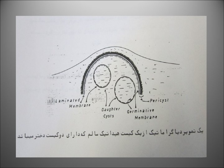

 Mother cyst (left) daughter cyst (right)")


. These eggs are virtually indistinguishable from other closely")
 Although")


 indirect fluorescent antibody (IFA) tests, and enzyme immunoassays (EIA)")

- Slides: 44
Pulmonary hydatidosis Dr. mohammadzadeh Thorasic surgeon
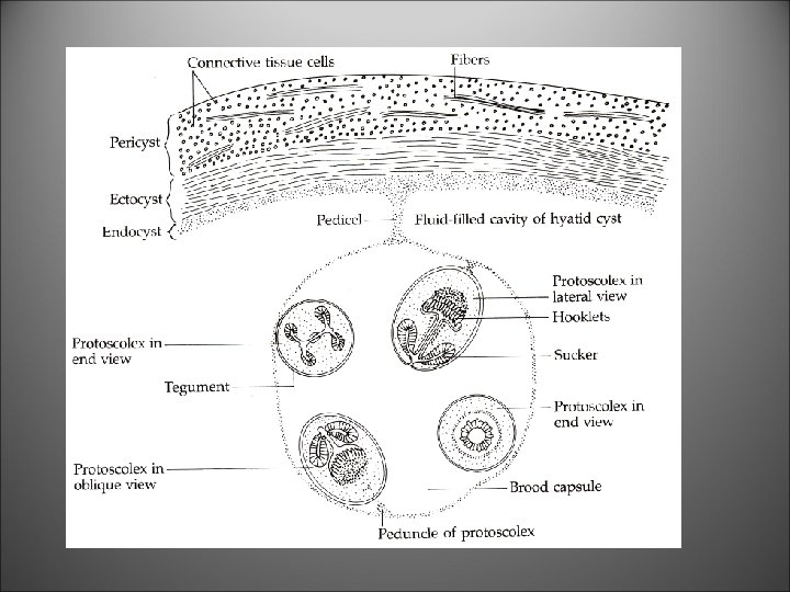
A section of a hydatid cyst The cyst consists of a thick outer layer (*), several thinner internal layers, and many protoscolices. The protoscolices are often called "hydatid sand. "
Hydatid cyst (Hydatid disease) Mother cyst (left) daughter cyst (right)
Higher magnification of the protoscolices
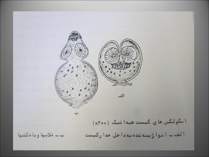
protoscolex. Note the "hooks" that will form the hooks associated with the adult worm's armed rostellum. Protoscolices with double row hooklets
Hydatid cyst showing a row of brood capsules attached to the germinal layer.
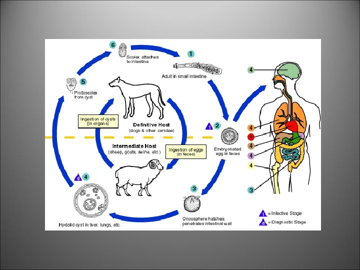
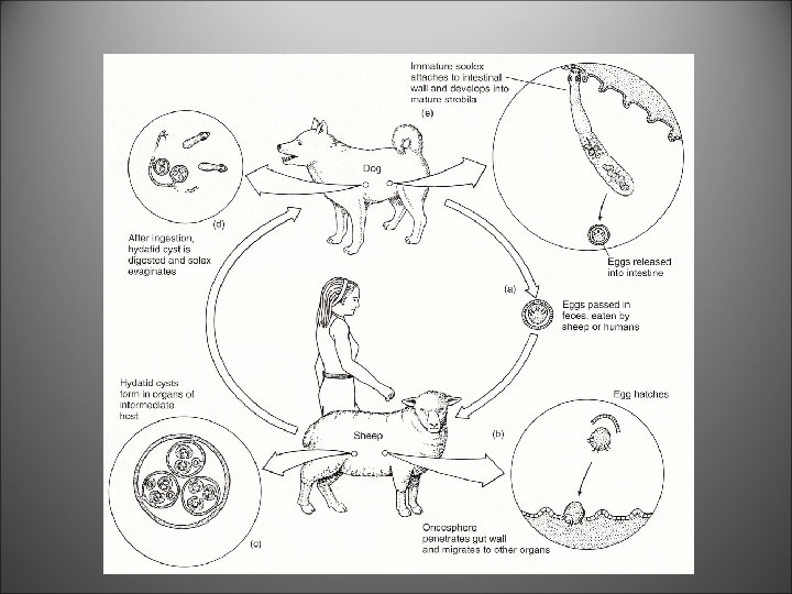
Egg An egg of Echinococcus granulosus(upper). These eggs are virtually indistinguishable from other closely related species of tapeworms such as Taenia.
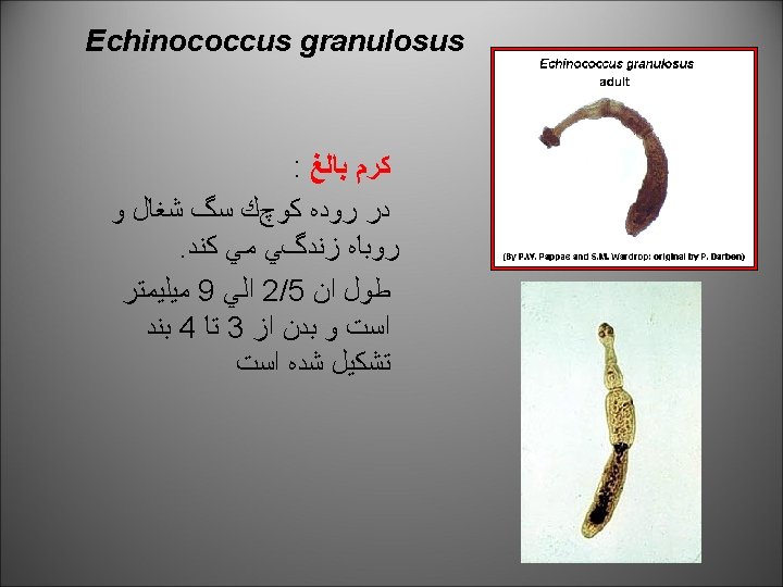
Echinococcus granulosus in the small intestine of a dog (the small, white objects) Although these tapeworms are quite small, a single dog can be infected with many of them.
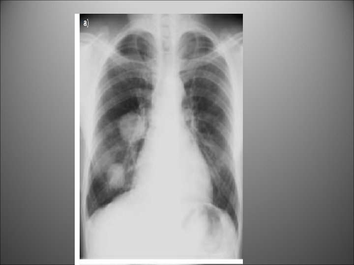
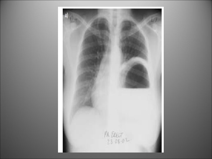
Hydatid cyst in lung. This patient had a single large cyst in the left lung.
An intact hydatid cyst
An intact hydatid cyst
Clinical Features of ruptured cyst i. Quiescent type ii. Abscess type iii. Progressive type
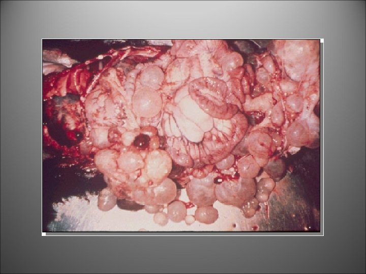
A patient with bilateral hydatid cyst
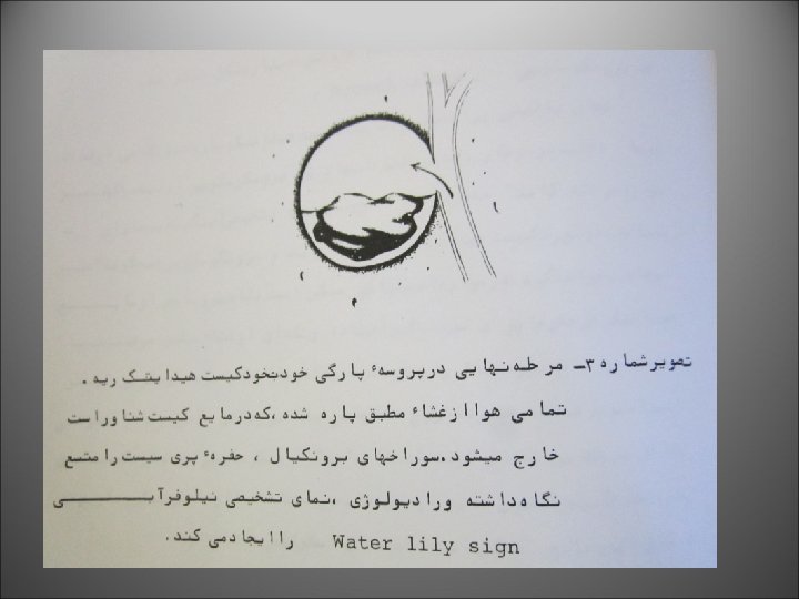
Water lily sign
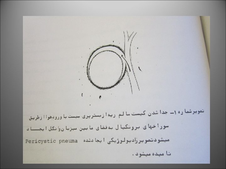
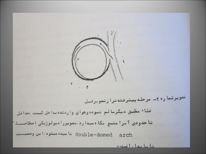
Allergic reactions and sometimes anaphylactic shock may occur if the cyst ruptures. Ruptures of the cyst may also lead to the escape of protoscolices into the surrounding tissues, these can then develop into further cysts.
Differential Diagnosis of hydatid cyst
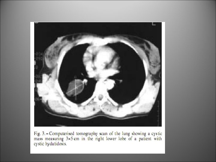
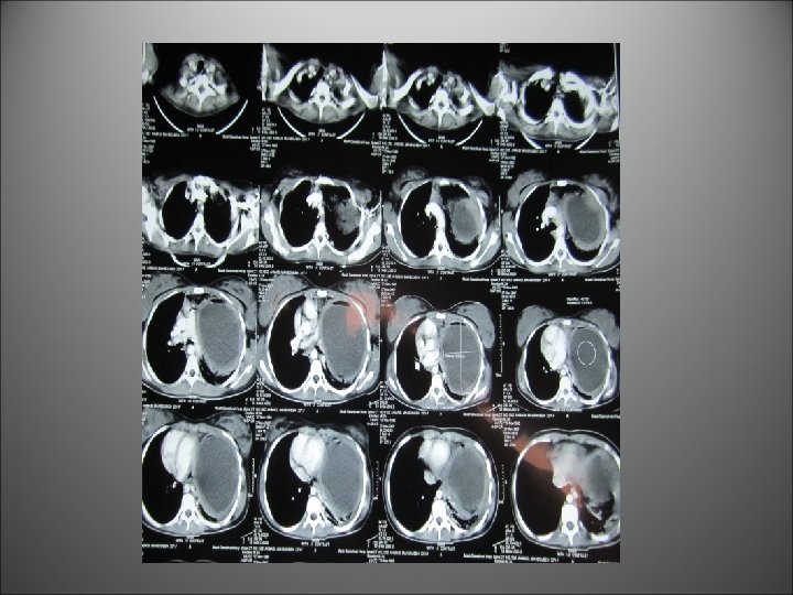
A Very huge hydatid cyst
Diagnosis Microscopy Fluid aspirated from a hydatid cyst will shows multiple protoscolices (size approximately 100 µm), each of which has typical hooklets. The protoscolices are normally invaginated (left), and evaginated (middle, then right) when put in saline.
Antibody Detection Indirect hemagglutination (IHA) indirect fluorescent antibody (IFA) tests, and enzyme immunoassays (EIA) are sensitive tests for detecting antibodies in serum of patients.
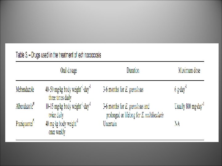
Treatment Surgery is the most common form of treatment for echinococcosis. The drug of choice for treatment is albendazole.