Pulmonary Edema Prepared by Dr Sanaa Saber Mohamed


Pulmonary Edema Prepared by Dr/ Sanaa Saber Mohamed Critical care nursing
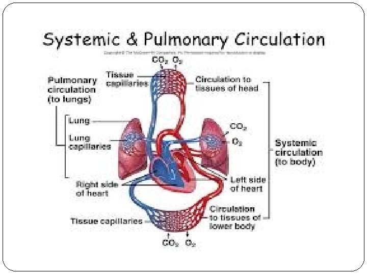

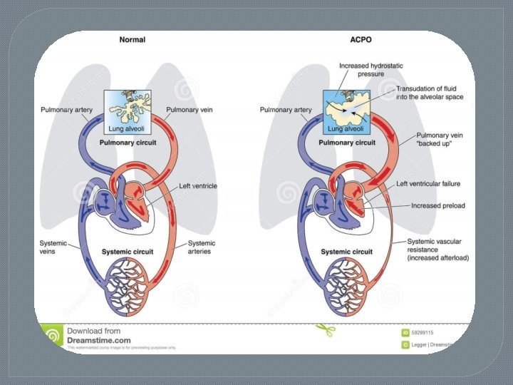
Introduction Pulmonary edema most commonly occurs as a result of increased micro vascular pressure from abnormal cardiac function. The backup of blood into the pulmonary vasculature resulting from inadequate left ventricular function causes an increased micro vascular pressure, and fluid begins to leak into the interstitial space and the alveoli.

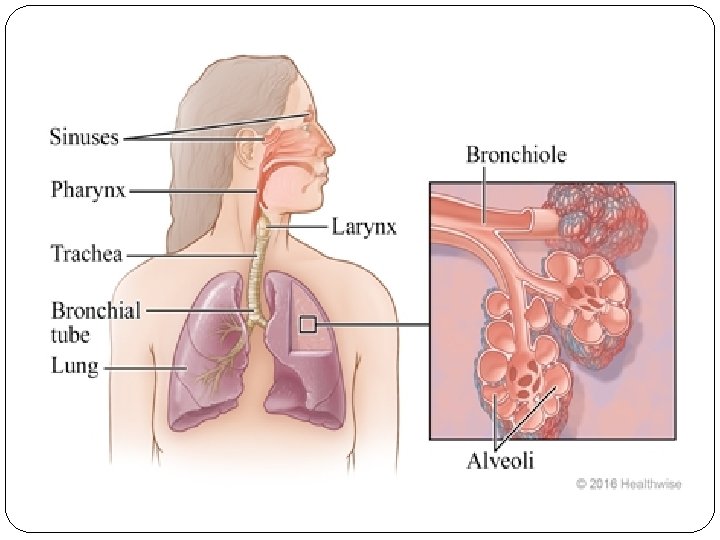
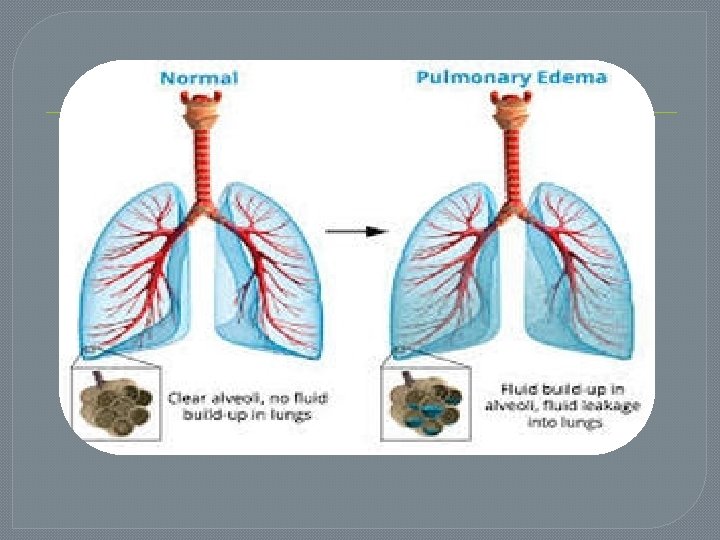
Definition Pulmonary edema is defined abnormal accumulation of fluid in the lung tissue. The fluid may accumulate in the interstitial spaces or in the alveoli. It is a severe, life threatening condition

Pathophysiology
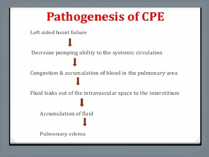

The backup of blood into the pulmonary vasculature resulting from inadequate left ventricular function causes an increased micro vascular pressure, and fluid begins to leak into the interstitial space and the alveoli.

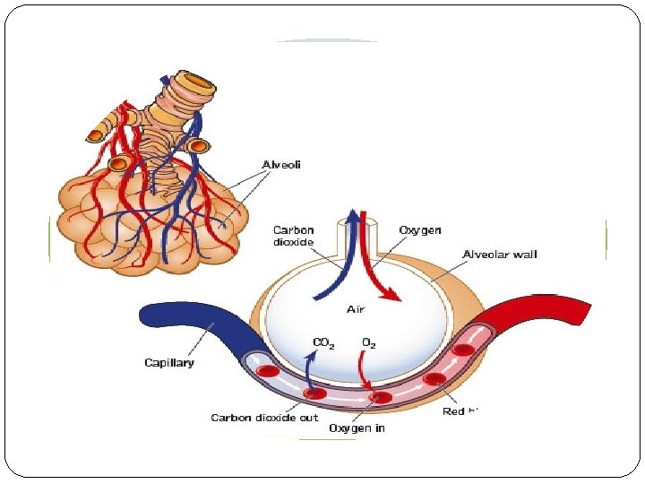
1 • Increased hydrostatic pressure in the pulmonary capillaries • elevated pulmonary venous pressure • increased left ventricular end diastolic pressure and 2 pressure • left Asatrial left atrial pressure rises further (>25 mm Hg) • edema fluid breaks through the lung epithelium • flooding the alveoli with protein poor fluid 3 • Noncardiogenic pulmonary edema • increase in the vascular permeability of the lung • resulting in an increased flux of fluid and protein into the lung interstitial and air spaces

Etiology 1 - Cardiogenic pulmonary oedema: Left ventricular failure as a complication of a myocardial infarction, mitral or aortic valve disease, cardiomyopathy, or other disorders characterized by cardiac dysfunction. Fluid overload, e. g. , from kidney failure or intravenous therapy which may cause dilatation and failure of the left ventricle.

2 - Non-cardiogenic Ø Hypertensive crisis; due to a combination of increased pressures in the right ventricle and pulmonary circulation and also increased systemic vascular resistance and left ventricle contractility increasing the hydrostatic pressure within the pulmonary capillaries leading to extravasation of fluid and edema. Ø Upper airway obstruction (negative pressure pulmonary edema ) Ø Neurogenic causes (seizures, head trauma, strangulation, and electrocution).
 Inhalation of hot or toxic gases 2) Pulmonary contusion, i.")
Other causes: 1) Inhalation of hot or toxic gases 2) Pulmonary contusion, i. e. , high-energy trauma 3) Aspiration, e. g. , gastric fluid 4) Re-expansion, i. e. post large volume thoracentesis, resolution of pneumothorax, removal of endobronchial obstruction. 5) Reperfusion injury, i. e. post lung transplantation 6) Immersion pulmonary edema 7) Multiple blood transfusions 8) Severe infection or inflammation, which may be local or systemic.

Clinical manifestation Shortness of breath with lying down, causing the patient to sleep with head propped up or using extra pillows Difficulty in breathing Wheezing Feeling of "air hunger" or "drowning" Grunting or gurgling sounds with breathing Cough, Anxiety, Restlessness, Excessive sweating Pale skin, Coughing blood Decrease in level of awareness, Rapid breathing and increased heart rate Crackles in the lungs and abnormal heart sound

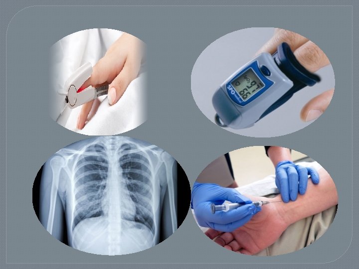
Assessment and Diagnostic Findings Chest X-ray: shows interstitial edema Echocardiogram to detect valvular disease Measurement of pulmonary artery wedge pressure by Swan-Ganz catheter (differentiates etiology of pulmonary edema: cardiogenic or altered alveolar -capillary membrane) Blood cultures in suspected infection: may be positive Cardiac markers in suspected MI: may be elevated Arterial blood gas (ABG) analysis: may show hypoxemia and impending respiratory failure

Medical management q Pharmacologic therapy 1. Oxygen therapy. Is administered to correct the hypoxemia and dyspnea. 2. Morphine is administered to reduce peripheral resistance and venous return so that blood can be redistributed from the pulmonary circulation to other parts of the body. 3. Diuretic therapy. It is used to increase rate of urine production and removal of excess extracellular fluid from the body. 4. Other intravenous medications. These include dobutamine, a catecholamine that increases myocardial contractility; amrinone, which dilates the arteries and increases CO; and digitalis, which increases contractility.

Nursing Management ü Position the patient to promote circulation ü Providing psychological support ü Monitoring medications

Nursing Diagnoses: Impaired Gas Exchange related to excess fluid in the lungs Nursing Interventions: Improving Oxygenation Give oxygen in high concentration: to relieve hypoxia and dyspnea. Take steps to reduce venous return to the heart. • Place patient in upright position; head and shoulders up, feet and legs hanging down: to favor pooling of blood in dependent portions of body by gravitational forces; to decrease venous return.

Nursing Diagnoses: Anxiety related to sensation of suffocation and fear Nursing Interventions: Stay with patient and display a confident attitude: the presence of another person is therapeutic, because the acute anxiety of the patient may tend to intensify the severity of patient's condition. (Arterial vasoconstriction diminishes as anxiety is relieved. ) Explain to patient in a calm manner all therapies administered and the reason for their use. Explain to patient importance of wearing oxygen mask. Assure patient that mask will not increase sensation of suffocation. Inform patient and family of progress toward resolution of pulmonary edema. Allow time for patient and family to voice concerns and fears

Nursing diagnosis: Decreased Cardiac Output Related to Alterations of Preload Nursing Interventions and Rationale 1. Collaborate with physician regarding the administration of oxygen to main tain a Spo 2 >92% to prevent tissue hypoxia. 2. Maintain surveillance for signs of decreased tissue perfusion and acidosis to facilitate the early identification and treatment of complications. 3. Monitor fluid balance and daily weights to facilitate regulation of the patient's fluid balance.

Nursing diagnosis: Decreased Cardiac Output Related to Alterations in Contractility Nursing Interventions and Rationale Collaborate with physician regarding the administration of oxygen to main tain a Spo 2 >92% to prevent tissue hypoxia. Monitor homodynamic and intake and output. Ensure electrolytes are optimized. Collaborate with physician regarding the administration of electrolyte replacement therapy to enhance cellular ionic environment. Collaborate with physician regarding the administration of inotropes to enhance myocardial contractility. Monitor ST segment continuously to determine changes in myocardial tissue perfusion.
 as directed. •")
Give morphine in small, titrated intermittent doses (I. V. ) as directed. • Morphine usually is not given if pulmonary edema is caused by stroke or occurs with chronic pulmonary disease or cardiogenic shock. • Watch for excessive respiratory depression. • Monitor BP because morphine may intensify hypotension. • Have morphine antagonist available: naloxone (Narcan).

Give I. V. injections of diuretic. • Insert an indwelling catheter: large urinary volume will accumulate rapidly. • Watch for falling BP, increasing heart rate, and decreasing urinary output: Indications that the total circulation is not tolerating diuresis and that hypovolemia may develop. • Check electrolyte levels because potassium loss may be significant. • Watch for signs of urinary obstruction in men with prostatic hyperplasia. Administer vasodilator if patient fails to respond to therapy.

- Slides: 29