PULMONARY ARTERY BANDING Dr DG Buys Department of

PULMONARY ARTERY BANDING • • Dr DG Buys Department of paediatric cardiology Sunday 5 June 2011 T: 051 401 9111 info@ufs. ac. za www. ufs. ac. za

OVERVIEW • • • Introduction History Pathophysiology Pulmonary hypertension / pulmonary vascular resistance Diagnosis Indications Formulas/ how tight Future Discussion

INRODUCTION • • Banding in Africa Palliative – not curative Performed as stage approach Purpose to maintain balanced pulmonary-to-systemic blood flow (Qp/Qs) • Not to distort the pulmonary arteries • Facilitate future surgical interventions

HISTORY OF BANDING • • • Muller and Dammonn – UCLA – 11 July 1951 (1952) Patient 5/12 infant with large VSD To create PS and prevent Qp Used 1 cm umbilical tape Started in period when surgical repair not available 25 patients – 1951 -1955 - 9 operative deaths – 5 before surgery 1 late death Kron et al Ann Surg May 1989

HISTORY • Describe – banding 1955 -1988 170 Total mortality rate 45% - did not vary from different decades • Remains preferred palliation to delay surgery • Later used for more complex lesions • Materials – tape, nylon, PTFE , non-stretchable Gore-Tex • Devices and dilatable bands • Although use decreased it continues to play role in management of some CHD – up to 2% of congenital cardiac cases in current surgical databasis

PATHOPHYSIOLOGY • 6 weeks drop in PVR • Pulmonary overflow • Medial hypertrophy of pulmonary arterioles and fixed pulmonary hypertension – Eisenmenger • Creating PS – decreased flow – decreased return to LV – improved LV function • PHPT: m. PA pressure >25 mm. Hg in rest and >30 mm. Hg with exercise

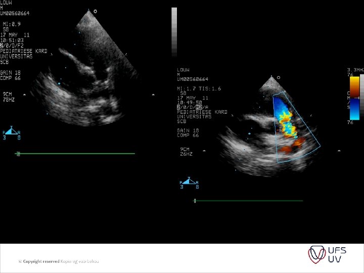
DIAGNOSIS • Clinical - AP, Load P 2 , RVHT, HTS • ECG/CXR - RVHT , p-pulmonale, decreased flow, RVHT, PA • Echo - usually indirect - variable - patient / songrapher / machine dependant - RVPSP - needs TR - PIG – needs shunt - BP - can be inacurate

PVR = PAP/ PAflow Substitude PA pressure with TR jet Substitude PA flow by RVOT VTI (velosity time integral) And we get PVR = TR jet velocity/ RVOT VTI x 10
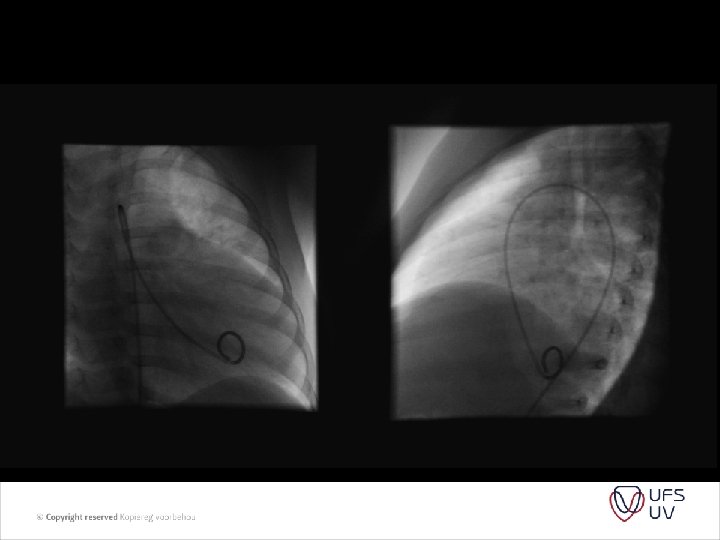
 and right")
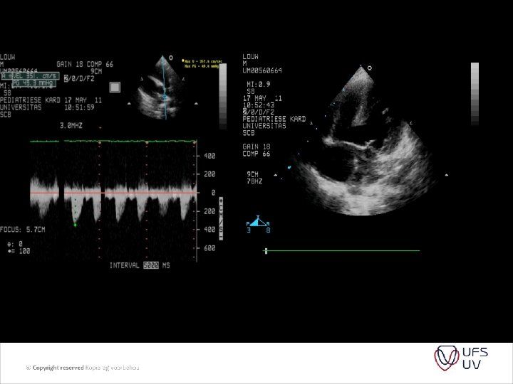
• • Figure 1 Images showing peak tricuspid regurgitant velocity (TRV) and right ventricular outflow time-velocity integral (TVIRVOT) in a patient with normal pulmonary vascular resistance (PVR). (A) TRV is 2. 86 m/s. (B) TVIRVOT is 20. 8 cm. The ratio of TRV/TVIRVOT = 2. 86/20. 8 = 0. 1375. . This patient’s invasive PVR measurement was within 0. 4 WU of the echocardiographic value (PVRCATH = 1. 3 WU). PVRECHO = PVR in WU calculated based on the linear regression equation in which a value for PVR in WU was modeled based on TRV/TVIRVOT. PVRCATH = invasive PVR.

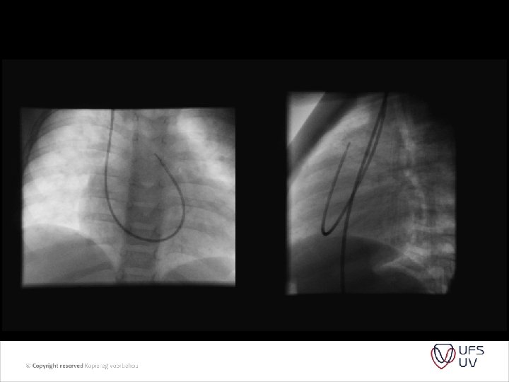
• Figure 2 Images showing TRV and TVIRVOT in a patient with elevated PVR. (A) TRV is 3. 64 m/s. (B) TVIRVOT shows a clear deceleration of pulmonary flow before the pulmonic valve closure click and is calculated at 6. 5 cm. The ratio of TRV/TVIRVOT = 3. 64/6. 5 = 0. 56. . This patient’s invasive PVR measurement is also within 0. 4 WU of the echocardiographic value (PVRCATH = 6. 0 WU). Abbreviations as in Figure 1. • • J Am Coll Cardiol, 2003; 41: 10211027

• Cath – more accurate, but still uses Fick’s principle Qp/Qs = Ao – RA(SVC) / LA – PA Many variables

WHO SHOULD WE BAND? • Indications: 3 Groups - A: Pulmonary over circulation – L-R shunting who require reduction in PBF B: TGA/VSD C: Hybrid

• Group A: VSD, AVSD, TA type 1 C, DURV without PS, Truncus arteriosus, absent pulmonary valve syndrome ext. - Prevent Pulmonary over circulation / reduction in pulmonary hypertension • Group B: d. TGA with initial late presentation - To train LV for arterial switch • Group C: HLHS – ductal stent and branch PA banding
 Difficulty in determining tightness of band b)")
• Limited by several factors a) Difficulty in determining tightness of band b) Several peri-operative variables – anaesthesia, p. H , PPV c) Age-related variability of ventricular adaptive response d) Repeat banding to adjust the band parameters – overbanding / underbanding e) Long periods of meds and ICU to control pulmonary bloodflow f) Need for reconstruction of PA at time of debanding

• Caption: Picture 4. Pulmonary artery banding. Circumferential banding of a dilated pulmonary artery can acutely lead to internal infolding of the arterial wall. Later resorption of the infoldings and remodeling of the arterial wall restore a greater internal crosssectional area.

HOW TIGHT SHOULD THE BAND BE? • Trusler formula - early 1972 • A method of banding the pulmonary artery for large isolated ventricular septal defect with and without transposition of the great arteries. Trusler GA, Mustard WT. I - noncyanotic nonmixing lesions - 20 mm + 1 mm/kg II - Mixing lesions (TGA+VSD) - 24 mm + 1 mm/kg III - Single ventricle for Fontan - 22 mm + 1 mm/kg

• Intra-op pressure and saturation monitoring , aim to lower PAP to normal or ½ of systemic without desaturation or bradycardia - many variable factors - GA - Mechanical ventilation - Open chest - Days after op when hematocrit / p. H ext. • Determine Qp/Qs after Trusler formula was used. • Site of placement – mid MPA trunk

COMPLICATIONS • Migration of band - impingement and stenosis of branch PA • To proximal placement – PV distortion • Inadequate banding – Pulm overflow/CCF • Over banding • Erotion of PA • Distortion of PA • Mortality rate assosiated with complexity of lesion and overall condition of the patient. • Early day as high as 25% - now 3 -5%

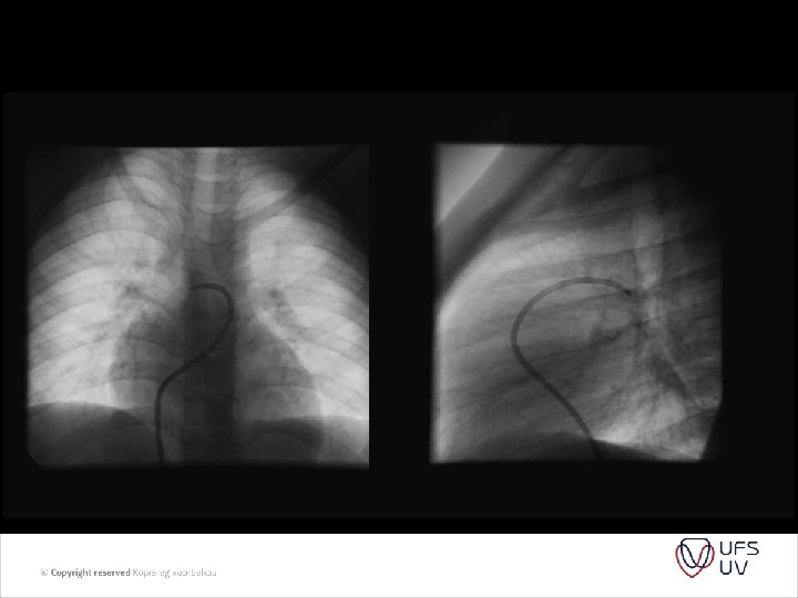
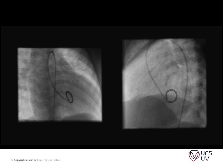
FUTURE • Intraluminal • Thoracoscopically implantable • Adjustable bands – Flo. Watch-R-PAB(Endoart SA, Lausanne, Switserland) – clinical trials • Devices – not option for Africa

• General View of the Flo. Watch-PAB implant: the four main functional parts are: • 1) The case (body of the device) • 2) The silicone membrane • 3) The piston • 4) The counter-piece • 5) The clip (a) with the place for the attachment to the case (b)

ALTERNATIVES • Dilatable bands – may postpone to more desirable weight - S Brown et al. / EJCTS 37 (2010) - 2003 – 2009 (20) - non-resorbable 2 mm nylon with vascular clips, 6/0 prolene - open ring 3. 0 -4. 0 mm Gore-Tex , polypropylene 7/0 - not exceeded 120% - Handmade, cheap , already available - Allows surgeon to make band tighter - Pulmonary artery pressures can progressively increased
")
S Brown et al. / EJCTS 37 (2010)

WHEN AND WHO TO BAND IN AFRICA ? • Timing – important - lesion • How will one decide to band – Echo / Cath / Other - What will be the minimum diagnostic equipment be • How will these patient be followed • What will the future hold for these patients

T: 051 401 9111 info@ufs. ac. za www. ufs. ac. za
- Slides: 30