Pterygopalatine Fossa Objectives 1 Describe the boundaries and

Pterygopalatine Fossa

Objectives 1. Describe the boundaries and communications of the pterygopalatine fossa 2. Describe the course, anatomical relations, and branches of the maxillary nerve 3. Name the various types of fibers that the pterygopalatine ganglion receives and their sources 4. Name the branches of the pterygopalatine ganglion and describe their course and the structures they supply 5. Name the branches of the 3 rd part of the maxillary artery and describe their course and the structures they supply

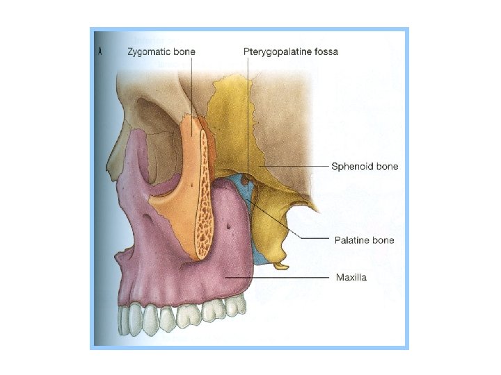
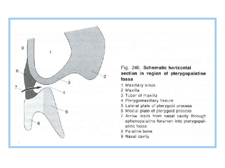
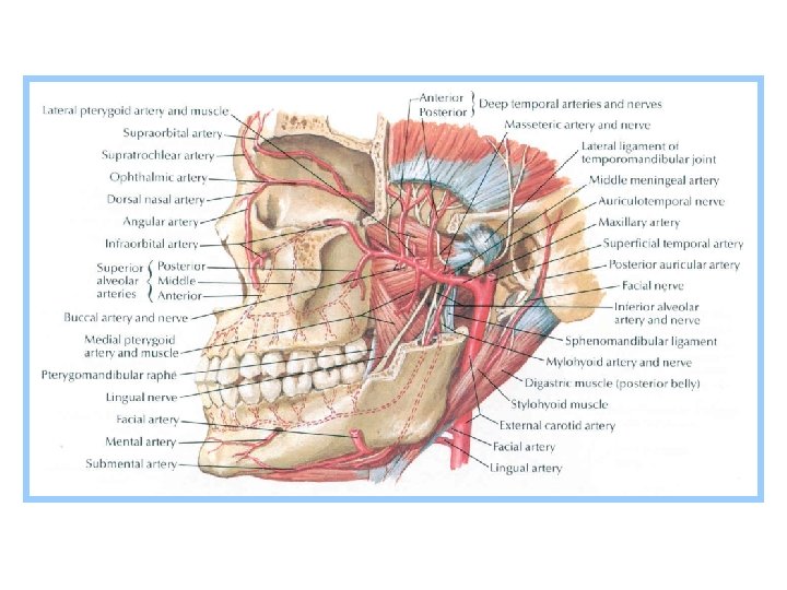
Pterygopalatine Fossa • small pyramidal space located medial to infratemporal fossa • boundaries: • anterior: posterior surface of body of maxilla • posterior: pterygoid process • superior: greater wing of sphenoid • medial: perpendicular plate of palatine • communications: • with infratemporal fossa via pterygomaxillary fissure • with orbit via inferior orbital fissure • with middle cranial fossa via foramen rotundum
: • with nasal cavity via sphenopalatine foramen •")
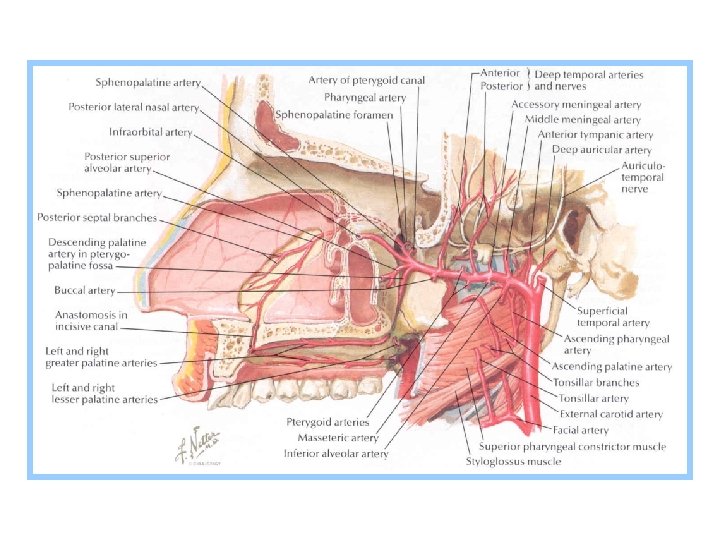
Pterygopalatine Fossa • communications (cont. ): • with nasal cavity via sphenopalatine foramen • pterygoid canal: runs in sagittal direction (anterior to posterior) through upper part of medial pterygoid plate posterior opening is located in anterior border of foramen lacerum anterior opening is located in posterior wall of pterygopalatine fossa, medial and inferior to foramen rotundum • greater and lesser palatine canals: begin at inferior end of pterygopalatine fossa and terminate on inferior aspect of hard palate • contents: maxillary nerve, pterygopalatine (3 rd) part of maxillary artery, and pterygopalatine ganglion


")
(Pterygoid)

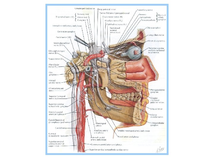
Maxillary Nerve • contains only general sensory fibers • leaves middle cranial fossa and enters pterygopalatine fossa via foramen rotundum passes anteriorly through upper part of pterygopalatine fossa & enters the orbit via inferior orbital fissure where it becomes the infraorbital nerve. • infraorbital nerve lies first in infraorbital groove and then in infraorbital canal &emerges onto face through infraorbital foramen.

Maxillary Nerve • Intracranial branch: • meningeal branch: supplies dura mater of middle cranial fossa • branches in pterygopalatine fossa: • ganglionic branches: connect maxillary nerve with pterygopalatine ganglion (parasympathetic ganglion located in pterygopalatine fossa, inferior to maxillary nerve) The ganglionic branches contain mostly sensory fibers (also contain a few postganglionic parasympathetic fibers for lacrimal gland) • zygomatic nerve: enters orbit via inferior orbital fissure & runs anteriorly, along lateral wall of orbit, and divides into zygomaticofacial and zygomaticotemporal nerves
: • zygomatic nerve (cont. ):")
Maxillary Nerve • branches in pterygopalatine fossa (cont. ): • zygomatic nerve (cont. ): • zygomaticofacial nerve: supplies skin over prominence of cheek • zygomaticotemporal nerve: supplies skin over anterior temporal region • posterior superior alveolar nerve: supply maxillary molar teeth and mucosa of maxillary sinus

Maxillary Nerve • branches from infraorbital part: • middle superior alveolar nerve supplies maxillary premolar teeth and mucosa of maxillary sinus • anterior superior alveolar nerve supplies maxillary incisors and canine and mucosa of maxillary sinus • branches in face: • infraorbital nerve emerges onto face via infraorbital foramen & divides into branches that supply skin and conjunctiva of lower eyelid, skin of side of nose, and skin and mucosa of upper lip
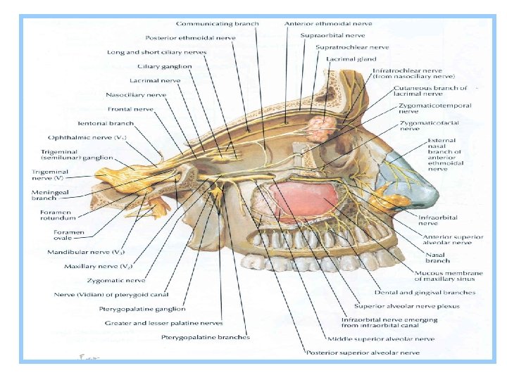


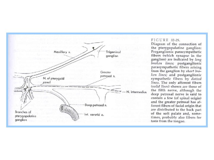
Pterygopalatine Ganglion • parasympathetic ganglion located in pterygopalatine fossa, inferior to maxillary nerve • receives three types of fibers (sensory, preganglionic parasympathetic and postganglionic sympathetic), but only preganglionic parasympathetic fibers synapse in ganglion • source of general sensory fibers: maxillary nerve • source of preganglionic parasympathetic fibers: greater petrosal nerve • source of postganglionic sympathetic fibers: deep petrosal nerve • branches that originate from pterygopalatine ganglion contain sensory, postganglionic parasympathetic and postganglionic sympathetic fibers

Pterygopalatine Ganglion Greater petrosal nerve: • branch of facial nerve • contains preganglionic parasympathetic fibers for lacrimal glands of nasal cavity, palate and nasopharynx • joins deep petrosal nerve (branch of internal carotid plexus) to form nerve of pterygoid canal • deep petrosal nerve contains postganglionic sympathetic fibers (from neurons located in superior cervical ganglion of sympathetic trunk)

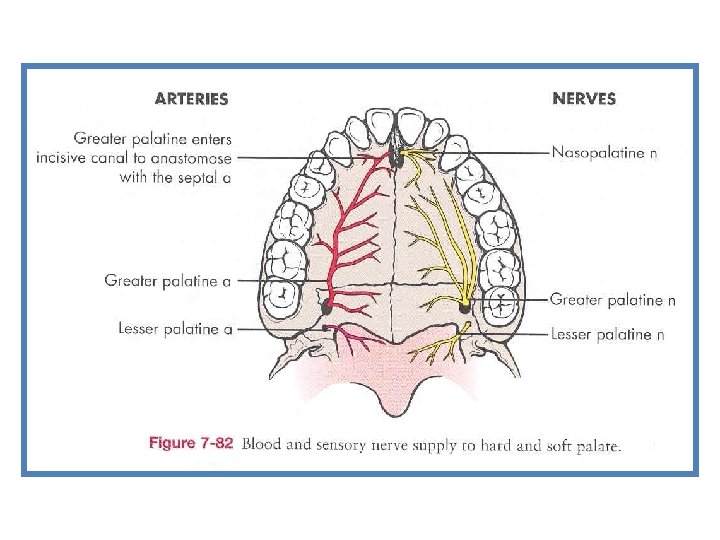
Pterygopalatine Ganglion • nerve of pterygoid canal passes anteriorly, through pterygoid canal, and terminates in pterygopalatine ganglion • preganglionic parasympathetic fibers synapse in pterygopalatine ganglion • sensory and postganglionic sympathetic fibers do not synapse in ganglion pass without interruption into branches of ganglion • branches: • greater palatine nerve: emerges on inferior surface of hard palate via greater palatine foramen runs anteriorly on inferior surface of hard palate supplies mucosa of posterior part of hard palate
: • lesser palatine nerve: emerges on inferior surface")
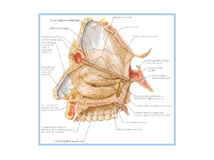
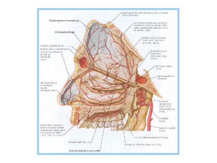
Pterygopalatine Ganglion • branches (cont. ): • lesser palatine nerve: emerges on inferior surface of hard palate via lesser palatine foramen supplies mucosa of soft palate and tonsil • nasopalatine (long sphenopalatine) nerve: enters nasal cavity via sphenopalatine foramen passes anteriorly and inferiorly along nasal septum and supplies its mucosa passes through incisive canal and emerges on inferior surface of hard palate via incisive fossa supplies mucosa of anterior part of hard palate (behind incisor teeth)
: • posterolateral nasal branches: originate from pterygopalatine ganglion")
Pterygopalatine Ganglion • branches (cont. ): • posterolateral nasal branches: originate from pterygopalatine ganglion and greater palatine nerve supply mucosa of posterior part of lateral wall of nasal cavity • pharyngeal nerve: supplies mucosa of sphenoidal sinus and nasopharynx • course of postganglionic parasympathetic fibers for lacrimal gland: pterygopalatine ganglion maxillary nerve zygomaticotemporal nerve communicating branch with lacrimal nerve lacrimal gland

Innervation of the Lacrimal Gland
 Part of Maxillary Artery • maxillary artery leaves infratemporal fossa and")
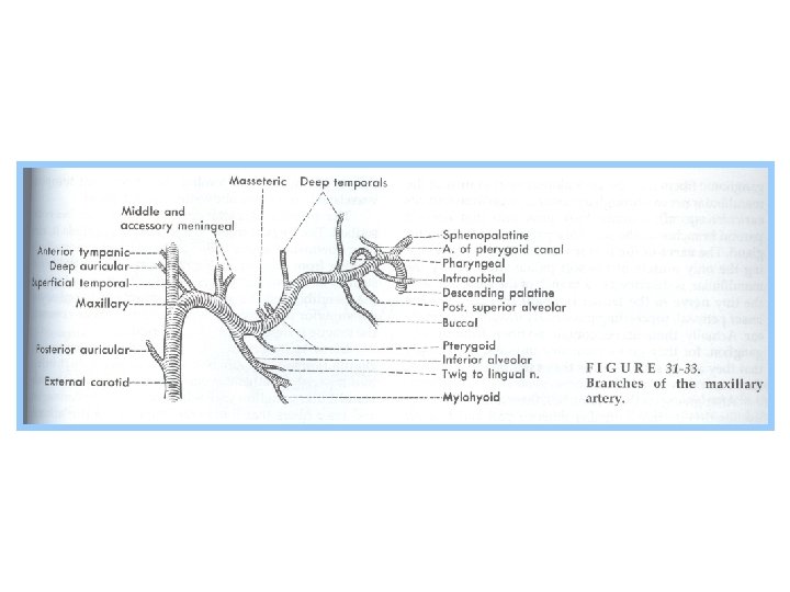
Pterygopalatine (3 rd) Part of Maxillary Artery • maxillary artery leaves infratemporal fossa and enters pterygopalatine fossa via pterygomaxillary fissure • its branches follow branches of maxillary nerve and pterygopalatine ganglion • branches: • posterior superior alveolar artery: accompanies posterior superior alveolar nerve descends on posterior surface of maxilla divides into branches which pass through alveolar foramina of maxilla supplies maxillary molar and premolar teeth and mucosa of maxillary sinus
 Part of Maxillary Artery • branches (cont. ): • infraorbital artery:")
Pterygopalatine (3 rd) Part of Maxillary Artery • branches (cont. ): • infraorbital artery: accompanies infraorbital nerve enters orbit through inferior orbital fissure runs anteriorly in infraorbital groove and infraorbital canal emerges onto face via infraorbital foramen gives following branches: • anterior superior alveolar artery supplies maxillary incisor and canine teeth and mucosa of maxillary sinus (sometimes there is also a middle superior alveolar artery) • terminal branches (on face) supply lower eyelid, side of nose and upper lip
 Part of Maxillary Artery • branches (cont. ): • descending palatine")
Pterygopalatine (3 rd) Part of Maxillary Artery • branches (cont. ): • descending palatine artery: divides into lesser and greater palatine arteries lesser palatine artery emerges through lesser palatine foramen to supply soft palate and tonsil greater palatine artery reaches inferior surface of hard palate via greater palatine foramen passes forward to supply mucosa of hard palate gives off a branch that passes through incisive canal and anastomoses with a branch of sphenopalatine artery • pharyngeal artery: runs with pharyngeal branch of pterygopalatine ganglion supplies mucosa of nasopharynx and sphenoidal sinus • artery of pterygoid canal: travels posteriorly in pterygoid canal supplies walls and contents of pterygoid canal and mucosa of nasopharynx, auditory tube and tympanic cavity
 Part of Maxillary Artery • branches (cont. ): • sphenopalatine artery:")
Pterygopalatine (3 rd) Part of Maxillary Artery • branches (cont. ): • sphenopalatine artery: terminal branch of maxillary artery enters nasal cavity via sphenopalatine foramen gives off posterolateral nasal branches and posterior septal branches • posterolateral nasal branches: supply mucosa of posterior part of lateral wall of nasal cavity • posterior septal branches: supply mucosa of posterior part of nasal septum one of these branches anastomoses with a branch of greater palatine artery
- Slides: 33