PSYCHOLOGY Chapter 3 BIOPSYCHOLOGY Power Point Image Slideshow

PSYCHOLOGY Chapter 3 BIOPSYCHOLOGY Power. Point Image Slideshow

CHAPTER TOPICS • Genetics and DNA • Evolution by Natural Selection • The Genetics and Evolution of Behavior • The Experience Dependent Brain • Building Blocks of the Nervous System and Brain • Communication among Neurons

CHAPTER TOPICS • Methods for Studying the Nervous System • The Architecture of the Nervous System • The Cerebral Cortex • Plasticity • Some Final Thoughts: Do All Psychological Questions Have Biological Answers? • Summary
 In 1859, Charles Darwin proposed his theory of evolution by")
FIGURE 3. 3 (a) In 1859, Charles Darwin proposed his theory of evolution by natural selection in his book, On the Origin of Species. (b) The book contains just one illustration: this diagram that shows how species evolve over time through natural selection.

DARWIN AND EVOLUTION • Charles Darwin hypothesized that all modern organisms: • are descended from a small set of shared ancestors. • have emerged over time through the process of evolution. • An enormous amount of evidence has confirmed these proposals.

DARWIN AND EVOLUTION • The key mechanism is natural selection. • If individuals with certain traits are more likely to survive and reproduce, • their genes will be better represented in the next generation. • And if the genes gave rise to the advantageous traits, • those traits will be more common in the next generation.

NATURALISTIC FALLACY • It is important to avoid the naturalistic fallacy, however—it does not follow that evolution somehow improves organisms or that anything natural is good. • This means the genotypes and phenotypes that are passed on to survive allow the organisms to survive. It does not necessarily mean this is good or bad. • Can you think of examples?

QUIZ 1 q. Please define natural selection. q. What is the naturalistic fallacy?

GENETICS AND DNA • The nucleus of each biological cell contains chromosomes, which each contain a single molecule of DNA. • Within this molecule, genes govern the cell’s functioning by providing detailed instructions for making proteins. This a very complex process.

MAKING PROTEINS q. The genes contain the information needed to make proteins. This allows the information in the gene to be expressed for the genotype. q. A genotype is your complete heritable genetic identity. It is written in code (your DNA). This is the “nature” meaning what you inherit from your parents. It is the way your genes are “expressed”.

GENOTYPE AND PHENOTYPE q. Phenotype: This is the description of your actual physical characteristics, temperament, personality, etc. This is influenced by your genotype and by your environment. q. The phenotype and genotype are in constant interaction. q. Why do we need to know this information for the study of psychology?
 Genotype refers to the genetic makeup of an individual based")
FIGURE 3. 4 (a) Genotype refers to the genetic makeup of an individual based on the genetic material (DNA) inherited from one’s parents. (b) Phenotype describes an individual’s observable characteristics, such as hair color, skin color, height, and build. (credit a: modification of work by Caroline Davis; credit b: modification of work by Cory Zanker)

DOMINANT VS. RECESSIVE • The genes may or may not be the same allele. Ex: blue eyes (allele) from mom and brown eyes (allele) from dad. • Geneticists use the terms dominant and recessive to describe the inheritance patterns of certain traits. In other words, how likely is it for a certain phenotype to pass from parent to offspring. • These terms are used to predict the probability of someone inheriting a phenotype.

DOMINANT AND RECESSIVE q. Organisms have two alleles for each trait. q. One allele may be dominant (so it is expressed) and the other is recessive (so it is masked). q. Co-dominant is when both alleles are expressed (AB blood type). q. Incomplete dominant is when both alleles mix, as in flowers. This means both traits are shown and not masked.
 A Punnett square is a tool used to predict how")
FIGURE 3. 5 (a) A Punnett square is a tool used to predict how genes will interact in the production of offspring. The capital B represents the dominant allele, and the lowercase b represents the recessive allele. In the example of the cleft chin, where B is cleft chin (dominant allele), wherever a pair contains the dominant allele, B, you can expect a cleft chin phenotype. You can expect a smooth chin phenotype only when there are two copies of the recessive allele, bb. (b) A cleft chin, shown here, is an inherited trait.

GENE EXPRESSION • In each cell, some genes are expressed, or activated in some way, at any point in time and others are not. • Gene expression is controlled by the biochemical environment inside the cell, which is influenced by the organism’s: • Overall environment. • Experiences and Behaviors.

QUIZ 2 q. Please define Genome? q. What is a genotype? q. What is a phenotype? q. What does “gene expression” mean? q. Please define the naturalistic fallacy.

FIGURE 3. 7 Nature and nurture work together like complex pieces of a human puzzle. The interaction of our environment and genes makes us the individuals we are. (credit “puzzle”: modification of work by Cory Zanker; credit “houses”: modification of work by Ben Salter; credit “DNA”: modification of work by NHGRI)

THE DIATHESIS STRESS MODEL qhttps: //youtu. be/ay. WVkm. YH 6 TY q. A person’s genetic predisposition, combined with psychological stressors, are what cause someone to become ill.

GENETICS IN PSYCHOLOGY q. Heritability refers to a statistic of proportion (the part in relation to the whole) to explain how likely we are to inherit something through our genetics. q. The numbers range from 0. 0, where we see that genes do not contribute at all to phenotypic individual difference or the overt traits and behaviors, to 1. 0 where genes are the only reason for these differences.

GENETIC EPIDEMIOLOGY OF SELECTED MENTAL HEALTH ISSUES Prevalence Paternal Age onset Mortality Fertility Heritability (%) age effect 0. 30 1 2. 0 0. 05 0. 90 1. 4 Autism Anorexia 0. 60 nervosa Schizophre 0. 70 nia Bipolar affective 1. 25 disorder Unipolar 10. 22 depression Anxiety 28. 80 disorders 15 6. 2 0. 33 0. 56 — 22 2. 6 0. 40 0. 81 1. 4 25 2. 0 0. 65 0. 85 1. 2 32 1. 8 0. 90 0. 37 1 11 1. 2 0. 90 0. 32 — Molecular Psychiatry (2009) 14, 1072– 1082; doi: 10. 1038/mp. 2009. 85; published online 25 August 2009 The role of genetic variation in the causation of mental illness: an evolution-informed framework. By R. Uher.

GENETIC PREDISPOSITION q. The likelihood of inheriting something based on genetic makeup. q. Prevalence refers to proportion of the population found to have a condition. This one is number of cases per 100 people. q. Incidence means the number of new cases in a population at a given time. q. What do you believe are the factors involved that create the illness when someone has a genetic predisposition?

OUTSIDE FORCES ILLNESS Anorexia Autism Schizophrenia Bipolar Disorder Major Depression Generalized Anxiety OCD OTHER CAUSES Related to being white, female, and in a Western Culture. Diagnosis has increased with time. Increased risks living in urban environment and being an ethnic minority. Depression 1 in 10 people. MTHFR, 1/3 in lifetime.

MENTAL HEALTH BOOSTER

CELLS OF THE NERVOUS SYSTEM q. Identify the basic parts of the Neuron q. Describe how neurons communicate with each other q. Explain how drugs act as agonists or antagonists for a given neurotransmitter system

NEURONS AND GLIA CELLS q. THE MAIN CELLS IN YOUR CENTRAL NERVOUS SYSTEM. q. NEURONS ARE THE INFORMATION PROCESSING STRUCTURES WITHIN YOUR BRAIN AND CNS. q. WHAT IS INFORMATION PROCESSING? q. GLIA CELLS SUPPORT THE NEURONS IN THEIR EFFORT TO PROCESS, SEND, PROCESS, AND RECEIVE INFORMATION. THEY ARE THE FOUNDATION AND STRUCTURES THAT ALLOW NEURONS TO DO THEIR JOB. q. NEURONS RECEIVE INPUT FROM OTHER NEURONS, PROCESS AND MAKE SENSE OF THIS COMMUNICATION, AND SEND OUTPUT TO ANOTHER NEURON(S) BASED ON THE COMMUNICATIONS THEY RECEIVED. q. NEURONS CONTROL THE FLOW FOR ALL MOTOR, SENSORY, AND COGNITIVE INFORMATION TO RUN THE UNIQUE BRAIN AND BODY THAT IS YOU. q. THIS WHOLE PROCESS IS VERY CHAOTIC! q. NOW FOR BACKGROUND INFORMATION….

THE STRUCTURE OF THE NEURON • THERE ARE OVER 10, 000 TYPES OF NEURONS. • THERE ARE MOTOR NEURONS CONTROLLING MOVEMENTS, SENSORY NEURONS, AND COGNITVE NEURONS. • YOUR NEURONS CONTAIN FOUR PARTS: • A CELL BODY THAT FUNCTIONS AS THE METABOLIC MANAGING CENTER. • PROCESSES THAT ARE CONDUITS TO INFORMATION FLOW: THOUSNDS OF DENDRITES THAT RECEIVE SIGNALS FROM OTHER NEURONS and ONE AXON THAT SENDS AN OUTGOING SIGNAL. • AXON TERMINALS THAT CONTAIN THE NEUROTRANSMITTERS THAT SEND CHEMICAL SIGNALS BETWEEN NEURONS.

FIGURE 3. 8 This illustration shows a prototypical neuron, which is being myelinated.

QUIZ 3 q. What are the four parts of the neuron? q. What do each of these four parts do? q. Draw a picture of a neuron. Please label each part.

NEUROTRANSMISSION: COMMUNICATION BETWEEN NEURONS q. THIS BEGINS WHERE ELECTRICAL CONDUCTION IS LEFT OFF WHICH IS AT THE AXON TERMINALS. q. NEUROTRANSMISSION HAPPENS AT THE SYNAPSES. q. THERE ARE TWO TYPES OF TRANSMISSIONS: CHEMICAL SYNAPSES AND ELECTRICAL SYNAPSES. MOST TRANSMISSIONS ARE CHEMICAL.

NEUROTRANSMISSION VIA THE SYNAPSES q. NEURONS HAVE NO CONTINUTITY BETWEEN NEURONS MEANING THEY ARE NOT CONNECTED. q. A SYNAPSE IS THE GAP BETWEEN EACH NEURON. THIS BOUNDARY IS SELECTIVE WHICH CAN BE BAD OR GOOD. q. NEUROTRANSMITTERS FUNCTION AS THE MESSENGER ACROSS THIS GAP. q. THEY ARE SPECIALIZED CHEMICALS THAT ALLOW THE NEURONS TO COMMUNICATE WITH EACH OTHER. THEY EITHER EXCITE OR INHIBIT ACTION BETWEEN THE NEURONS. q. EXAMPLES ARE SEROTONIN, DOPAMINE, NOREPINEPHRINE, AMINO ACIDS, HISTAMINE, ETC. q. THEY TELL YOUR HEART TO BEAT, YOUR STOMACH TO DIGEST, AND AFFECT YOUR MOOD, SLEEP, WEIGHT, ETC.
 The synapse is the space between the terminal button of")
FIGURE 3. 9 (a) The synapse is the space between the terminal button of one neuron and the dendrite of another neuron. (b) In this pseudo-colored image from a scanning electron microscope, a terminal button (green) has been opened to reveal the synaptic vesicles (orange and blue) inside. Each vesicle contains about 10, 000 neurotransmitter molecules. (credit b: modification of work by Tina Carvalho, NIH-NIGMS; scale-bar data from Matt Russell)

SYNAPTIC TRANSMISSION q. CHEMICAL TRANSMISSION OF INFORMATION ALLOWS THE NEURONS MORE CONTROL OVER THE COMMUNICATION. q. THE FOLLOWING PROCESS OCCURS: q. THE ACTION POTENTIAL ARRIVES AT THE AXON TERMINAL (THE END OF THE AXON). q. ION CHANNELS OPEN FOR COMMUNICATION TO OCCUR. THE IONS ENTER THE PRESYNAPTIC NEURON. q. THE IONS BIND WITH OTHER CHEMICALS. THIS ALLOWS THE VESICLES TO MERGE WITH THE PRESYNAPTIC MEMBRANE. THIS MERGE ALLOWS THE NEUROTRANSMITTERS TO BE RELEASED IN THE SYNPTIC CLEFT.

q. AS THE NEUROTRANSMITTERS RELEASE INTO THE SYNAPTIC CLEFT, THEY DIFFUSE THROUGH THE MEMBRANE (THE MEMBRANE IS SEMI PERMEABLE). q. NOW THE NEUROTRANSMITTER BINDS WITH THE MEMBRANES AT THE ENDS OF BOTH CELLS (THE SENDER CELL WHICH IS PRESYNAPTIC AND THE RECEIVER CELL THE POST SYNAPTIC). THIS IS EITHER DIRECT (JUST HAPPENS) OR INDIRECT (CAUSES THE RELEASE OF A SECONDARY MESSENGER). q. THIS CAN CAUSE TWO RESPONSES FROM THE RECEIVER CELL (POST SYNAPTIC). THE RESPONSE IS EITHER AN EXCITATORY OR INHIBITORY.
 is more highly concentrated outside")
FIGURE 3. 10 At resting potential, Na+ (blue pentagons) is more highly concentrated outside the cell in the extracellular fluid (shown in blue), whereas K+ (purple squares) is more highly concentrated near the membrane in the cytoplasm or intracellular fluid. Other molecules, such as chloride ions (yellow circles) and negatively charged proteins (brown squares), help contribute to a positive net charge in the extracellular fluid and a negative net charge in the intracellular fluid.

QUIZ 5 q. Please explain chemical neural communication.

FIGURE 3. 11 During the action potential, the electrical charge across the membrane changes dramatically.

FIGURE 3. 12 Reuptake involves moving a neurotransmitter from the synapse back into the axon terminal from which it was released.
 the")
FIGURE 3. 13 The nervous system is divided into two major parts: (a) the Central Nervous System and (b) the Peripheral Nervous System.
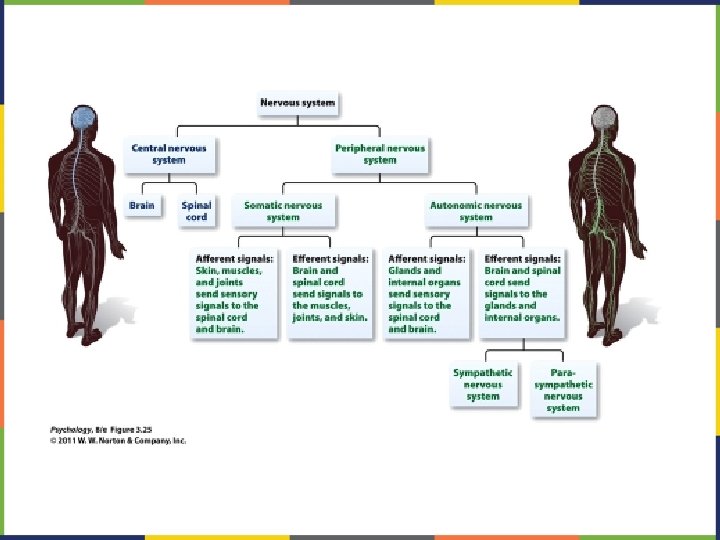

FIGURE 3. 15 The surface of the brain is covered with gyri and sulci. A deep sulcus is called a fissure, such as the longitudinal fissure that divides the brain into left and right hemispheres. (credit: modification of work by Bruce Blaus)
 The corpus callosum connects the left and right hemispheres")
FIGURE 3. 16 (a, b) The corpus callosum connects the left and right hemispheres of the brain. (c) A scientist spreads this dissected sheep brain apart to show the corpus callosum between the hemispheres. (credit c: modification of work by Aaron Bornstein)

THE STRUCTURE AND THE FUNCTION OF THE BRAIN qhttps: //youtu. be/k. MKc 8 nf. PATI q. Discussion of the CNS and the PNS q. The CNS consists of the brain and spinal cord. q. The PNS consists of the nerves and ganglia (nerve cell clusters) outside of the brain and spinal cord.

FIGURE 3. 17 The brain and its parts can be divided into three main categories: the forebrain, midbrain, and hindbrain.

FIGURE 3. 18 The lobes of the brain are shown.
 Phineas Gage holds the iron rod that penetrated his skull")
FIGURE 3. 19 (a) Phineas Gage holds the iron rod that penetrated his skull in an 1848 railroad construction accident. (b) Gage’s prefrontal cortex was severely damaged in the left hemisphere. The rod entered Gage’s face on the left side, passed behind his eye, and exited through the top of his skull, before landing about 80 feet away. (credit a: modification of work by Jack and Beverly Wilgus)

QUIZ 1 q. How do scientists learn about the inner workings of the human brain? q. Who is Phinneas Gage? How is what happened to him significant for neuroscience? q. You will need to know the structures and their functions of the brain as discussed in the video. What study methods will you use to remember this information?

FIGURE 3. 20 Spatial relationships in the body are mirrored in the organization of the somatosensory cortex.

FIGURE 3. 21 Damage to either Broca’s area or Wernicke’s area can result in language deficits. The types of deficits are very different, however, depending on which area is affected.

FIGURE 3. 22 The thalamus serves as the relay center of the brain where most senses are routed for processing.

FIGURE 3. 23 The limbic system is involved in mediating emotional response and memory.
 are located in")
FIGURE 3. 24 The substantia nigra and ventral tegmental area (VTA) are located in the midbrain.

FIGURE 3. 25 The pons, medulla, and cerebellum make up the hindbrain.

FIGURE 3. 1 Different brain imaging techniques provide scientists with insight into different aspects of how the human brain functions. Left to right, PET scan (positron emission tomography), CT scan (computed tomography), and f. MRI (functional magnetic resonance imaging) are three types of scans. (credit “left”: modification of work by Health and Human Services Department, National Institutes of Health; credit “center”: modification of work by “Aceofhearts 1968”/Wikimedia Commons; credit “right”: modification of work by Kim J, Matthews NL, Park S. )
 The")
FIGURE 3. 26 A CT scan be used to show brain tumors. (a) The image on the left shows a healthy brain, whereas (b) the image on the right indicates a brain tumor in the left frontal lobe. (credit a: modification of work by “Aceofhearts 1968”/Wikimedia Commons; credit b: modification of work by Roland Schmitt et al)

FIGURE 3. 27 A PET scan is helpful for showing activity in different parts of the brain. (credit: Health and Human Services Department, National Institutes of Health)

FIGURE 3. 28 An f. MRI shows activity in the brain over time. This image represents a single frame from an f. MRI. (credit: modification of work by Kim J, Matthews NL, Park S. )

FIGURE 3. 29 Using caps with electrodes, modern EEG research can study the precise timing of overall brain activities. (credit: SMI Eye Tracking)

BRAIN ILLNESS/INJURY AND DEPRESSION q. Stroke can cause paralysis, speech problems, and an inability to complete daily tasks. q. Depression and stroke and heart attack are linked. q. Seizures happen when the brain cells, which communicate through electrical signals, send out the wrong signals.

SEIZURES q. In about half the cases the cause is not known. q. Causes include head injury, genes, dementia, brain injury, lupus, meningitis, stroke, heart attack. q. Treatment may include brain removal.

BRAIN INJURY q. Post Concussive Syndrome qhttps: //youtu. be/N 9 S_Hdkqf 08 q. Other Traumatic Brain Injury qhttps: //youtu. be/Hxa. Ne. E 7 Qzn 4 q. Due to illness of medications qhttp: //www. cbsnews. com/videos/u-s-troops-given-anti-malaria-drugdespite-concern-of-side-effects

QUIZ 6 q. What is an experience-dependent brain? What is plasticity? q. What is an experience-expectant brain? q. What are two types of brain injury? q. Can your brain be injured due to medications or illegal drug use?

PLASTICITY IN ACTION qhttp: //bigthink. com/think-tank/brain-exercise q. Another example of neuroplasticity has been found in London taxi drivers. A cab driver's hippocampus -- the part of the brain that holds spatial representation capacity -- is measurably larger than that of a bus driver. By driving the same route every day, the bus drivers don't need to exercise this part of the brain as much. The cabbies, on the other hand, rely on it constantly for navigation.

FIGURE 3. 30 The major glands of the endocrine system are shown.

This Power. Point file is copyright 2014 -2015, Rice University. All Rights Reserved.
- Slides: 67