PSY 368 Human Memory Neuropsychology Memory Announcements Experiment


- Slides: 68

PSY 368 Human Memory Neuropsychology & Memory

Announcements • Experiment 2 due today • Focus Questions for Weldon and Roediger (1987) Due Monday March 26 th • Exam 2 a week from Wednesday (March 28)

Neuropsychology of Memory • Where is memory? The Brain • Methods of study • Neurons and Brains • Role of the hippocampus • Memory Disorders • Amnesia • Alzheimer’s Disease Memory Mapping memory in 3 D

Neurons and Memory • Connections between neurons change based on experiences • Brain is less ‘hard-wired’ than we used to believe • Lashley provided evidence of plasticity in monkeys in the 1920’s but not widely accepted until 1960’s • Neuroplasticity is fundamental property of brain (nervous system) • Capacity of nervous system to modify its organization • Changes in structure and function as a result of experience • Changes largely within the synapses

Neurons and Memory • Synaptic changes that could store memories • Current dominant theory: Long-term potentiation (LTP) • Persistent increase in synaptic strength following high-frequency stimulation • “the molecular and cellular changes mediating the induction of LTP in the hippocampus are widely considered to provide a basis for memory” (Mc. Gaugh, 2000) • Not all learning-related changes involve changes in synaptic strength (Martin & Morris, 2002) • Neurogenesis – new evidence suggest that new neurons are formed in some regions of the brain • Changes in neuronal excitability – changes in the firing threshold

The Brain: networks of neurons • “There are many steps between synaptic change and behavioral memory. ” Squire (pg 8, 1987) • Lots of interesting questions • Does “memory” reside in single neurons, or in networks of neurons? • Are all of the networks the same, or are there differences (i. e. , do different regions of the brain deal with different kinds of things)?

The Brain Vital Statistics • Adult weight: about 3 pounds • Adult size: a medium cauliflower • Number of neurons: 100, 000, 000 (100 billion) • Number of synapses (the gap between neurons): 100, 000, 000 (100 trillion) • These neurons are connected, organized into networks of neurons

Structure of the brain • Cortex - four lobes • Occipital - vision Frontal Lobe • Parietal - sensation • Temporal – memory, hearing • Frontal - reasoning, memory Parietal Lobe Occipital Lobe Temporal Lobe

Brain and Memory Other Crucial Parts • Limbic system: controls emotions and instinctive behavior (includes the hippocampus and parts of the cortex) • Thalamus: receives sensory and limbic information and sends to cerebral cortex • Hypothalamus: monitors certain activities and controls body’s internal clock • Hippocampus: where short-term memories are converted to long-term memories

The Brain: networks of neurons • Networks of neurons hold memories Neurobiological systems regulating the consolidation of memory • “… memory consolidation involves interactions among neural systems as well as cellular changes within specific systems, and that the amygdala is critical for modulating consolidation in other brain regions”Mc. Gaugh, 2000

The Brain: networks of neurons • So where is memory? It is complicated • Multiple brain regions are involved in encoding (as shown by f. MRI) -term memory.

Brain and Memory • So where is memory? It is complicated • Multiple brain regions are involved in encoding (as shown by f. MRI) • For recalling pictures, the right prefrontal cortex and parahippocampal cortex in both hemispheres are activated. • For recalling words, the left prefrontal cortex and the left parahippocampal cortex are activated. • Consolidation of memory involves the hippocampus but the hippocampal system does not store long-term memory. • LTM storage occurs in the cortex, near where the memory was first processed and held in short-term memory.

Brain and Memory • So where is memory? It is complicated • Seven Sins of Memory • Hippocampus and nearby structures related to sin of transience • Parts of the frontal lobe related to transience, but even more central to absent-mindedness and misattribution (and maybe suggestibility) • Area near front of temporal lobe related to blocking • Amygdala closely related to persistence • Not much is known about bias

Brain and Memory • Hippocampus • Important formation of new episodic memories • Important for encoding perceptual aspects of memories • Novel events, places, and stimuli • Important for declarative memory • Especially as part of medial temporal lobe • Supported by case of HM • Video (location, 1 min)

Brain and Memory • Hippocampus • Recollection vs. Knowing (familiarity) • Eldridge et al have shown the hippocampus is selectively involved in R, not with K. • Verfaelle & Treadwell (1993), using process dissociation procedure showed similar pattern (discussed in detail in your textbook) (Eldridge et al. , Nature Neuroscience 2000)

Brain and Memory: Amnesia • Diencephalic amnesia damage to the medial thalamus and mammillary nuclei • Medial temporal lobe amnesia - damage to the hippocampal formation, uncus, amygdala, and surrounding cortical areas • Other implicated regions include Anterior Lateral Temporal Lobe and Frontal Lobes

Amnesia • Loss of memory ability - usually due to lesion or surgical removal of various parts of the brain • Relatively spared performance in other domains • A pure amnesia is relatively rare • video (#18, 10 mins) • Video (~ 7 mins) • Video 3 (Clive Wearing, 7 mins)

Amnesia • Loss of memory ability - usually due to lesion or surgical removal of various parts of the brain • Three different kinds of classifications • Source of the disease (e. g. , illness, injury) • Location of the area of damage • Functional deficit (i. e. , what kind of memory is impaired) • This mixed way of categorizing amnesia causes some difficulties

Amnesia • Loss of memory ability - usually due to lesion or surgical removal of various parts of the brain • Two broad categories: • Retrograde: loss of memories for events prior to damage • Anterograde: loss of ability to store new memories of events after damage Injury Time

Causes of Amnesia • • Korsakoff’s syndrome Traumatic Brain Injury (TBI) (Concussion) Alzheimer’s disease Other causes include • Specific brain lesions (i. e. surgical removal) • Psychological • Dissociative Fugue • Psychogenic • Migraines • Hypoglycemia • Epilepsy • Electroconvulsive shock therapy • Drugs (esp. anesthetics) • Infection • Nutritional deficiency

Amnesia • Korsakoff’s syndrome: • Results from chronic alcoholism and consequent thiamine deficiency • Lesions to Medial Thalamus • Neuropathology: most sources attribute the amnesia to combined lesions in two diencephalic structures: the dorsomedial nucleus of the thalamus and the mammillary bodies of the hypothalamus

Amnesia • Korsakoff’s syndrome • Generally preserved IQ, including a normal digit span. • Personality changes, the most common of which is apathy, passivity and indifference • inability to formulate and follow through a series of plans • Lack of insight into their condition. • How can someone with a shattered memory remember that he has become unable to remember?

Amnesia • Korsakoff’s syndrome • Retrograde amnesia with a temporal gradient • Anterograde amnesia • Confabulation, which is a tendency to "fill in the gaps" of one's memories with plausible made-up stories. • confabulations are rare among chronic Korsakoff patients who've had the disease for more than 5 years. Patients in the chronic stage are more likely to say "I don't know" or remain silent when faced with memory failures rather than to invent stories.

Amnesia • Korsakoff’s syndrome • Worst impairments are on episodic memory tests, including list learning of words, figures, or faces, paragraph recall. • Relatively preserved semantic memory, including normal verbal fluency, vocabulary, rules of syntax, and basic arithmetic operations • Intact sensori-motor memory (mirror tracing, mirror reading, pursuit rotor) • Intact performance on perceptual tasks (e. g. , perceptual identification, generating category exemplars)

Amnesia • Post-traumatic amnesia • Damage due to lesions as well as twisting and tearing of microstructure of brain • Symptomology • After severe TBI, individuals typically lose consciousness • After they begin to regain consciousness, there is often a gradual recovery during which patients have difficulty keeping tracking of and remembering on-going events, though there may be islands of lucidity and memory • In the news • Football (ESPN video) • Soldiers (6 part video series)

Amnesia Injury Time • Retrograde amnesia • Refers to difficulty remembering events that occurred prior to injury • The duration of amnesia varies but can extend back for several years • Rare, short-lived • Typically due to brain trauma • Case Study: Doug Bruce (Unknown White Male) • His case is exceptional (the extent and persistence of the memory loss)

Amnesia Injury Time • Retrograde amnesia • Duration of retrograde amnesia typically shrinks as time passes • e. g. , Russell (1959) described case of TBI as a result of a motorcycle accident • 1 week post accident patient had lost 11 years of memory extending back from injury • 2 weeks post accident patient had last 2 years of memory • about 10 weeks post injury memories of the last two years gradually returned • This pattern of results suggests that retrograde amnesia is a retrieval problem • The pattern of damage/recovery -- from most distant to most recent -- has been argued by some to reflect a failure of consolidation (Ribot’s Law)

Amnesia Injury Time • Retrograde amnesia • Butters & Cermak (1986) reported a case study of an eminent scientist (born 1914) who had written his autobiography only two years prior to becoming amnesic • Tested him by asking him questions all drawn from his autobiography

Amnesia • Anterograde amnesia • Refers to problems of learning new facts • Specific to episodic memories • Procedural memories intact • Implicit memory performance normal • Famous Cases: • H. M. • N. A. • Clive Wearing • Video 3 a, b, c, d (each ~10 mins) Injury Time

Amnesia Case Study: HM • Henry Molaison (Patient H. M. ) (brief news video following his death) • Suffered from extreme epilepsy • Bilateral mesial temporal lobe resection extending 8 cm. back from the temporal tips, including the uncus and amygdala, and destroying the anterior two-thirds of the hippocampus and hippocampal gyrus • Scoville & Milner (1957)

Amnesia Case Study: HM • Henry Molaison (Patient H. M. ) • prototype of amnesia attributable to hippocampal damage • Surgery led to a permanent, severe anterograde amnesia, limited retrograde amnesia, and normal intelligence.

Amnesia Case Study: HM • Henry Molaison (Patient H. M. ) • Functional characteristics • Declarative and nondeclarative memories • Although patients can learn other tasks, they cannot recall ever learning them • Learning and memory involve different processes • 2 major categories of memories • Declarative memories – memory that can be verbally expressed, such as memory for events, facts, or specific stimuli; this is impaired with anterograde amnesia • Nondeclarative memories – memory whose formation does not depend on the hippocampal formation; a collective term for perceptual, stimulusresponse, and motor memory; not affected by anterograde amnesia; these control behavior; cannot always be described in words

Amnesia Case Study: HM • Henry Molaison (Patient H. M. ) • Functional characteristics • Episodic memory is impaired • Both autobiographical and nonautobiographical episodic memory • even for emotionally charged information such as the death of his favorite uncle • Verbal learning is disrupted in anterograde amnesia • e. g. H. M. did not learn any new words after his surgery (biodegradable = “two grades”)

Amnesia Case Study: HM • Henry Molaison (Patient H. M. ) • Functional characteristics • Perceptual learning • e. g. recognize broken drawings; also faces and melodies • Stimulus-response learning • Can acquire a classical conditioned eyeblink response • Working memory is intact • Essentially normal STM, seen on the Brown-Peterson task, digit span, and conversation • Semantic memory is spared • Procedural memory is intact

Amnesia Case Study: HM • HM shows normal procedural and implicit memory despite extensive declarative and explicit memory deficits. • In particular, he shows normal motor priming on pursuit-rotor and mirror tracing tasks • (Milner video start 5: 45)

Amnesia • Anatomy of anterograde amnesia • Damage to the hippocampus or to regions that supply its inputs and receive its outputs causes anterograde amnesia • How does the hippocampus form new declarative memories? • Hippocampus receives info about what is going on from sensory and motor assc. cortex and from some subcortical regions • It processes this info and then modifies the memories being consolidated by efferent connections back to these regions • Experiences that lead to declarative memories activate the hippocampal formation • The hippocampal formation enables us to learn the relationship between the stimuli that were present at the time of an event (i. e. context) and then events themselves

Amnesia • Anatomy of anterograde amnesia • Damage to other subcortical regions that connect with the hippocampus can cause memory impairments • Limbic cortex of the medial temporal lobe • Semantic memories – a memory of facts and general info; different from episodic memory • Destruction of hippocampus alone disrupts episodic memory only; must have damage to limbic cortex of medial temporal lobe to also impair semantic memory (and thus all declarative memory) • Fornix and mammillary bodies • Patients with Korsakoff’s syndrome suffer degeneration of the mammillary bodies where the efferent axons of the fornix terminate in the mammillary bodies • Damage to any part of the neural circuit that includes the hippocampus, fornix, mammillary bodies and anterior thalamus cause memory impairments

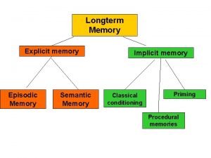
Amnesia • Theoretical implications of amnesia • Provides evidence for STM versus LTM distinction • Supports the notion that there are different systems mediating explicit (episodic) and implicit (procedural memory) • May indicate that semantic and episodic memory can be fractionated • May provide insight into nature of consciousness

Alzheimer’s Disease • Alzheimer’s disease (video clip # 19, ~7 mins) • cortical, progressive dementia • disease is associated with the development of neurofibrillary tangles and plaques • The brain has billions of neurons, each with an axon and many dendrites. • To stay healthy, neurons must communicate with each other, carry out metabolism, and repair themselves. • AD disrupts all three of these essential jobs.

Alzheimer’s Disease • Alzheimer’s disease Preclinical AD • Signs of AD are first noticed in the entorhinal cortex, then proceed to the hippocampus. • Affected regions begin to shrink as nerve cells die. • Changes can begin 10 -20 years before symptoms appear. • Memory loss is the first sign of AD.

Alzheimer’s Disease Mild to Moderate AD • Alzheimer’s disease • AD spreads through the brain. The cerebral cortex begins to shrink as more and more neurons stop working and die. • Mild AD signs can include memory loss, confusion, trouble handling money, poor judgment, mood changes, and increased anxiety. • Moderate AD signs can include increased memory loss and confusion, problems recognizing people, difficulty with language and thoughts, restlessness, agitation, wandering, and repetitive statements.

Alzheimer’s Disease Severe ADs • Alzheimer’s disease • In severe AD, extreme shrinkage occurs in the brain. Patients are completely dependent on others for care. • Symptoms can include weight loss, seizures, skin infections, groaning, moaning, or grunting, increased sleeping, loss of bladder and bowel control. • Death usually occurs from aspiration pneumonia or other infections. Caregivers can turn to a hospice for help and palliative care.

Alzheimer’s Disease • Alzheimer’s disease

Alzheimer’s Disease Pet Scan of Normal Brain Pet Scan of Alzheimer’s Disease Brain

Alzheimer’s Disease • Criteria • deficit in two or more areas of cognition, at least one of which is memory • interferes with social or occupational functioning • decline from premorbid level • gradually progressive course • rule out other causes

Alzheimer’s Disease • The Hallmarks of AD • The brains of people with AD have an abundance of two abnormal structures: • Beta-amyloid plaques • Dense deposits of protein and cellular material that accumulate outside and around nerve cells An actual AD plaque • Neurofibrillary tangles • Twisted fibers that build up inside the nerve cell An actual AD tangle

Alzheimer’s Disease Beta-amyloid Plaques Amyloid precursor protein (APP) is the precursor to amyloid plaque. • Alzheimer’s disease 1. APP sticks through the neuron membrane. 2. Enzymes cut the APP into fragments of protein, including beta -amyloid. 3. Beta-amyloid fragments come together in clumps to form plaques. In AD, many of these clumps form, disrupting the work of neurons. This affects the hippocampus and other areas of the cerebral cortex.

Alzheimer’s Disease • Alzheimer’s disease • three types of memory problems • episodic memory impaired (e. g. , free recall) • executive function (Baddeley appears to be affected) • semantic memory is also impaired • note: pure amnesics do not have the latter two impairments

Alzheimer’s Disease • AD • semantic memory • system for storing, organizing, and manipulating information pertaining to the meaning of words, concepts, and their associations • conceptualized as a broadly distributed network • enables judgments about the properties and functions of items

Semantic Memory Performance in AD • naming and word generation to semantic cues both require semantic memory, both impaired in AD • Explanations: • degradation of the semantic network? • loss of access to preserved concepts? • both?

Recapitulation during retrieval • Introduction • Memory performance depends on the similarity of conditions at encoding to those at retrieval. This finding and memory others suggest that encoding and retrieval processes are closely related to each other • It is widely believed that the brain regions activated at encoding will tend to be activated at retrieval

Recapitulation during retrieval • Purpose (Wheeler et al. , 2000) • To identify regions of brain associated with the retrieval of vivid visual and auditory information • To determine the extent to which these regions are associated with the regions activated at the time of encoding

Recapitulation during retrieval • Method • Subjects a set of picture and sound items each of which was paired with a descriptive label • E. g. , half Ss presented DOG with picture of dog; half Ss presented DOG with sound of dog barking • Ss performed task in which they perceived the sounds and pictures or recalled the studied sounds and pictures from memory when shown the label

Recapitulation during retrieval • Results • Brain areas in the visual cortex are active during retrieval of memories with visual content • Brain areas in the auditory cortex are active during retrieval of memories with auditory content

Recapitulation during retrieval • Discussion • Sensory aspects of a multisensory event are stored in some of the brain regions that were activated at encoding

E. P. • Suffered acute viral disease in brain • Damage sustained in temporal lobes, notably the hippocampus • Displays anterograde amnesia • Short term memory intact • Lives in a permanent present • What else can we infer from the interview seen?

Hypermnesia - S. • “Photographic” extreme memory ability (a mnemonist)

Hypermnesia - S. • “Photographic” extreme memory ability (a mnemonist) • Able to recall complex test stimuli

Hypermnesia - S. • S. used two “strategies” or abilities typical of mnemonists: • Rich synesthesia-like quality to his perception of stimuli - leads to stronger associative links • Vivid and elaborate mental imagery of things he should remember

Hypermnesia - S. • “ Even numbers remind me of images. Take the number 1. This is a proud, well-built man; 2 is a high-spirited woman; 3 a gloomy person (shy, I don’t Know); 6 a man with a swollen foot. . . ” Luria, A. R. The mind of a mnemonist. 1968 Luria, A. R. The man with a shattered world. 1972

Neuropsychology of Memory • Consciousness • Tulving has proposed that different memory systems have associated with them different levels of consciousness • noetic -- awareness • episodic memory -- autonoetic, self awareness • semantic memory -- noetic, aware of the information, but not aware of event • procedural memory -- anoetic no conscious awareness

Neuropsychology of Memory Episodic Autonoetic Semantic Noetic Procedural Anoetic

Neuropsychology

The Neuron Nerual signal: The Action potential • Positive chemicals flow into axon, making it positively charged • Causes positive charge to flow down cell to terminal buttons • Myelin sheath helps

Neuropsychology Strong emotions can enhance memory formation and retrieval. Many compounds participate: acetylcholine, epinephrine, norepinephrine, vasopressin, the opioids, and GABA. Drugs that are agonists or antagonists of these can be involved.

Dissociations Good Neuropsychology studies look for dissociations: Task 1 Brain Area 1 Activation Brain Area 2 No Activation Task 2 No Activation Often found across patients Example: H. M. Intact Working Memory, poor ability to recall episodic memories K. F. no problem recalling daily episodes, but digit span of 1 (Shallice and Warrington, 1970)

Amnesia • Injury Time Basic description • Results from study with H. M. 1. The hippocampus is not the location of long-term memory (LTM); nor is it necessary for the retrieval of LTM 2. The hippocampus is not the location for short-term memory (STM) 3. The hippocampus is involved in converting STM into LTM § These results are too simple; anterograde amnesia is actually much more complex § Learning consists of at least 2 stages: § • STM – immediate memory for events, which may or may not be consolidated into LTM; can only hold a limited amount of info • LTM – relatively stable memory of events that occurred in the more distant past, as opposed to STM; no limit on amount of info Consolidation – the process by which STM are converted

Structure of the brain • Video 3 (damage) • Video 3 a, b, c, d (Clive Wearing, damage)
 Fundamentals of human neuropsychology
 Fundamentals of human neuropsychology
 Fundamentals of human neuropsychology
 Kolb whishaw
 Neuropsychology in south africa
 Broca's aphasia
 Hm study
 Dijana dominis prester
 Sta 302
 Eehor
Eehor Ece 368
 Ece 368
 Ece 368
 Ece 368
 Ece 465 uic
 Cs 368 uw madison
 Bc 368
 Pvu market cap
 R/announcements!
 Church announcements
 Fahrenheit 451 burning bright
 Kluver bucy syndrome
 General announcements
 Euro psy
 Psy 3024 ucf
 Psy2055
 Psy 2055
 Psy 2055
 Psy
Psy Daniel sanin
 Psy
Psy Psy 421
 Phonological loop
 Psy
Psy Psy 226
 Suppose twin teenagers are vying
 Alla kulikova
 Nukleus caudatus
 Psy walsh
 Pes charakteristika
 Túlavé psy vzor
 11 psy
 Regression psy
 Psy
Psy Psy 335 purdue
 Spreading activation psychology
 Implicit memory
 Long term memory vs short term memory
 Internal memory and external memory
 Primary memory and secondary memory
 Logical versus physical address space
 Which memory is the actual working memory?
 Page fault
 Virtual memory in memory hierarchy consists of
 Eidetic memory vs iconic memory
 Shared vs distributed memory
 Human memory management
 Types of human memory
 Human vs non human bones
 Chapter 8 human needs and human development
 Chapter 8 human needs and human development
 Human development index definition ap human geography
 Human nouns
 Why does the coin jump
 Stanley milgram obedience experiment unethical
 One factor experiment
 Independent variable science
 Classical conditioning examples
 Terrella experiment
