Protozoa Lab 13 Intestinal Protozoa Commensal amoeba Endolimax

Protozoa

Lab 13 Intestinal Protozoa Commensal amoeba

Endolimax nana A. Trophozoites: karyosome large, centric or eccentric, without peripheral chromatin

Endolimax nana B. cyst: ovoid in shape; nuclei 1, 2, or 4; karyosome large with little or no peripheral duration.
 stain Endolimax nana Cyst (I. H. ) Stain Endolimax")
Dimethyl sulfuxide – modified (DMSO) stain Endolimax nana Cyst (I. H. ) Stain Endolimax nana Cyst in Iodine Endolimax nana Trophozoite Endolimax nana from stool smear

Iodamoeba butschlii A. Trophozoites nuclei with large central karyosome; karyosome surrounded by small chromatin granules;

Iodamoeba butschlii B. cyst: large glycogen vacuole; nucleus as in trophozoites.
")
Iron hematoxylin stain Iodine stain Iodamoeba butschlii Cyst ( 7 – 18 µm ) 31 Iodine stain Iodamoeba butschlii Trophozoite Iodamoeba butschlii from stool smear

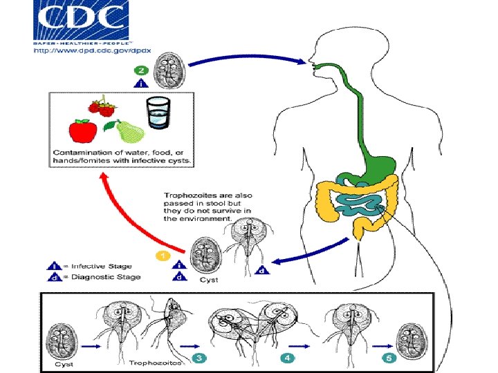
LAB : 13 Flagellates of digestive tract and genital organs Giardia lamblia Chilomastix mesnili Trichomonas vaginalis
; nuclei 2, sucking disc 2; axonemes central;")
Giardia lamblia A. trophozoite: pyriform shape (pear-shape); nuclei 2, sucking disc 2; axonemes central; flagella 4 pairs.
 evident • bilateral symmetry §Pair of claw-shaped median bodies §Adhesive disk")
Trophozoites §Fibrils (axonemes) evident • bilateral symmetry §Pair of claw-shaped median bodies §Adhesive disk (not always evident) § 4 pair flagella • motility likened to falling leaf § inhabit the small intestine mucosa

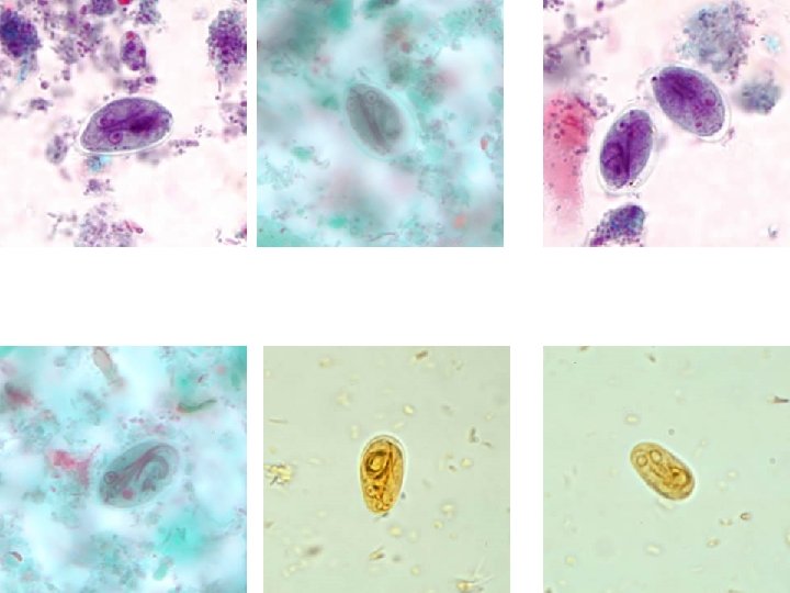
44 Fig 45 Saline s. Iodine stain Giardia lamblia Trophozoite, stool smear ( 12 – 16 µm ) Note : the motile in saline, twisting – jerky

Giardia intestinalis in culture. In preparations the flagellae ( four pairs. per cell are clearly visible A B C A, B, C: three trophozoites of Giardia intestinalis. Stained with trichrome (A) and stained with iron hematoxyline (B and C) each cell has two nuclei with a large, central karyosome. Cell size: 9 to 21 µm in length ( duodenal aspiration smear )

Giardia lamblia B. cyst: ovoid; 1 -4 nuclei; axonemes crossing.
 Fig 50 : Giardia lamblia Cyst in a")
Iodine stain ( X 40 ) Fig 50 : Giardia lamblia Cyst in a wet preparation and stained with iron – hematoxylin, these cysts have two nuclei. Each ( more mature ones will have four ) ( 10 – 13 µm ) ( X 1000 )

Chilomastix mesnili A. trophozoite: pyriform shape; flagella 3; anterior cytostome present, with in-curved flagellum.
 stain DMSO STAINE 11 – 14 µm ( X")
Dimethyl sulfuxide – modified (DMSO) stain DMSO STAINE 11 – 14 µm ( X 1000 ) Iron – hematoxylin stain Chilomastix mesnilli (Trophozoite), stool smear Note : the motile in saline spiral – jerky

Chilomastix mesnili B. cyst: shape lemon-like; nucleus single; cytostome with flagellum.

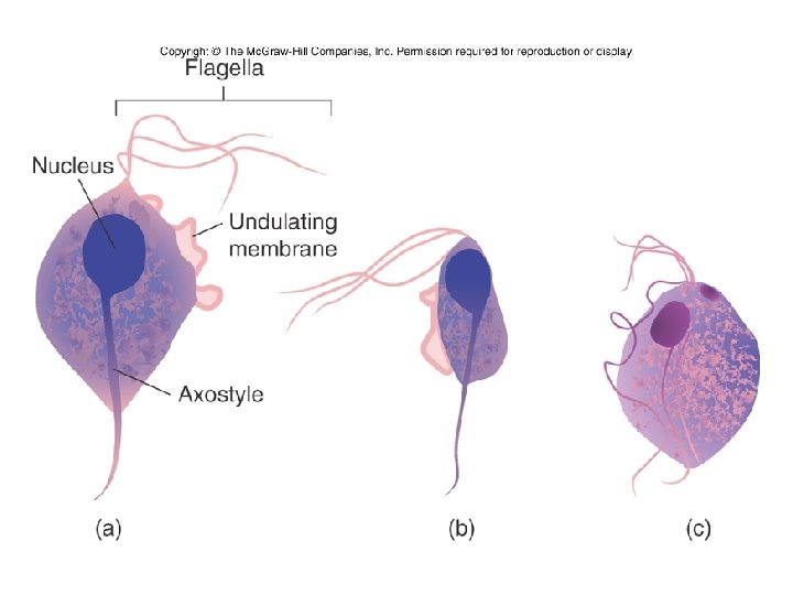
TRICHOMONADS • 3 -5 anterior flagella • one undulating membrane • axostyle
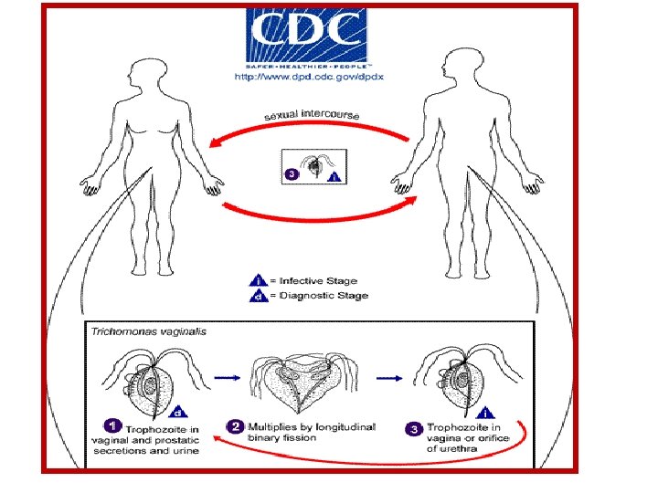

Trichomonas vaginalis trophozoite only shape pyriform; flagella 4 anterior, 1 trailing along undulating membrane; axostyle present.
 Note")
Trichrome stain Trichomonas vaginalis Trophozoite 15 – 30 µm ( X 1000 ) Note : the motile, rotary – clock – wise for all Trichomonas spp.

A B Methylene blue S. Trichomonas vaginalis, trophozoite A smear from swab of vagina from infected female, B smear from urtheral discharge from infected male
 trophozoite of Trichomonas hominis, stool smear ( 8 – 20")
Trichrome stain (X 1000) trophozoite of Trichomonas hominis, stool smear ( 8 – 20 µm ) trophozoite of Trichomonas tenax, smear of scraping from mouth ( 5 – 12 µm )
- Slides: 27