Protozoa Lab 12 Intestinal Protozoa Entamoeba histolytica Entamoeba

Protozoa

Lab 12 Intestinal Protozoa Entamoeba histolytica

Entamoeba histolytica A. Trophozoites §Irregular shape. §Transparent ectoplasm §Granulated endoplasm small nucleus with perinuclear chromatin dots, regular in size & spacing.

Trophozoites of Entamoeba histolytica
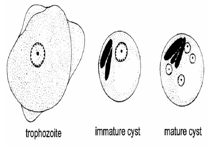

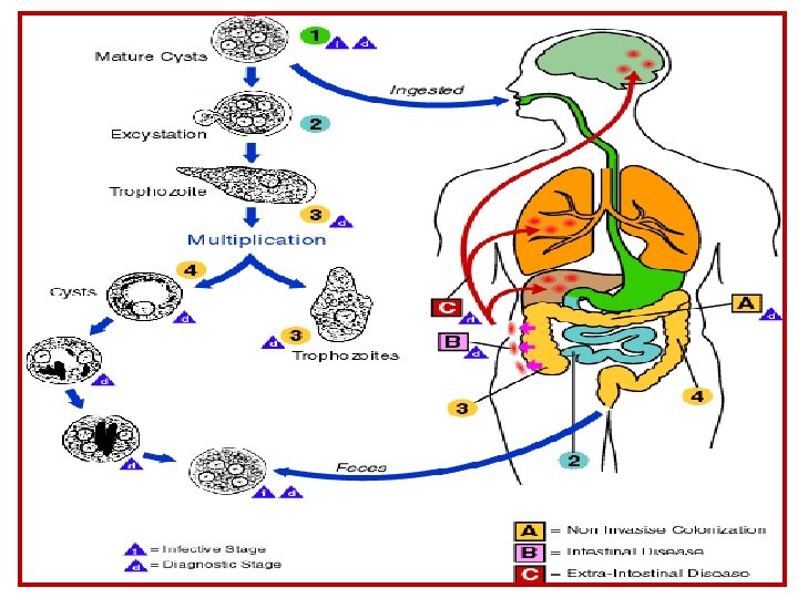
1. Entamoeba histolytica b. cyst, note: round in shape outer cyst wall nuclei 1, 2 or 4 chromatin bars may be present.

Cigar shape chromatoid body Cysts of E. histolytica

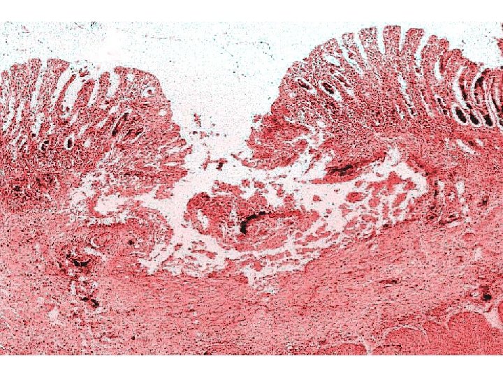
1. Entamoeba histolytica c. Section in infected intestine- note: v v v Columnar epithelium, Lamina propria, Muscularis mucosae, Sub-mucosa, muscularis and serosa in intact mucosa Clusters of amebae extending from epithelium through lamina propria and muscularis mucosa and muscularis. Some amebae show their nuclei and / or ingested RBCs if the section passes through these structures.

Flask like ulcer caused by E histolytica

ulcer of E. histolytica in large intestine Fig 23 -A Fig 23 -B

Digested RBCs Hematophagous’ trophozoites

Iodamoeba butschlii A. Trophozoites nuclei with large central karyosome; karyosome surrounded by small chromatin granules;

Iodamoeba butschlii B. cyst: large glycogen vacuole; nucleus as in trophozoites.
")
Iron hematoxylin stain Iodine stain Iodamoeba butschlii Cyst ( 7 – 18 µm ) 31 Iodine stain Iodamoeba butschlii Trophozoite Iodamoeba butschlii from stool smear

Intestinal Protozoa Commensal amoeba

Entamoeba coli A. Trophozoites: v. Irregular shape but more compact than E. histolytica; v. Ectoplasm thin; v. Granulate endoplasm; v. Small nucleus with perinuclear chromatin dots irregular in size & spacing.

Entamoeba coli B. Cyst Large; nuclei 1, 2, 4 or 8; Chromatoid bars with pointed end.
 Entamoeba Coli Trophozoite ( 20")
Entamoeba histolytica Trophozoite ( 18 – 60 µm ) Entamoeba Coli Trophozoite ( 20 – 50 µm ) Entamoeba histolytica Cyst ( 5 – 20 µm ) Entamoeba Coli Cyst ( 15 – 33 µm ) trophozoite and cysts of E. histolytica and E. coli form stool smear
- Slides: 20