Protozoa Lab 1 Intestinal Protozoa Entamoeba histolytica Entamoeba

Protozoa

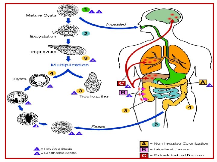
Lab: 1 Intestinal Protozoa Entamoeba histolytica

Entamoeba histolytica A. Trophozoites §Irregular shape. §Transparent ectoplasm §Granulated endoplasm small nucleus with perinuclear chromatin dots, regular in size & spacing.

Trophozoites of Entamoeba histolytica
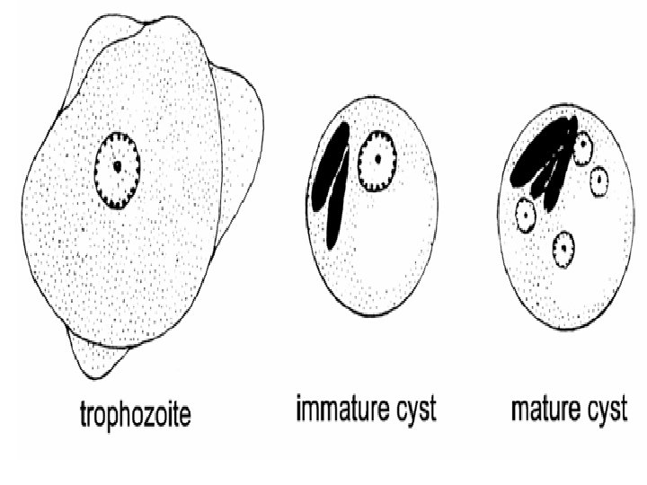

1. Entamoeba histolytica b. cyst, note: round in shape outer cyst wall nuclei 1, 2 or 4 chromatin bars may be present.

Cigar shape chromatoid body Cysts of E. histolytica

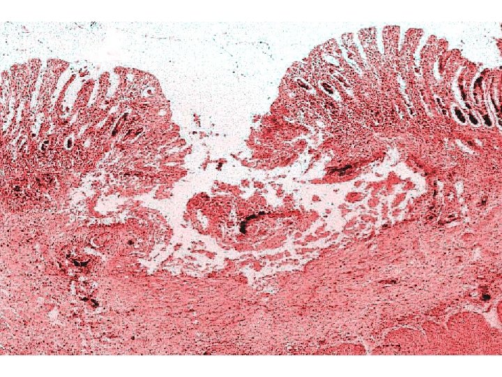
1. Entamoeba histolytica c. Section in infected intestine- note: v v v Columnar epithelium, Lamina propria, Muscularis mucosae, Sub-mucosa, muscularis and serosa in intact mucosa Clusters of amebae extending from epithelium through lamina propria and muscularis mucosa and muscularis. Some amebae show their nuclei and / or ingested RBCs if the section passes through these structures.

Flask like ulcer caused by E histolytica

ulcer of E. histolytica in large intestine Fig 23 -A Fig 23 -B

Digested RBCs Hematophagous’ trophozoites

Intestinal Protozoa Commensal amoeba

Entamoeba coli A. Trophozoites: v. Irregular shape but more compact than E. histolytica; v. Ectoplasm thin; v. Granulate endoplasm; v. Small nucleus with perinuclear chromatin dots irregular in size & spacing.

Entamoeba coli B. Cyst Large; nuclei 1, 2, 4 or 8; Chromatoid bars with pointed end.
 Entamoeba Coli Trophozoite ( 20")
Entamoeba histolytica Trophozoite ( 18 – 60 µm ) Entamoeba Coli Trophozoite ( 20 – 50 µm ) Entamoeba histolytica Cyst ( 5 – 20 µm ) Entamoeba Coli Cyst ( 15 – 33 µm ) trophozoite and cysts of E. histolytica and E. coli form stool smear
- Slides: 17