
PROTEINS The Building Blocks INTRODUCTION Proteins are the

PROTEINS The Building Blocks
INTRODUCTION • Proteins are the main building blocks from which cells are assembled. • They constitute most of the cell’s dry mass. • Providing the cell with I. Shape and II. Structure, III. Proteins also execute nearly all its innumerable functions.
PROTEIN STRUCTURE The levels of organization
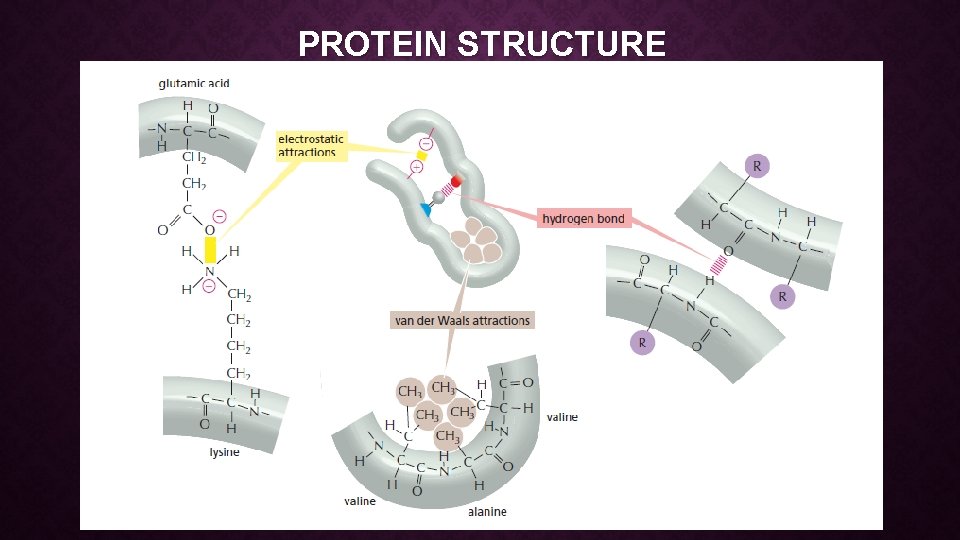
PROTEIN STRUCTURE • WEAK NONCOVALENT CHEMICAL BONDS • Organic molecules can interact with other molecules through three types of short-range attractive forces known as noncovalent bonds: 1. van der Waals attractions, 2. electrostatic attractions, and 3. hydrogen bonds. • The repulsion of hydrophobic groups from water is also important for these interactions and for the folding of biological macromolecules.
PROTEIN STRUCTURE • VAN DER WAALS ATTRACTIONS • If two atoms are too close together they repel each other very strongly. • For this reason, an atom can often be treated as a sphere with a fixed radius. • The characteristic “size” for each atom is specified by a unique van der Waals radius. • The contact distance between any two non covalently bonded atoms is the sum of their van der Waals radii.
PROTEIN STRUCTURE • VAN DER WAALS ATTRACTIONS • At very short distances, any two atoms show a weak bonding interaction due to their fluctuating electrical charges. • The two atoms will be attracted to each other in this way until the distance between their nuclei is approximately equal to the sum of their van der Waals radii. • Although they are individually very weak, such van der Waals attractions can become important when two macromolecular surfaces fit very close together, because many atoms are involved.
PROTEIN STRUCTURE • HYDROGEN BONDS • Hydrogen bonds form when a hydrogen atom is “sandwiched” between two electron-attracting atoms (usually oxygen or nitrogen). • Hydrogen bonds are strongest when the three atoms are in a straight line:
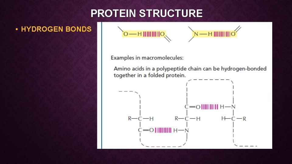
PROTEIN STRUCTURE • HYDROGEN BONDS
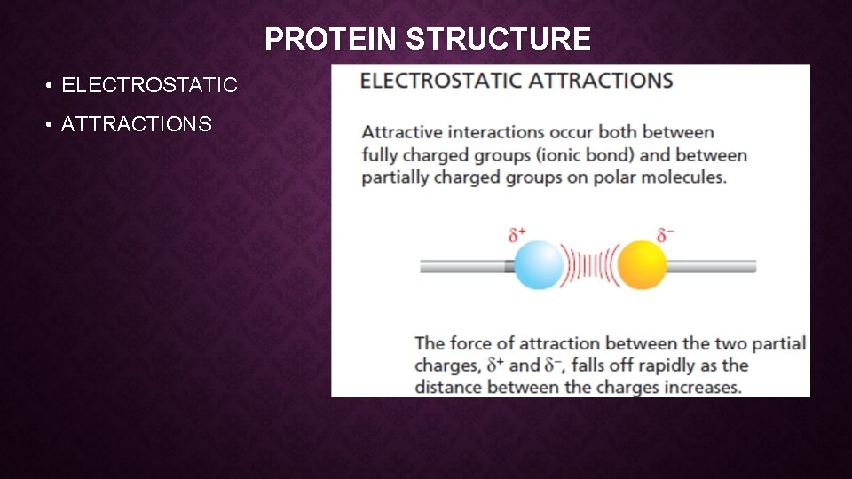
PROTEIN STRUCTURE • ELECTROSTATIC • ATTRACTIONS
PROTEIN STRUCTURE

PROTEIN STRUCTURE
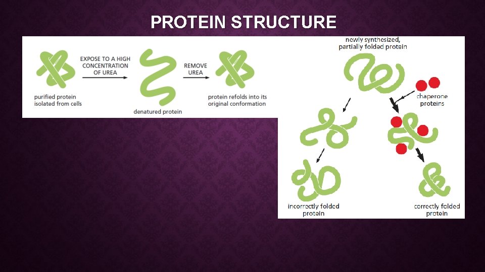
PROTEIN STRUCTURE • The final folded structure is called conformation • Which is determined by energetic considerations: • A protein generally folds into the shape in which its free energy (G) is minimized. • The folding process is thus energetically favorable, as it releases heat and increases the disorder of the universe (see Panel 3– 1, pp. 96– 97). • The protein often refolds spontaneously into its original conformation—a process called renaturation.
PROTEIN STRUCTURE • More than 60 years ago scientists studying hair and silk present in many different proteins. • The first α helix, was found in the protein α-keratin, which is abundant in skin and its derivatives—such as hair, nails, and horns. • Within a year of the discovery of the α helix a second folded structure, called a β sheet, was found in the protein fibroin, the major constituent of silk.
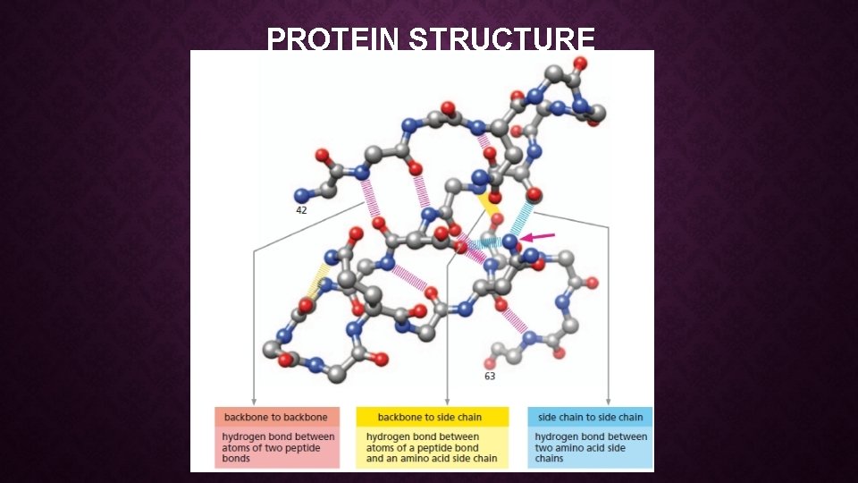
PROTEIN STRUCTURE • These two folding patterns are particularly common. • because they result from hydrogen bonds that form between the N–H and C=O groups in the polypeptide backbone (see Figure 4– 6). • Because the amino acid side chains are not involved in forming these hydrogen bonds, α helices and β sheets can be generated by many different amino acid sequences. • In each case, the protein chain adopts a regular, repeating form.
PROTEIN STRUCTURE
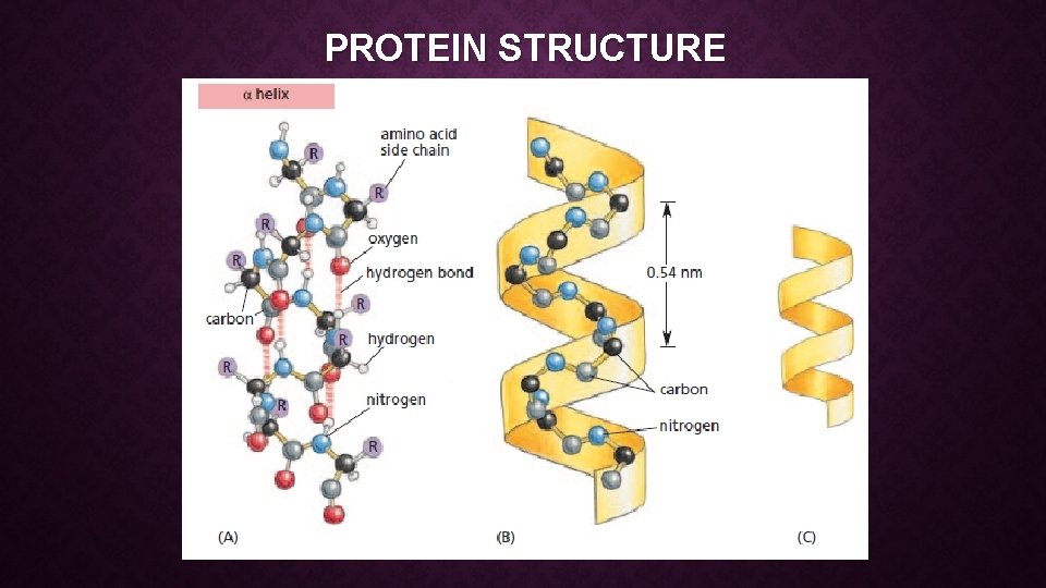
PROTEIN STRUCTURE • A helix is a regular structure that resembles a spiral staircase. • An α helix is generated when a single polypeptide chain turns around itself to form a structurally rigid cylinder. • A hydrogen bond is made between every fourth amino acid, linking the C=O of one peptide bond to the N–H of another (see Figure 4– 13 A). • This gives rise to a regular right handed helix with a complete turn every 3. 6 amino acids.
PROTEIN STRUCTURE
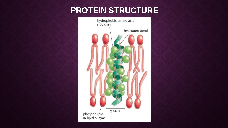
PROTEIN STRUCTURE • Short regions of α helix are especially abundant in proteins that are embedded in cell membranes, such as transport proteins and receptors. • Those portions of a transmembrane protein that cross the lipid bilayer usually form an α helix that is composed largely of amino acids with nonpolar side chains. • The polypeptide backbone, which is hydrophilic, is hydrogen-bonded to itself in the α helix, and it is shielded from the hydrophobic lipid environment of the membrane by its protruding nonpolar side chains (Figure 4– 15).
PROTEIN STRUCTURE
PROTEIN STRUCTURE • Sometimes two (or three) α helices will wrap around one another to form a particularly stable structure known as a coiled-coil. • This structure forms when the α helices have most of their nonpolar (hydrophobic) side chains on one side, so that they can twist around each other with these side chains facing inward—minimizing their contact with the aqueous cytosol (Figure 4– 16). • Long, rodlike coiled-coils form the structural framework for many elongated proteins. • Examples include α-keratin,

PROTEIN STRUCTURE
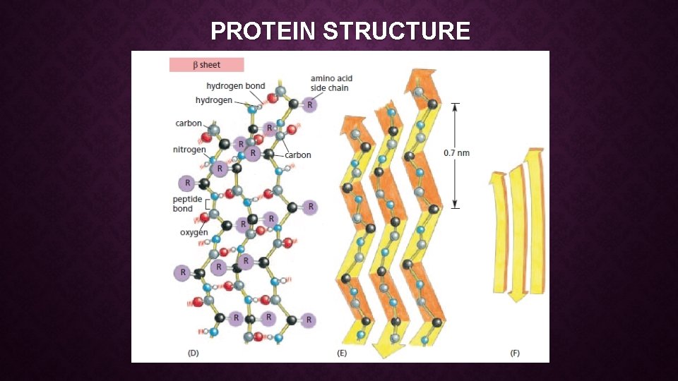
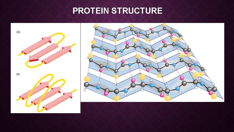
PROTEIN STRUCTURE • BETA (β) SHEET • A β sheet is made when hydrogen bonds form between segments of a polypeptide chain that lie side by side (see Figure 4– 13 D). • When the neighboring segments run in the same orientation (say, from the Nterminus to the C-terminus), the structure is a parallel β sheet; when they run in opposite directions, the structure is an antiparallel β sheet (Figure 4– 17). • Both types of β sheet produce a very rigid, pleated structure, and they form the core of many proteins. • Even the small bacterial protein HPr (see Figure 4– 12) contains several β sheets.
PROTEIN STRUCTURE
PROTEIN STRUCTURE
PROTEIN STRUCTURE • All of the information necessary for folding the peptide chain into its “native” structure is contained in the amino acid sequence of the peptide. • The arrangement of all atoms of a single polypeptide chain in threedimensional space is referred to as its tertiary structure. • The first determinations of the tertiary structure of a protein were by John Kendrew and Max Perutz. • Kendrew’s structure of myoglobin and Perutz’s structure of hemoglobin, reported in the late 1950 s, were each the result of more than 20 years of work.
PROTEIN STRUCTURE • The full, three-dimensional conformation formed by an entire polypeptide chain—including A. the α helices, B. β sheets, C. random coils, and D. any other loops and folds that form between the N- and C-termini— • is sometimes referred to as the TERTIARY STRUCTURE. • Finally, if the protein molecule is formed as a complex of more than one polypeptide chain, then the complete structure is designated its quaternary structure.
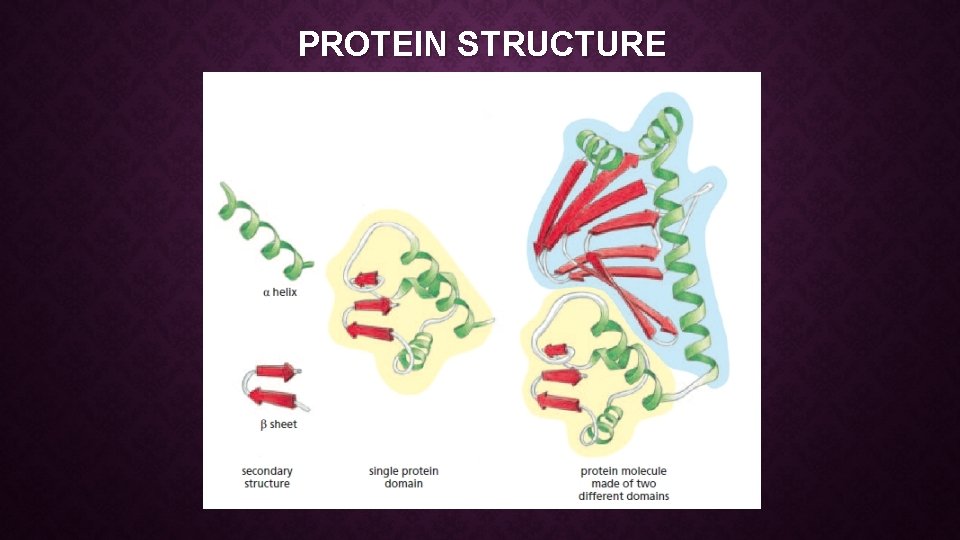
PROTEIN STRUCTURE • Studies of the conformation, function, and evolution of proteins have also revealed the importance of a level of organization distinct from the four just described. • This organizational unit is the protein domain, which is defined as any segment of a polypeptide chain that can fold independently into a compact, stable structure. • A protein domain usually contains between 40 and 350 amino acids—folded into α helices and β sheets and other elements of secondary structure—and it is the modular unit from which many larger proteins are constructed.
PROTEIN STRUCTURE • The different domains of a protein are often associated with different functions. • For example, the bacterial catabolite activator protein (CAP), illustrated in Figure 4– 19, has two domains: • the small domain binds to DNA, while the large domain binds cyclic AMP, a small intracellular signaling molecule. • When the large domain binds cyclic AMP, it causes a conformational change in the protein that enables the small domain to bind to a specific DNA sequence and thereby promote the expression of an adjacent gene.
PROTEIN STRUCTURE
PROTEIN STRUCTURE
PROTEIN STRUCTURE I. Secondary structures—helices and sheets—form whenever possible as a consequence of the formation of large numbers of hydrogen bonds. II. Helices and -sheets often associate and pack close together in the protein. No protein is stable as a single-layer structure, for reasons that become apparent later. There a few common methods for such packing to occur. III. Because the peptide segments between secondary structures in the protein tend to be short and direct, the peptide does not execute complicated twists and knots as it moves from one region of secondary structure to another.
PROTEIN STRUCTURE IV. Proteins generally fold so as to form the most stable structures possible. • The stability of most proteins arises from • (1) the formation of large numbers of intramolecular hydrogen bonds and • (2) the reduction in the surface area accessible to solvent that occurs upon folding. • Two factors lie at the heart of these four “principles. ” First, proteins are typically a mixture of hydrophilic and hydrophobic amino acids. Why is this important? • Imagine a protein that is composed only of polar and charged amino acids. • In such a protein, every side chain could hydrogen bond to water. • This would leave no reason for the protein to form a compact, folded structure.
DEFENSE PROTEINS • Plant defensins are defense-related proteins with antimicrobial activity. • This growing family of basic cysteine-rich peptides with a molecular mass below 7 k. Da is of particular interest for three reasons. First, unlike SAmediated defense responses, plant defensin accumulation in vegetative plant tissues is controlled by a combination of JA and ethylene. Second, JA levels rise in systemic tissues. Third, insects, birds, and mammals produce structurally and functionally similar defensin peptides following microbial attack. For example, the fruit fly Drosophila melanogaster produces a peptide called drosomycin that resembles plant defensins from radish. Both peptides exhibit potent antifungal activity (Fig. 21. 55). This conserved structure/function relationship indicates an ancient conserved defense strategy against microbial attack involving extracellular peptide production.
BIOLOGICAL ROLE OF PROTEINS • Proteins are the agents of biological function. Virtually every cellular activity is dependent on one or more particular proteins. Thus, a convenient way to classify the enormous number of proteins is to group them according to the biological roles they serve. • Figure 5. 26 summarizes the classification of proteins found in the human proteome according to their function. • Proteins fill essentially every biological role, with the exception of information storage. The ability to bind other molecules (ligands) is common to many proteins.
BIOLOGICAL ROLE OF PROTEINS • Binding proteins typically interact noncovalently with their specific ligands. Transport proteins are one class of binding proteins. Transport proteins include membrane proteins that transport substances across membranes, as well as soluble proteins that deliver specific nutrients or waste products throughout the body. Scaffold proteins are a class of binding proteins that uses protein–protein interactions to recruit other proteins into multimeric assemblies whose purpose is to mediate and coordinate the flow of information in cells. Catalytic proteins (enzymes) mediate almost every metabolic reaction.
BIOLOGICAL ROLE OF PROTEINS • Regulatory proteins that bind to specific nucleotide sequences within DNA control gene expression. Hormones are another kind of regulatory protein in that they convey information about the environment and deliver this information to cells when they bind to specific receptors. • Switch proteins such as G-proteins can switch between two conformational states—an “on” state and an “off” state—and act via this conformational switching, as regulatory proteins. Structural proteins give form to cells and subcellular structures. The great diversity in function that characterizes biological systems is based on attributes that proteins possess.
- Slides: 37