PROTEINS STRUCTURE AND FUNCTIONS Svjetlana Kalanj Bognar svjetlana

PROTEINS - STRUCTURE AND FUNCTIONS Svjetlana Kalanj Bognar svjetlana. kalanj. bognar@mef. hr

OUTLINES § What are proteins? § What is a protein structure? § In which way the structural organisation of proteins determines protein functions? § How disorders in protein structure cause disease? § Slides 3 -7 give a few historical remarks on protein research, and diversity of protein functions. § Slides 8 -10 remind on structural features of amino acids as protein building blocks. § Slides 9 -17 present classification of amino acids and proteins. § Other slides are dealing with explaining protein structural organization. § The presentation ends with several review questions.
 suggested the term protein.")
PROTEINS - the first 1838. Jöns J. Berzelius (1779 -1848) suggested the term protein.

DIVERSITY AND SIGNIFICANCE OF PROTEIN BIOLOGICAL FUNCTIONS Enzyme catalysis: all enzymes are structurally the proteins with extraordinary catalytic power enabling acceleration of chemical reactions up to million and more times. (Michaelis-Menten constant) Structure of acetylcholinesterase

Transport and storage of various other molecules and ions hemoglobin

Movement coordination - actin, myosin and other muscle tissue proteins; also cellular movement by protein motor molecules Mechanical rigidity - collagen in the skin and bone structure

Immunity of the organism antibodies are highly-specific proteins Generation and propagation of nerve impulses - receptor proteins Nicotinic receptor Control of growth and differentiation proteins associated with DNA replication machinery; growth factors, hormones, etc. DNA polymerase

PROTEINS ARE POLYMERS OF AMINO ACIDS General structure of an amino acid: §Amino group (-NH 2) §Carboxyl group (-COOH) bonded to the same carbon atom (α carbon) §R-group (side chain) varying in structure, size and electric charge α

STRUCTURAL FEATURES OF AMINO ACIDS In protein molecules, amino acid residues are exclusively L-stereoisomers.
. Amino")
Amino acid dissolved in a water solution are present as dipolar ion (zwitterion). Amino acids are amphoteric molecules (ampholytes). Amino acid can act as an acid (proton donor) or a base (proton acceptor) :
 amino acids Non-proteinogenic")
§ § Proteinogenic amino acids Essential proteinogenic amino acids Modified (uncommon) amino acids Non-proteinogenic amino acids – ornithine and citrulline Proteins of all species (from bacteria to human) consist of the same 20 amino acids – proteinogenic amino acids!

10 PROTEINOGENIC AMINO ACIDS ARE ESSENTIAL AMINO ACIDS – THESE AMINO ACIDS CANNOT BE SYNTHESIZED BY HUMAN ORGANISM Threonine, Tryptophan, Valine, Arginine, Histidine, Lysine, Phenylalanine, Leucine, Isoleucine, Methionine These Ten Valuable Amino Acids Have Long Preserved Life In Man

Several uncommon amino acids with important biological functions - modifications of these amino acids occur after their incorporation into the polypeptide chain (posttranslational modifications). In prothrombin In collagen In ellastin In myosin In several enzymes
 ornitine citrulline")
Non-proteinogenic amino acids (Ornitine and citrulline participate in urea cycle) ornitine citrulline

CLASSIFICATION OF PROTEINS according to “shape” and chemical properties § FIBRILLARY/FIBROUS proteins are insoluble in water; mostly structural proteins § GLOBULAR proteins are soluble in water § COMPLEX PROTEINS, contain other nonprotein groups, prosthetic groups etc.

Water-soluble globular proteins fold into compact structures with nonpolar cores. Hydrophobic amino acids in the interior of the myoglobin structure (in yellow). Membrane proteins are situated within hydrophobic environment. Inverse position of the amino acids in membrane protein porin.
 PROTEINS")
CLASSIFICATION OF COMPLEX (CONJUGATED) PROTEINS

LEVELS OF PROTEIN STRUCTURAL ORGANISATION PROTEINS ARE POLYMERS OF AMINO ACIDS

PRIMARY STRUCTURE -amino acid sequence in a polypeptide chain AMINO ACID MOLECULES ARE JOINED THROUGH AMIDE LINKAGE -PEPTIDE BONDS Peptide bond is formed between -carboxyl group of one amino acid and -amino group of another amino acid.
 than single C-N bond (0. 149")
Peptide C-N bond is shorter (0. 132 nm) than single C-N bond (0. 149 nm) but longer than double C=N bond (0. 127 nm) → Linus Pauling and Robert Corey suggested a partial double-bond character of the peptide bond.

Peptide bond is planar and has the properties of double bond; the rotation around the bond is limited - peptide bond is rigid - which determines the conformation of the peptide backbone.

Two possible configurations of peptide bond – trans configuration predominates.
 and C - C")
Rotation is permitted about the N -C (Φ rotation angle) and C - C (ψ rotation angle). Many combinations of rotations are not possible due to steric hindrance between atoms.

Amino acids associate in dipeptides, tripeptides, tetrapeptides…oligopeptides…. polypeptides…proteins. Distinction between polypeptides and proteins is not strict (approximately, proteins contain more than 100 amino acids). STRUCTURE OF A TRIPEPTIDE

It is accepted to designate the amino acid sequence in the polypeptide chain starting from amino terminal (N-terminal) residue to carboxyl terminal (C-terminal) residue. (example shown in picture: leu-enkephalin, opioid peptide)
 is formed from –SH groups of two")
Glutathione – tripeptide γ-Glu-Cys-Gly Disulfide bridge (-S-S-) is formed from –SH groups of two cysteinyl residues leading to formation of cystine dimer.

SECONDARY STRUCTURE -local conformation of amino acid residues in a polypeptide chain 1. -helix 2. -pleated sheet 3. collagene helix

-helical structure §polypeptide backbone is wound around an imaginary axis §each helical turn includes 3. 6 amino acid residues

Structure of the α-helix is stabilized by hydrogen bonds between NH and CO groups of the polypeptide chain.

In α-helix, CO-group of one amino acid residue is linked to NH-group of n-residue by hydrogen bond (n+4).

Alfa helices are mostly right handed helices.

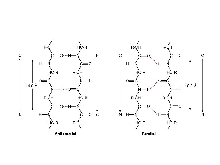
-pleated sheets • Antiparallel and parallel -sheets • Backbone of the polypeptide chain is extended

N C H O H N O C ANTIPARALLEL PLEATED SHEET Adjacent polypeptide chains are oriented in the opposite direction. Hydrogen bonding is present between NH i CO groups.

PARALLEL PLEATED SHEET Polypeptide chains are strechted in one direction; hydrogen bonds between NH and CO groups link one amino acid of one chain with two amino acids of the neighboring chain.

Combination of parallel and antiparallel sheets

-Polypeptide chains may change the direction. -Many proteins have globular shape due to change in chain direction. -Structural elements enabling the change in direction are: -turn (hairpin) and -loop. -Turns and loops are usually situated at the protein surface and participate in interactions with other proteins and molecules. Antibody

Proline has cyclic form – its side chain is linked to both amino-group and C atom; proline can not fit in the helix thus proline may cause the helical turn.

Relative probabilities for a given amino acid to occur in one of the three common types of secondary structure.

Collagen helix is a specific secondary structure found in a fibrillary protein collagen. Collagen helix – left helix containing 3. 3 amino acid residues per turn; hydrogen bonds are not possible in this secondary structure.

Collagen protein is formed by 3 peptide chains of the same size, supertwisted about each other; several collagen helices form collagen fibres.

TERTIARY STRUCTURE Three-dimensional conformation of a protein determined by amino acid sequence myoglobin

PROTEIN CONFORMATION IS STABILIZED MAINLY BY WEAK INTERACTIONS – hydrogen bonds, hydrophobic and ionic interactions Stabilization of structural organisation of proteins: -hydrogen bonds (secondary structure) -disulfide bonds (tertiary structure) -electrostatic forces (between side chains; tertiary structure) -hydrophobic interactions of non-polar groups and residues (tertiary structure)

Interactions stabilising tertiary structure
 is formed")
Insulin structure – mature peptide, stabilized by disulfide bridges Disulfide bridge (-S-S-) is formed from –SH groups of two cysteinyl residues leading to formation of cystine dimer.

Structure of hair – helical structural elements of keratin.

Keratin in nature • • hair nails wool claws hooves horns feather • Keratin structure strength depends on disulfide bonds. • Keratin of the rhinoceros’ horn contains approx. 18% of cystine residues within disulfide bonds.

Disulfide bridges in hair keratin structure See Box 4 -2, in Lehninger Biochemistry – “Permanent Waving is Biochemical Engineering”

SECONDARY AND TERCIARY STRUCTURE DETERMINE AND ENABLE PROTEIN FUNCTIONALITY. Amino acid sequence determines tertiary structure Christian Anfinsen’s experiment with ribonuclease showing denaturation and renaturation of the protein molecule.

QUATERNARY STRUCTURE ASSOCIATION OF TWO OR MORE CHAINS IN SUBUNITS (for example: dimers, trimers, tetramers etc. ) 2 2 tetramer of human hemoglobin

Methods used for analysis, separation and purification of proteins 1. According to size of molecule 2. According to solubility 3. According to charge 4. According to specific binding affinity
; 3.")
Chromatographic methods used in protein purification: 1. ion-exchange chromatography; 2. size-exclusion chromatography (gel-filtration); 3. affinity chromatography.

Electrophoresis
 AND MISFOLDING OF PROTEINS MAY")
DISORDER IN AMINO ACID SEQUENCE IN POLYPEPTIDE CHAIN (PROTEIN) AND MISFOLDING OF PROTEINS MAY CAUSE A DISEASE Examples: Ø Sickle cell anemia (replacement of glutamate with valine at the 6 th position of hemoglobin beta chain) Ø Some neurological diseases are characterized by disturbed protein processing and consequently altered protein conformation: prion diseases, Alzheimer’s disease

COLLAGEN – the extracellular matrix protein §Most mammalian cells are located in tissues, surrounded by a complex extracellular matrix (ECM), a connective tissue. §Extracellular matrix is complex network of proteins and polysaccharides which strenghten the structure and function of connective tissue. Protein collagen is the major component of most connective tissues, which constitutes approx. 25% of mammalian proteins: liver: 4% lung: 10% aorta: 12 -24% bone: 23% skin: 74%
 -")
TROPOCOLLAGEN is a structural unit of collagen. - 3 polypeptide chains (collagen helices) - approx. 1000 amino acid residues - triple helix stabilized by hydrogen bonds -being composed of more than one polypeptide chain, collagen is showing a quaternary structural organization Tropocollagen structure ensures the strength of a collagen fibre: – collagen fibre of 1 mm diameter will break under 10 kg weight!

Amino acid composition of collagen 33% 10% 1% Gly Pro Hyp Hyl Amino acid sequence: each 3. aa in collagen helix is Gly

The interior of the helical structure of collagen is extremely compact: – glycine is the only amino acid which suits to internal part of the collagen helical structure.
 ensures the stability of the triple collagen helix. Proline")
Hydroxylation of prolin (and lysine) ensures the stability of the triple collagen helix. Proline residues are modified in collagen to hydroxyproline by the catalytic activity of a prolyl-hydroxylase, while part of lysine residues is hydroxylated (hydroxylysine) by activity of a lysil-hydroxylase Prolyl-hydroxylase contains Fe(II)ion in the active site, and for normal activity, the enzyme needs the presence of a reducing agent (ascorbic acid). SCURVY - this disease is the consequence of C avitaminosis

SUMMARY – Proteins 1. Proteins are made of amino acids. All naturally occurring proteins consist of the same 20 amino acids. Side chains of amino acids are of variable sizes, shapes and contain different functional groups, thus many diverse structures are possible. 2. Levels of protein structure are: primary, secondary, tertiary and quaternary. 3. Amino acid sequence of protein (primary structure) determines its threedimensional structure (secondary and tertiary structure) and the function of a protein.

FROM PROTEIN STRUCTURE TO PROTEIN FUNCTION! Literature: 1. D. L. Nelson i M. M. Cox: Lehninger Principles of Biochemistry, 5 th edition, Worth Publishers, USA, 2008. 2. J. M. Berg, J. L. Tymoczko i L. Stryer: Biochemistry, 7 th edition, W. H. Freeman and Company, USA, 2012

QUIZ! 1. Represent by structural formula the dipeptide alanyl-glycine. 2. Consider the following molecule: a) Name it. b) Using the three-letter symbols for the amino acids, how would this molecule be represented? 3. Define the following terms: a) α-carbon b) isoelectric point c) peptide bond 4. Indicate the level(s) of protein structure to which each of the following contributes: a) amino acid sequence b) β–pleated sheet c) hydrogen bond d) disulfide bond 5. List six functions of proteins in the body.
- Slides: 62