Protein Synthesis Learning Outcome B 7 Learning Outcome

Protein Synthesis Learning Outcome B 7

Learning Outcome B 7 • Demonstrate an understanding of the process of protein synthesis

Student Achievement Indicators Students who have fully met the prescribed learning outcome are able to: Identify the roles of DNA, messenger RNA (m. RNA), transfer RNA (t. RNA), and ribosomes in the processes of transcription and translation, including initiation, elongation, and termination. Determine the sequence of amino acids coded for by a specific DNA sequence (genetic code), given a table of m. RNA codons. Identify the complementary nature of the m. RNA codon and the t. RNA anti-codon

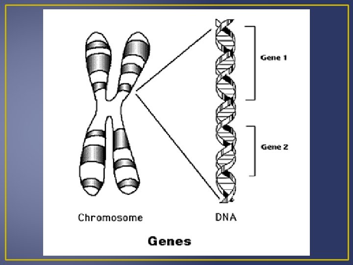
Gene Expression Genetic Disorders have a missing or malfunctioning enzymatic or structural protein Example – Huntingtons’s Disease The protein for Huntington’s disease is altered and not able to carry out normal functions. Proteins are the link between genotypes (genetic codes) and phenotypes (appearance) Genes are segments of DNA that specifies the amino acid sequence of proteins. Differences in base sequences cause a difference in protein structure. A gene does NOT directly control proteins synthesis; it passes genetic information on to RNA which is involved in protein synthesis.

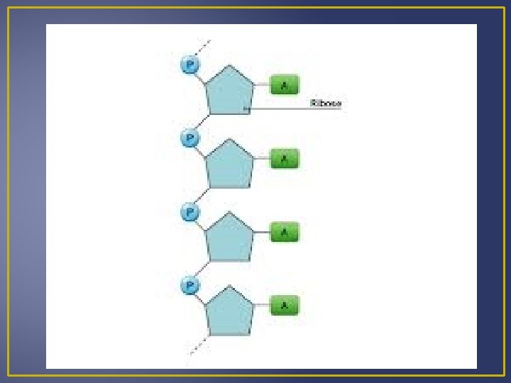
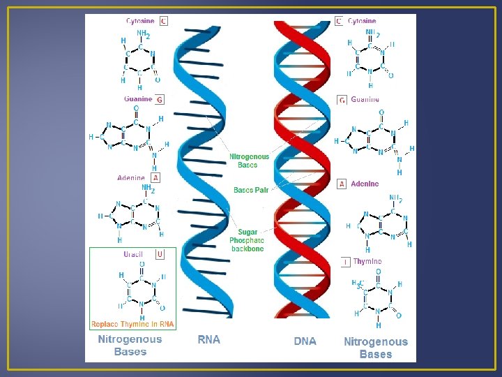
RNA Contains a ribose sugar Has the nitrogen base uracil instead of thymine Single-stranded nucleic acid Three Classes of RNA: Messenger RNA (m. RNA) – takes messages form DNA to ribosomes Ribosomal RNA (r. RNA) – along with proteins make up ribosomes, ribosomes are where proteins are synthesized. Transfer RNA (t. RNA) – transfers amino acids to ribosomes
 1. Transcription")
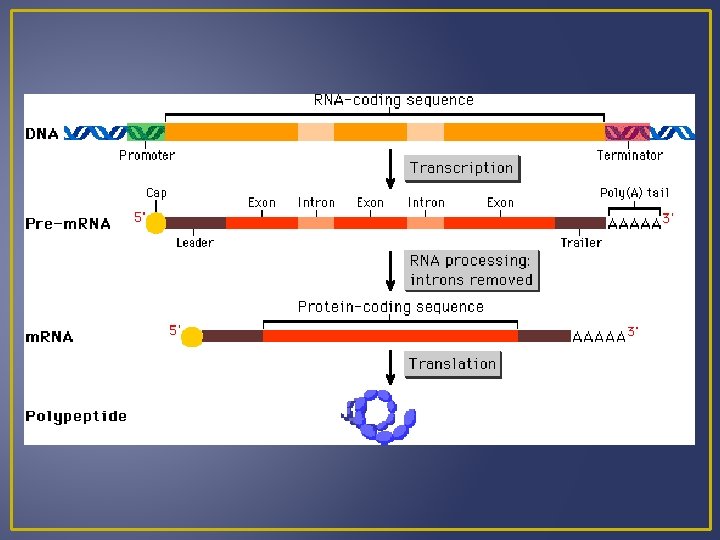
Protein Synthesis There are 2 Steps in Protein Synthesis (AKA Gene Expression) 1. Transcription (TXN) Information is transferred from DNA to RNA 2. Translation (TSL) An RNA transcript directs the sequence of amino acids. This means that a nucleotide sequence from DNA directs amino acids sequences.

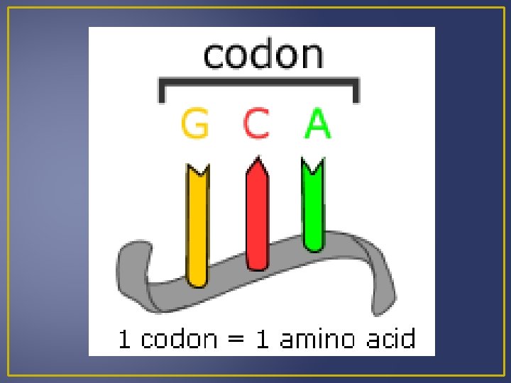
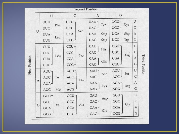
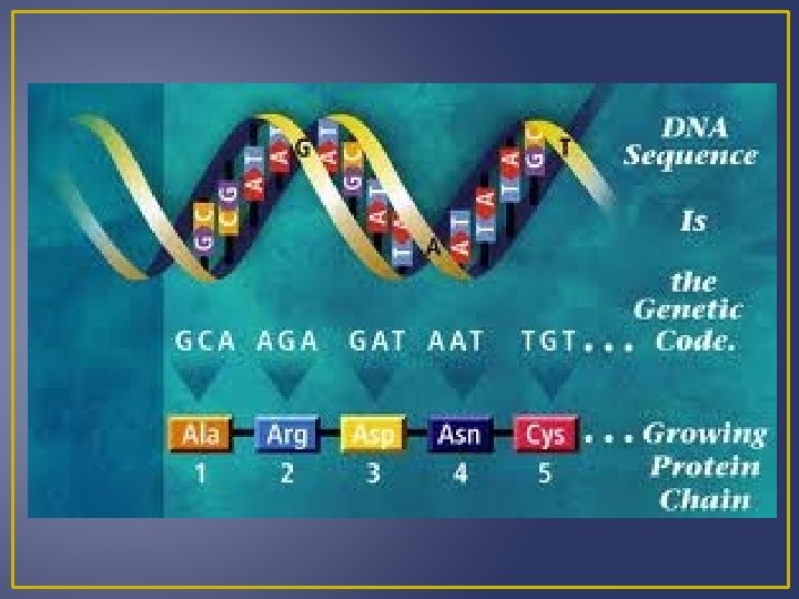
Genetic Code is a triplet; 3 bases stand for one amino acid. Each three-letter unit of an m. RNA molecule is called a codon. 64 m. RNA codons have been determined 61 m. RNA codons code for a particular amino acids 3 m. RNA codons are known as stop codons Stop codon single polypeptide termination

 also is an initiation codon Most amino")
Genetic Code The codon for methionine (AUG) also is an initiation codon Most amino acids have more than one codon. WHY? Example – Leucine Having more than one codon offers some protection against harmful mutations that change base sequences.
 RNA nucleotides pair complementary with")
Transcription A segment of the DNA helix unwinds (“unzips”) RNA nucleotides pair complementary with the DNA nucleotides of one strand. The nucleus contains an RNA nucleotide pool. The RNA nucleotide joins to the DNA by the enzyme RNA polymerase. When m. RNA forms it has sequences of bases complementary to DNA. These bases are grouped in threes and are known as codons
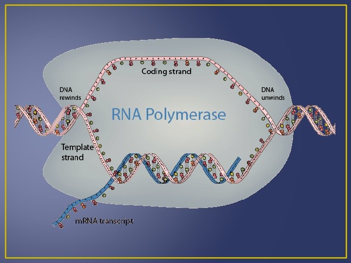
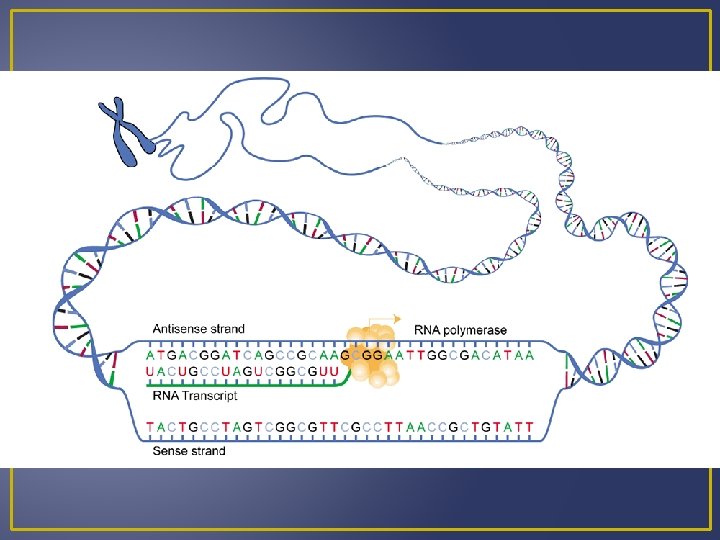
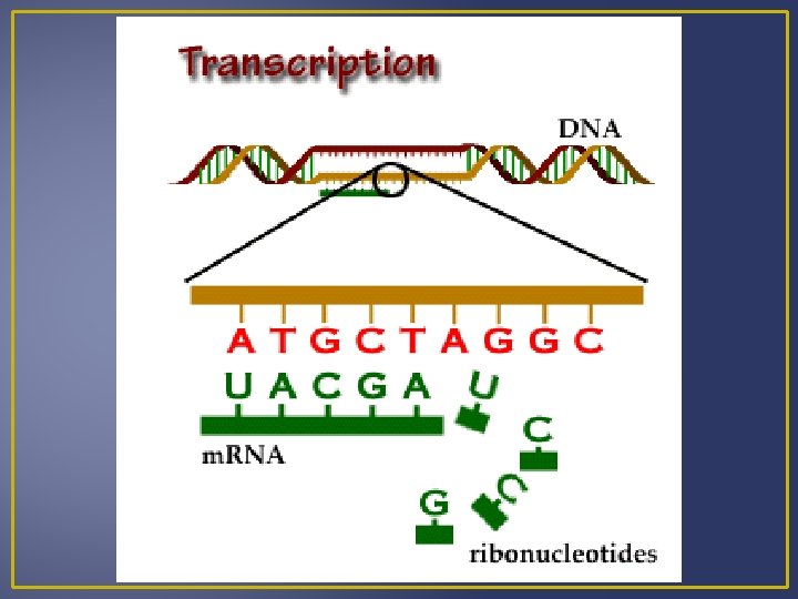

Transcription DNA RNA A A G G C C and T and U Example DNA m. RNA A C G C T A A C T U G C G A U U G A m. RNA is processed and then moves out of the nucleus into the cytoplasm where it can meet a ribosome for translation.

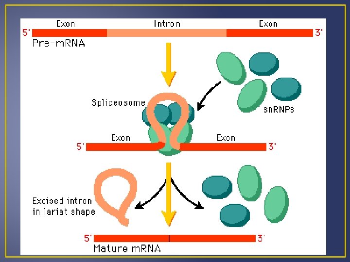
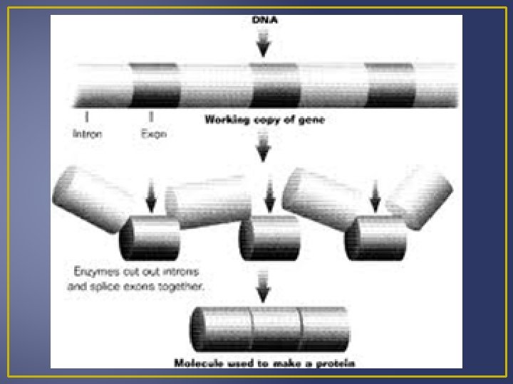
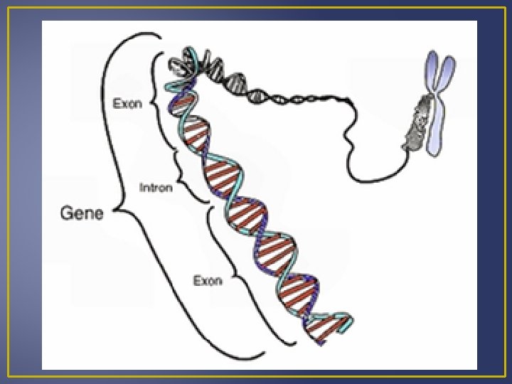
Transcription Processing of m. RNA Introns are segments of DNA that are not part of the gene Exons are part of the gene that code for proteins When DNA is transcribed to m. RNA, the m. RNA contains both exons and introns. Before m. RNA can move out of the nucleus it must be processed to get rid of the introns. Splicing m. RNA to remove introns is done by ribozymes which are organic catalysts composed of proteins not RN Processed m. RNA is called mature m. RNA and exits the nucleus and enters the cytoplasm where it can become associated with ribosomes.

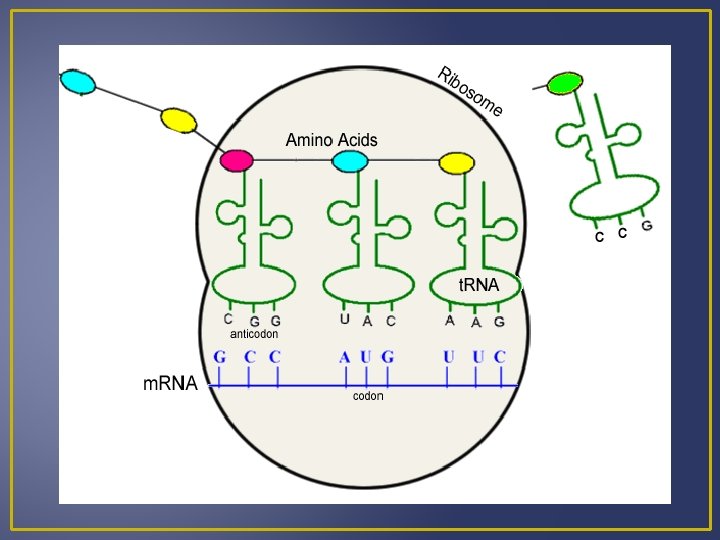
Translation Codons in m. RNA specify the order of amino acids in a protein Requires several enzyme and two types of RNA
 Brings amino acids to the ribosomes (strands of")
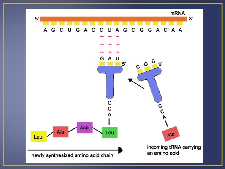
Translation 1. Transfer RNA (t. RNA) Brings amino acids to the ribosomes (strands of DNA) Single-stranded nucleic acid than doubles back on itself to create regions where complementary base pairing results in a boot-like shape. One t. RNA molecule for each of the 20 amino acids found in proteins. Amino acids bind at one end (matches the m. RNA strand) An anticodon is on one end of the t. RNA molecule

Translation An anticodon is a group of 3 bases that are complementary to a specific condon of m. RNA. Amino acids and t. RNA with anticodons are known as a t. RNA-amino acid complex. When a t. RNA-amino acid complex comes into contact with a ribosome, its anticodon pairs with the m. RNA codon. Example: Codon – ACC Anticodon – UGG Codes for amino Acid – threonine The m. RNA codon determines which t. RNA-amino acid complex must come to the ribosome and therefore determine the sequence of amino acids in a protein.
 Produced in nucleus Joins with proteins and is")
Translation 2. Ribosomal RNA (r. RNA) Produced in nucleus Joins with proteins and is manufactured in the cytoplasm Composed of two ribosomal subunits There is 1 large and 1 small ribosomal subunit which contains r. RNA and many different proteins. These subunits leave the nucleus and join together in the cytoplasm just before protein synthesis begins. r. RNA has a binding site for m. RNA and two for t. RNA.
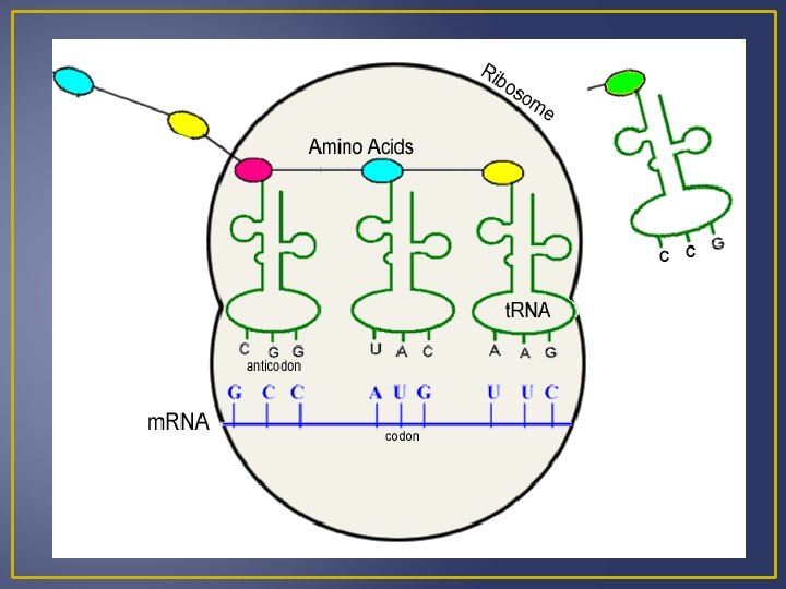

Translation Binding sites facilitate bonding of the t. RNA anticodons and m. RNA codons. As the ribosome moves down the m. RNA molecule; new t. RNA’s arrive and they polypeptide grows. Translation terminates once the peptide is formed and the ribosome subunits dissociate and fall of the m. RNA molecule. Several ribosomes may be attached on a single m. RNA, this is known as a polyribosome.
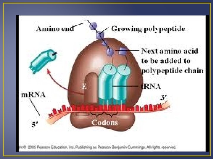

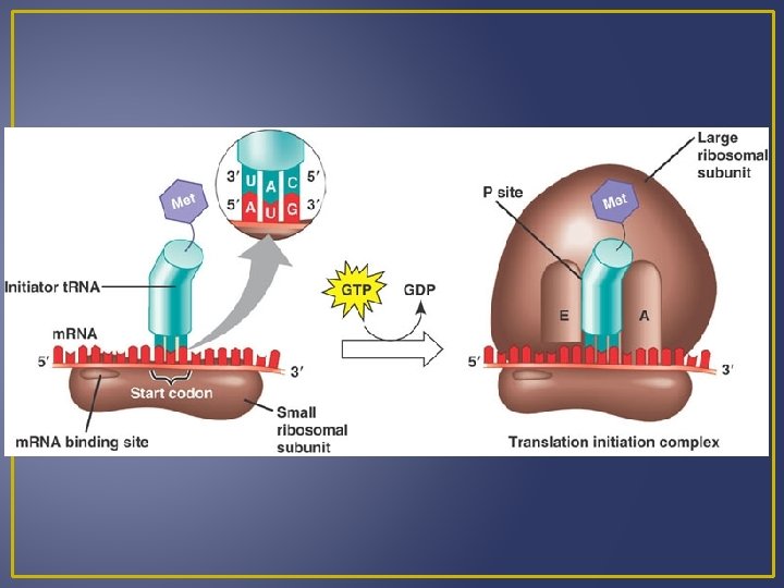
Steps in Translation 1. Chain Initiation Small ribosomal subunits, m. RNA, initiator t. RNA and large ribosome subunits all come together. Small ribosomal subunits attaches to m. RNA near the start codon AUG The anticodon on the t. RNA called the initiatior codon pairs with the m. RNA codon. The large ribosomal subunits join the small subunit; there are now two binding sites for t. RNA’s.

Steps in Translation 2. Chain Elongation t. RNA at first binding site contains an attached peptide The initiator t. RNA passes its amino acids to a t. RNA – amino acid complex that has come to the second binding site. Ribosomes move forward and the t. RNA in the second site is now the first site. This process is known as translocation.

Steps in Translation Translocation occurs again and again Growing peptide is transferred to a newly arrived amino acid. • This requires energy and ribozyme • Ribozyme is a large part of the ribosomal subunit. • After translocation, outgoing t. RNA will pick up another amino acid before it returns to the ribosome

Translocation • http: //www. phschool. com/science/biology_place/biocoach /translation/elong 1. html
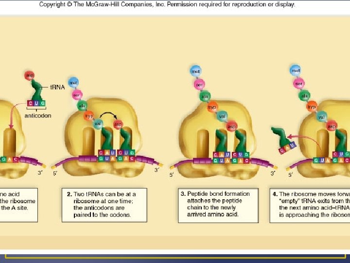

Steps in Translation 3. Chain Termination End of protein synthesis; stop codon (does not code for an amino acid) Polypeptide is enzymatically cleaved form the last t. RNA by a release factor. t. RNA and polypeptide leave ribosome and it dissociates. Newly synthesized polypeptide may function alone or become part of a protein that have more than one polypeptide.
 in humans and are known as")
Control of Gene Expression Cells undergo mitosis (growth) in humans and are known as totipotent. This means that each cell has the potential to develop into a new organism because it still contains the same gene that were present in the original cell. Differences in gene expression account for specializations within the cell. Gene expression is how much of the gene is being expressed. Genes are turned off and on at different times, so that each gene’s protein is only made when it is needed. Example - skin cells turn on genes that make keratin and red blood cell precursors turn on genes to make hemoglobin.

Control of Gene Expression Many steps in transcription and translation are required for gene expression and regulation of the gene can occur at any of these steps. There are positive and negative controls Positive controls increase the final amount of protein being made. Negative controls decrease the final amount of the protein being made. Many genes code for essential metabolic enzymes. These are known as “housekeeping genes” and are not finely regulated because they are always needed. Cells also all express a set of genes necessary to maintain general cellular function such as transport and acquiring energy.

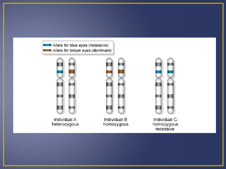
Levels of Gene Control Gene expression is controlled at 6 different levels: 1. Chromatic Structure Chromatic packing is a way to keep genes turned off. Genes with darkly stained areas of the chromosome on a micrograph have highly condensed chromatin and are known as heterochromatin. Heterochromatin is inactive This occurs on the XX chromosome in mammalian females. Females are XX and males are XY

Levels of Gene Control So females each have one inactive X chromosome in every cell of their body. Each inactivated X chromosome is known as Barr body and appears as a darkly stained mass of condensed chromatin along the inner edge of the nuclear envelope. During the pre-natal development, one X chromosome is shut off and it is turned back on at different times in different individuals.
 for black")
Levels of Gene Control Example – calico/tortoise-shell cats Alleles (sets of genes) for black or orange coats are carried on the X chromosome Random X-inactivation occurs in females and therefore in a heterozygous (two different alleles) individual some of the cells express the alleles for a black coat and some express alleles for an orange coat. If X-inactivation occurred early in fetal development; large patches would result. This pattern is known as a calico pattern.
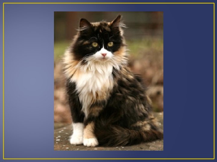

Levels of Gene Control If X-inactivation occurred late in fetal development, small patches would develop. This pattern is known as a tortoise shell pattern.

Levels of Gene Control White color in calicos is due to another gene. All cells formed in cell division will not have the same X chromosome inactivated; therefore patches of tissue will differ in which X chromosome is being expressed. If a female is heterozygous for a particular X-linked gene she will be a mosaic containing patches of cells expressing different alleles. Active genes are associated with loosely packed chromatin known as euchromatin. Even euchromatin must be “unpacked” before it is transcribed.
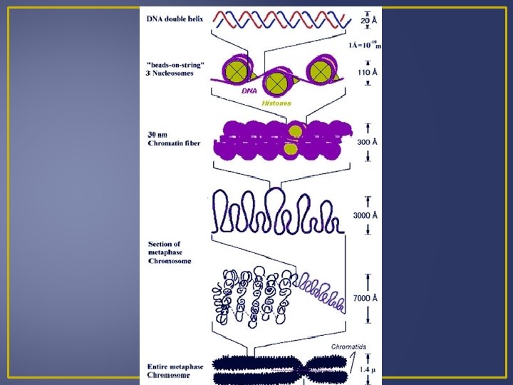

Levels of Gene Control Nucleosomes limit access to DNA by the transcription machinery

Levels of Gene Control 2. Transcriptional Control Most important level of control Interactions of proteins with particular DNA sequences. These proteins are known as transcription factors and activators, while the DNA sequences are known as promoters and enhancers. Transcription factors are proteins that help RNA polymerase bind to a promoter. Several transcription factors per gene form a transcriptioninitiation complex that helps pull double-stranded DNA apart and even acts to release RNA polymerase so transcription can begin. Some transcription factors can be used over and over again at different promoters.
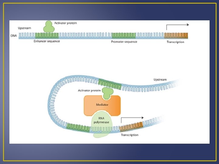

Levels of Gene Control If one transcription factor malfunctions it can be disastrous. Transcription activators are proteins that speed up transcription dramatically They bind to DNA regions known as enhancers; which may be quite a distance away from the promoter. A loop in DNA can bring the transcription activator into contact with the transcription-initiation complex. A group of 4 related transcription factors called the Myo-D family are responsible for the development and differentiation of muscle cells by controlling the expression of muscle specific proteins. The 4 transcriptions factors appear to act at different times during the development of muscle cells, allowing cells to become fully differentiated muscle cells.

Levels of Gene Control 3. Post-Transcriptional Control Some proteins are not active immediately after synthesis. Example – after translation insulin is folded into a 3 -D shape that is inactive. Then a sequence of 30 amino acids is enzymatically removed from the middle of the molecule leave two polypeptide chains that are bonded together by a disulphide bond. The disulphide bond activates the protein. Many proteins only function for a short period of time before they are degraded or destroyed.

Levels of Gene Control 4. Signalling between Cells are constantly communicating with one another. Generally a cell produces a signalling molecule that binds to a receptor protein in the target cell’s plasma membrane. A signaling molecule causes the receptor protein to initiate a series of reactions. These reactions will change the receiving cell’s behavior.

Levels of Gene Control This series of reactions is called signal transduction pathway. This pathway results in a stimulation of transcription activators. Activators will help turn a gene on and this allows a specialized cell to come on and is coordinated so the cell usually assumes a structure and function suitable to their location in the body.

Levels of Gene Control 5. Post-Transcriptional Control After transcription mr. RNA is processed before it leaves the nucleus to the cytoplasm. Primary m. RNA mature m. RNA Mature m. RNA has the addition of a poly-A tail and guanine cap. Introns are removed and exons are spliced back together. Introns are the nucleotide sequences that are removed as m. RNA reaches its mature state.

Levels of Gene Control m. RNA can be sliced in different ways to get slightly different products (proteins) in different tissues Example – both the hypothalamus and thyroid gland produce the hormone calcitonin but the calcitonin m. RNA that exits the nucleus contains different combination of exons in both the two tissues. Speed of m. RNA movement form the nucleus to cytoplasm affects the amount of gene production following transcription (amount of protein produced). m. RNA exits the nucleus via the nuclear pore, and m. RNA varies in the amount of time it needs to exit the nucleus.

Levels of Gene Control 6. Translational Control The longer the m. RNA is in cytoplasm before it is broken down the more protein that can be translated. Differences in poly-A tail and guanine caps can determine how long a particular transcript can remain active in the cytoplasm before it is destroyed by ribonuclease. Ribonuclease is an enzyme associated with ribosomes. Hormones can cause the stabilization of certain m. RNA transcripts so they are not broken down. Example – m. RNA for vitelline (egg membrane protein) can persist for three weeks if exposed to estrogen as opposed to 15 hours without estrogen.
- Slides: 62