Protein Synthesis Chapter 10 1 10 19 Learning


 the copied DNA fragments together to")
 by dictating")


 conveys genetic message from DNA to")
 match amino acids to appropriate m.")
 combine with proteins to form ribosomes")


- Slides: 32
Protein Synthesis Chapter 10. 1 -10. 19
Learning Objectives Genetic Code • Explain DNA and RNA as polymers made of nucleotides. • Explain the significance of Rosalind Franklin, James D. Watson and Francis Crick in terms of understanding DNA. • Explain the basic structure of a DNA nucleotide (sugarphosphate backbone) • Name the nucleotides of DNA and RNA • Explain how DNA and RNA differ • Explain what is meant by the DNA double helix • Explain the role of base pairing in DNA replication (assigned readings and related class activities)
Learning Objectives Protein Synthesis • • • You are responsible for completing the protein synthesis practice questions handed out in class (also on Moodle). Answer key is available on Moodle. Use the m. RNA codon chart in textbook or on the last page of your protein synthesis problems. Explain nucleic acids as they relate to the double helix Explain how DNA genotypes come to be expressed as proteins that result in phenotypic (physical) traits. Understand the role of transcription and translation in this process Explain how genetic information is written as codons and translated into amino acid sequences (readings and class activity) Explain how genetic messages are translated in the cytoplasm of cells Explain the role of m. RNA (messenger RNA), t. RNA (transfer RNA) and r. RNA (ribosomal RNA) in protein synthesis Explain how a t. RNA molecule is able to locate beginning and end when decoding a strand of m. RNA Understand the flow of genetic information in the cell DNA to RNA to protein Explain the “one gene-one polypeptide” theory Explain the terms mutagen, mutagenesis and mutation as they apply to genetics. Explain two main events that begin the process of mutagenesis
DNA and RNA • DNA and RNA are polymers of nucleotides made of PO 4, pentose sugar, nitrogenous base
DNA and RNA • There two groups of nitrogenous bases 1. purines have a double-ring structure (Guanine and Adenine) 2. pyrimidines have a single-ring structure (Cytosine, Thymine, Uracil)
Dexoyribo. Nucleic Acid • DNA stores the genetic code for the cell • Has 4 different nitrogenous bases: Thymine, Adenine, Guanine, and Cytosine • complementary bases pair up to form the rungs of the ladder • PO 4 and deoxyribose form the rails of the ladder Video: The Discovery of DNA (2: 17) https: //www. youtube. com/w atch? v=V 6 b. Kn 34 n. Sbk
Ribo. Nucleic Acid • Used for protein synthesis • Has the same 3 bases as DNA, Guanine, Adenine, Cytosine but has Uracil instead of Thymine • Pentose sugar is ribose instead of deoxyribose • Usually single stranded • Three types: m. RNA, t. RNA and r. RNA
Complementary Base Pairing • Purines pair up with Pyrimidines • Adenine pairs with Thymine (or Uracil) by forming two hydrogen bonds • Guanine pairs with Cytosine by forming three hydrogen bonds • This ensures correct base pairing when DNA is used to make copies of DNA or RNA
Replication • Produces identical copies of DNA • Cell uses free-floating nucleotides in the nucleus to form complementary base pairs with DNA template • Results in semi-conservative replication where one half of the strand is the parental strand one half is the daughter strand
Step 1. Strand Separation • Proteins bind to stretches of DNA called origins of replications • The enzyme DNA helicase breaks the H-bonds between the base pairs to separate the double helix and open up “ replication bubbles”
Step 2. Base Pairing • The copying proceeds in two directions as complementary bases pair up in the replication bubbles • DNA polymerase adds complementary nucleotides to the 3’ end of the parental strand • Replication proceeds from 5’ to 3’ end of the parental strand
Step 3. Joining • DNA ligase links (ligates) the copied DNA fragments together to form one continuous strand Note: DNA polymerase also checks base pairing to ensure that no copying errors occur Video: DNA replication (2: 39 min. ) https: //www. youtube. com/watch? v=Vpm. T 7 Lw_4 v 0
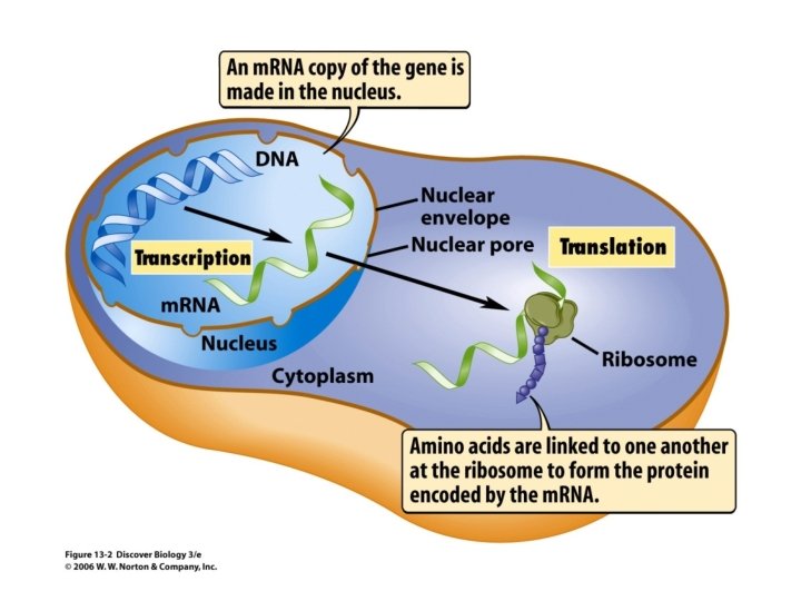
Genetic Flow from DNA to RNA • DNA specifies traits (ie. genotypes) by dictating protein synthesis • The gene instructions for protein synthesis are carried out in the form of RNA (Transcription) • RNA programs protein synthesis so that genes are expressed as phenotypes (Translation)
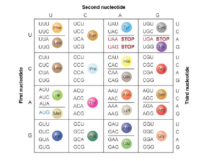
Transcription • Transcription is the synthesis of m. RNA under the direction of DNA • It transcribes the nucleic acid language of DNA into triplet code sequences of RNA called codons that specifies for amino acids
Transcription 1. Initiation – RNA polymerase attaches to promoter sequence on DNA template 2. Elongation - m. RNA strand grows longer as RNA synthesis continues 3. Termination – RNA polymerase detaches to release newly transcribed m. RNA strand Note: transcription also forms r. RNA and t. RNA
Transcription
Roles of RNA • Messenger RNA (m. RNA) conveys genetic message from DNA to the ribosomes at their codons • non-coding regions (introns) are cut out and coding regions (exons) are pasted together during in RNA splicing
Roles of RNA • Transfer RNA (t. RNA) match amino acids to appropriate m. RNA codons • t. RNA recognize matching codons in the m. RNA at the anticodon (complementary to m. RNA)
Roles of RNA • Ribosomal RNA (r. RNA) combine with proteins to form ribosomes • Ribosomes position m. RNA and t. RNA close together and catalyze protein synthesis
Translation - Initiation 1. m. RNA binds to the small ribosomal subunit at the start codon (AUG) for translation to begin 2. The large ribosomal subunit attaches to the smaller subunit The functional ribosome has two t. RNA binding sites: • P site holds the initiator t. RNA and will hold the growing polypeptide • A site is ready for the next amino-acid bearing t. RNA
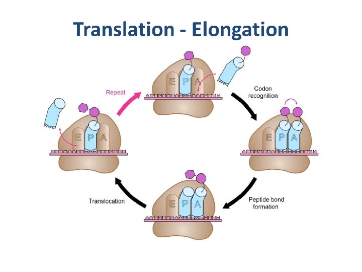
Translation - Elongation 1. The incoming t. RNA carrying the appropriate amino acid pairs with m. RNA in the A site 2. A new peptide bond forms between the amino acid in the A site and the polypeptide in the P site 3. t. RNA at the P site leaves and the ribosome translocates (moves) the t. RNA in the A site to the P site 4. The process repeats steps 1 -3 to add amino acids to a growing polypeptide
Translation - Termination • A stop codon reaches the ribosome A site • The stop codon (UAA, UAG, UGA) does not code for an amino acid and signals the end of translation • The polypeptide is freed from the last t. RNA and the ribosomal splits into two subunits Video: Translation (3: 32 min) https: //www. youtube. com/watch? v=5 b. LEDd-PSTQ
One Gene-One Polypeptide Theory • The specific sequence of DNA codes for one gene (genotype) • The gene serves as a template to dictate transcription of m. RNA • m. RNA dictates the linear sequence of amino acids to form a polypeptide • The polypeptide determines the appearance and function of the cell (phenotype)
One Gene-One Polypeptide Theory
Mutation • A mutation is any change in the nucleotide sequence of DNA • Mutations can involve large regions of chromosomes or a single-nucleotide pair • Divided into two types: nucleotide substitution or insertion/deletions
Nucleotide Substitution One nucleotide base pair is replaced with another nucleotide base pair (point mutation) 1. Silent – the substitution still codes for the same amino acid so there is no change in protein function 2. Missense – the substitution changes the amino acid and may affect protein function (eg. sickle cell anemia) 3. Nonsense – change an amino acid codon to a stop codon and the resultant shorter polypeptide will likely be non-functional (eg. Tay-Sachs disease)
Nucleotide Substitution
Insertion/Deletion Mutation • Inserting or deleting nucleotide pairs alters the codon reading frame • All the codons downstream for the mutation will be grouped into different codons • Usually results in a non-functional protein
Mutagenesis • The production of mutations is called mutagenesis • Physical, biological or chemical agents that cause mutations are called mutagens • High energy radiation – X-rays, UV light • Incorrect nucleotide pairing compounds (base analogs) – AZT blocks further replication of AIDs viral DNA Video: AZT blocks HIV reverse transcriptase (1: 45 min) https: //www. youtube. com/watch? v=1 so 7 D 5 tw. HSE