Protein Structure Foundation Block Dr Usman Ghani What
 Dr. Usman Ghani • What are proteins? • Four levels")
Protein Structure (Foundation Block) Dr. Usman Ghani • What are proteins? • Four levels of structure (primary, secondary, tertiary, quaternary) • Protein folding and stability • Protein denaturation • Protein misfolding and diseases

What are proteins? n Proteins are polymers of amino acids joined together by peptide bonds

Primary Structure n It is the linear sequence of amino acids

Voet Biochemistry 3 e Page 65 © 2004 John Wiley & Sons, Inc.

Voet Biochemistry 3 e Page 278 © 2004 John Wiley & Sons, Inc. Primary structure of proinsulin

Secondary Structure It is the local three-dimensional arrangement of a polypeptide backbone n Excluding the conformations (3 D arrangements) of its side chains n

a Helix n n n a helix is right-handed It has 3. 6 residues (amino acids) per turn The helix is stabilized by hydrogen bonding u Between carboxylic group and 4 th N–H group The amino acid side chains point outward and downward from the helix The core of the helix is tightly packed; its atoms are in van der Waals contact

Voet Biochemistry 3 e Page 224 © 2004 John Wiley & Sons, Inc. The right-handed a helix

Voet Biochemistry 3 e Page 65 © 2004 John Wiley & Sons, Inc.

b Sheets Two or more polypeptide chains form hydrogen bonding with each other n Also called pleated sheets n They appear as folded structures with edges n

Voet Biochemistry 3 e Page 228 © 2004 John Wiley & Sons, Inc. A two-stranded b antiparallel pleated sheet

Voet Biochemistry 3 e Page 65 © 2004 John Wiley & Sons, Inc.

Antiparallel b sheets Two or more hydrogen-bonded polypeptide chains run in opposite direction n Hydrogen bonding is more stable n

Voet Biochemistry 3 e Page 227 © 2004 John Wiley & Sons, Inc. b pleated sheets. (a) The antiparallel b pleated sheets

Parallel b sheets Two or more hydrogen-bonded polypeptide chains run in the same direction n Hydrogen bonding is less stable (distorted) n

Voet Biochemistry 3 e Page 227 © 2004 John Wiley & Sons, Inc. b pleated sheets. (b) The parallel b pleated sheets.
 n Loops n b bends n Random coils")
Other secondary structures Turns (reverse turns) n Loops n b bends n Random coils n

n Supersecondary structures or motifs: ubab motif: a helix connects two b sheets ub hairpin: reverse turns connect antiparallel b sheets uaa motif: two a helices together ub barrels: rolls of b sheets

Voet Biochemistry 3 e Page 249 © 2004 John Wiley & Sons, Inc. bab motif b hairpin aa motif Schematic diagrams of supersecondary structures

Voet Biochemistry 3 e Page 252 © 2004 John Wiley & Sons, Inc. b barrel

Voet Biochemistry 3 e Page 229 © 2004 John Wiley & Sons, Inc. Secondary structure of proteins (a-helix, b-sheets, loops, turns, random coils)

Tertiary Structure It is the three-dimensional structure of an entire polypeptide chain including side chains n It is the folding of secondary structure and side chains n Helices, sheets and side chains combined to form tertiary structure n

Voet Biochemistry 3 e Page 229 © 2004 John Wiley & Sons, Inc. Tertiary structure of proteins (Secondary structure + side chains)
 fold into two or more clusters")
n Domains u. Polypeptide chains (>200 amino acids) fold into two or more clusters known as domains u. Domains are units that look like globular proteins u. Domains are part of protein subunits

Voet Biochemistry 3 e Page 248 © 2004 John Wiley & Sons, Inc. One subunit with two domains

Voet Biochemistry 3 e Page 294 © 2004 John Wiley & Sons, Inc. One subunit with three domains

Quaternary Structure Many proteins contain two or more polypeptide chains n Each chain forms a three-dimensional structure called subunit n It is the 3 D arrangement of different subunits of a protein n

Voet Biochemistry 3 e Page 67 © 2004 John Wiley & Sons, Inc.
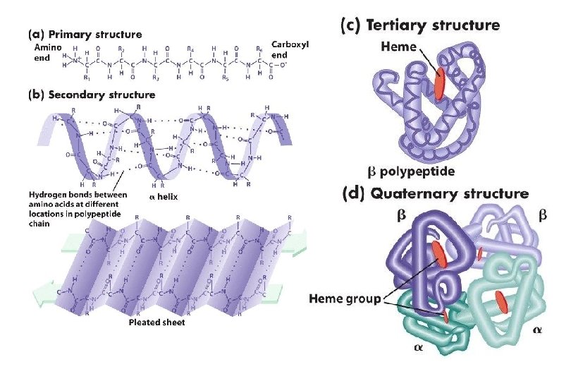

n Hemoglobin is a globular protein u. A multisubunit protein is called oligomer u. Composed of a 2 b 2 subunits (4 chains, 4 subunits) u. Two same subunits are called protomers

Protein folding Forces that stabilize proteins n Protein denaturation n

Forces that stabilize protein structure Hydrophobic effect: u. Nonpolar groups to minimize their contacts with water u. Nonpolar side chains are in the interior of a protein n Hydrogen bonding n
: u. Between positive and negative charges n van der Waals")
Electrostatic interactions (ion pairing): u. Between positive and negative charges n van der Waals forces (weak polar forces): u. Weak attractive or repulsive forces between molecules n

Protein denaturation n n Denaturation: A process in which a protein looses its native structure Factors that cause denaturation: u Heat: disrupts hydrogen bonding u Change in p. H: alters ionization states of aa u Detergents: interfere with hydrophobic interactions u Chaotropic agents: ions or small organic molecules that disrupt hydrophobic interactions

Protein misfolding Every protein must fold to achieve its normal conformation and function n Abnormal folding of proteins leads to a number of diseases in humans Alzheimer’s disease: n b-amyloid protein is a misfolded protein n It forms fibrous deposits or plaques in the brains of Alzheimer’s patients n

Creutzfeldt-Jacob or prion disease: n Piron protein is present in normal brain tissue n In diseased brains, the same protein is misfolded n Therefore it forms insoluble fibrous aggregates that damage brain cells

That’s all folks!
- Slides: 37