
Protein Structure and Function Proteins are the most
- Slides: 6
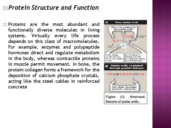
� Protein � Structure and Function Proteins are the most abundant and functionally diverse molecules in living systems. Virtually every life process depends on this class of macromolecules. For example, enzymes and polypeptide hormones direct and regulate metabolism in the body, whereas contractile proteins in muscle permit movement. In bone, the protein collagen forms a framework for the deposition of calcium phosphate crystals, acting like the steel cables in reinforced concrete Figure (1): Structural features of amino acids.
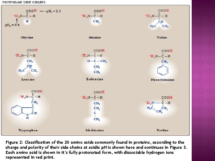
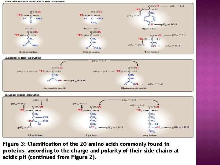
� Structure � � of amino acids Although more than 300 different amino acids have been described in nature, only 20 are commonly found as constituents of mammalian proteins. [Note: These are the only amino acids that are coded for by DNA, the gene tic material in the cell. ] Each amino acid has a carboxyl group, a primary amino group (except for proline, which has a secondary amino group), and a distinctive side chain (“R group”) bonded to the carbon atom (Figure 1 A). At physiologic p. H (approximately 7. 4), the carboxyl group is dissociated, forming the negatively charged carboxylate ion (– COO–), and the amino group is protonated (– NH 3+). In proteins, almost all of these carboxyl and amino groups are combined through peptide linkage and, in general, are not available for chemical reaction except for hydrogen bond formation (Figure 1 B). Thus, it is the nature of the s ide chains that ultimately dictates the role an amino acid plays in a protein. It is, the reform, useful to classify the amino acids according to the properties of their side chains, that is, whether they are nonpolar (have an even distribution of electrons) or polar (have an uneven distribution of electrons, such as acids and bases) as shown in Figures 2 and 3.
Figure 2: Classification of the 20 amino acids commonly found in proteins, according to the charge and polarity of their side chains at acidic p. H is shown here and continues in Figure 3. Each amino acid is shown in it’s fully protonated form, with dissociable hydrogen ions represented in red print.
Figure 3: Classification of the 20 amino acids commonly found in proteins, according to the charge and polarity of their side chains at acidic p. H (continued from Figure 2).
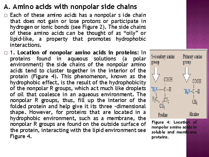
A. Amino acids with nonpolar side chains � � Each of these amino acids has a nonpolar s ide chain that does not gain or lose protons or participate in hydrogen or ionic bonds (see Figure 2). The side chains of these amino acids can be thought of as “oily” or lipid-like, a property that promotes hydrophobic interactions. 1. Location of nonpolar amino acids in proteins: In proteins found in aqueous solutions (a polar environment) the side chains of the nonpolar amino acids tend to cluster together in the interior of the protein (Figure 4). This phenomenon, known as the hydrophobic effect, is the result of the hydrophobicity of the nonpolar R groups, which act much like droplets of oil that coalesce in an aqueous environment. The nonpolar R groups, thus, fill up the interior of the folded protein and help give it its three -dimensional shape. However, for proteins that are located in a hydrophobic environment, such as a membrane, the nonpolar R groups are found on the outside surface of the protein, interacting with the lipid environment see Figure 4: Location of nonpolar amino acids in soluble and membrane proteins.