
Protein Structure and Function 1 2 3 4





- Slides: 22
Protein Structure and Function • 1 , 2 , 3 , 4 Structure • Viewing, interpreting structure • Protein Characterization BIO 520 Bioinformatics Jim Lund
Protein variety ~100 to >10, 000 aa • Soluble • Integral membrane proteins • Membrane-associated • Single domain, multiple domains • singular, multimeric, large complex
Protein Structure • 1 structure – aa sequence • 2 structure – regular local folding • 3 structure – packing and overall folding • 4 structure – polypeptide: polypeptide complexes
Modifications • Proteolysis/processing • Residues Modified – cysteine disulfides – phosphorylation – methylation • Heteroatoms – Metal ions, heme, cofactors….
Dogma Sequence=Structure
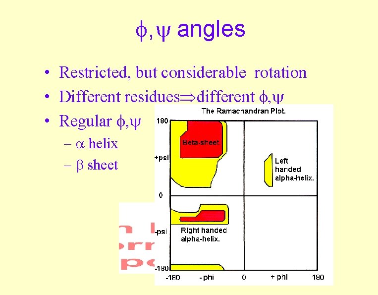
, angles • Restricted, but considerable rotation • Different residues different , • Regular , – helix – sheet
Experimental , angles Hovmöller et al. , 2002. Conformations of amino acids in proteins
An example Ramachandran plot Ideal a kinase
Forces holding proteins together • H-bonding – orientation (dielectric) • Hydrophobic effect – nonpolar to core • Ionic interaction – + to -, (dielectric) • Dipole effects – helix, N to C (+ to -)
-helix • right-handed • aa preferences – A, E, L, M (enriched) – P, G, Y, S (less likely) • 3. 6 residues/turn • helical wheel (amphipathic) • dipole N to C
-sheet • Parallel or antiparallel N-to-C – Mixed -sheets are rare: only ~20% of -sheets are mixed parallel/antiparallel. • “pleated” and “twisted” • aa preferences
-turn, loops • Length, conformation variable – 2 aa hairpins common • loops on surface • diverge rapidly
Simple Super-2 o Motifs • • hairpin ( -loop- ) Helix-loop-helix Greek key (4 antiparallel , wrap) - - motif ( -alpha helix-parallel )
Hairpin
Helix-loop-helix Helix-turn-helix
Helix-loop-helix Helix-turn-helix DNA binding EF hand (Ca++ binding)
Greek key (4 antiparallel , wrap)
- - motif ( -alpha helix-parallel )
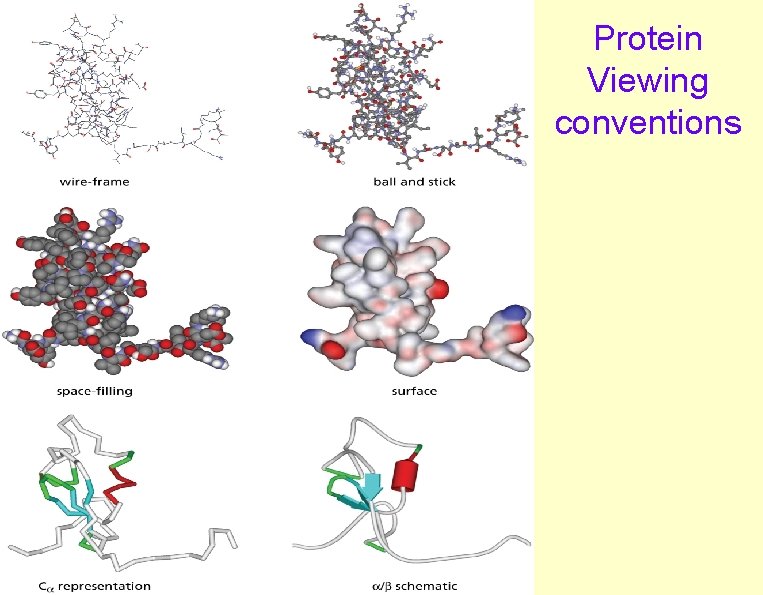
Protein Structure Viewers • Cn 3 D (NCBI) –. cn 3 files (MMDB, NCBI structures) • Ras. Mol – Protein. Explorer – CHIME • WWW compatible, animatable – Jmol • WWW compatible, animatable
Protein Viewing conventions
Protein Structure databases • MMDB, NCBI structures – Cn 3 D format (ASN 1) • Protein Data Bank (PDB) – PDB, Chime, other formats – (http: //www. pdb. org)