Protein Function Oxygen Binding Proteins CH 339 K

Protein Function: Oxygen Binding Proteins CH 339 K

Myoglobin • Sperm Whale Myoglobin was the first protein to have its 3 dimensional structure determined – John Kendrew(1958) – Shared the 1962 Nobel in chemistry • Solving the structure wasn’t hard, but getting the samples was a real achievement… Kendrew, JC; Bodo, G; Dintzis, HM; Parrish, RG; Wyckoff, H; Phillips, DC (1958). "A threedimensional model of the myoglobin molecule obtained by x-ray analysis". Nature 181 (4610): 662– 666.
 153 amino acids • 8 -helices,")
Myoglobin • Myoglobin - 17, 000 daltons (monomeric) 153 amino acids • 8 -helices, designated A - H • Conjugated protein - A conjugated protein has a nonprotein part in addition to a polypeptide component.

Myoglobin – naming of helices

Heme

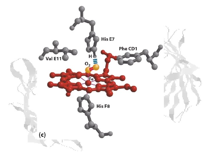
Heme Function • The heme group is responsible for the O 2 binding capacity of hemoglobin. • The heme group consists of the planar aromatic protoporphyrin made up of four pyrrole rings linked by methane bridges. • A Fe atom in its ferrous state (Fe+2) is at the center of protoporphyrin.

Heme Binding • • Fe+2 has 6 coordination bonds, four bonded to the 4 pyrrole N atoms. The nucleophilic N prevent oxidation of Fe+2. The two additional binding sites are on either side of the heme plane. One of these is occupied by the imidazole group of His F 8. (H 63 in (SWM) The second site can be reversibly occupied by O 2, which is hydrogenbonded to another His. (His E 7, H 94 in SWM)
, metmyoglobin (2) and")
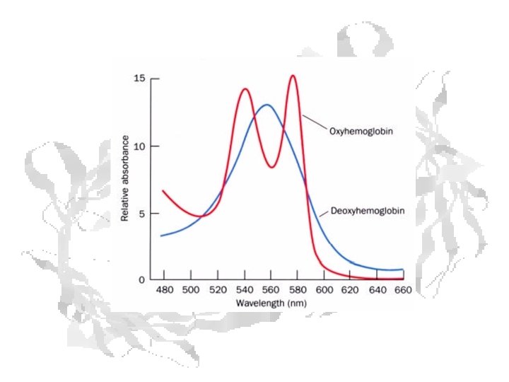
Oxygenation state can be measured spectrophotometrically Reflectance spectra of myoglobin (1), metmyoglobin (2) and oxymyoglobin (3).

Absorbance Curves for Mb

CO Poisoning Myoglobin’s affinity for carbon monoxide is ~ 60 x its affinity for O 2. Hemoglobin’s affinity for carbon monoxide is ~ 230 x its affinity for O 2. Autopsy photo showing characteristic skin discoloration

O 2 Binding Kinetics
 (2 (3) (4) (5)")
Reaction: Mb + O 2 ⇌ Mb O 2 (1) (2 (3) (4) (5)
 (6) (7) (8) Dr. Ready finally gets to the point!")
(5) (6) (7) (8) Dr. Ready finally gets to the point!

Remember Dalton’s Law – the concentration of a gas in a liquid … … is proportional to the partial pressure of that gas over the liquid

So: Converts to:

Hyperbolic Binding Kinetics

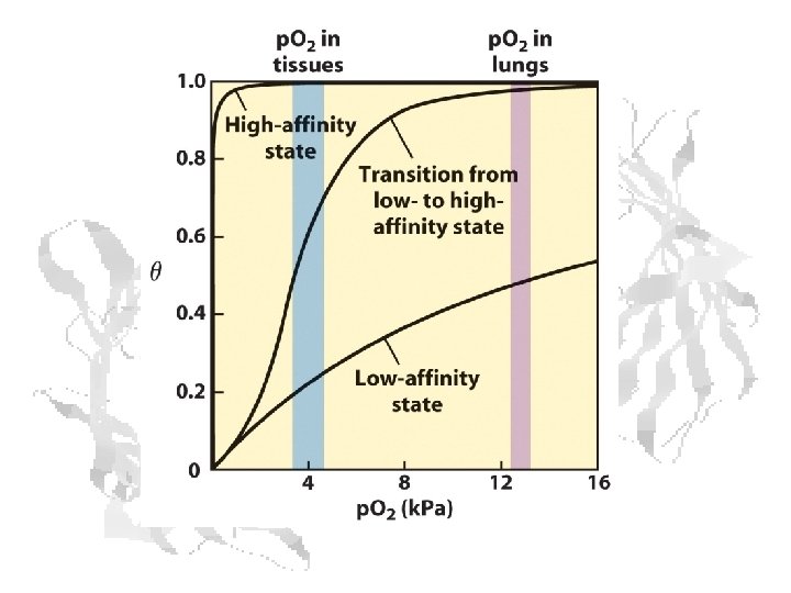
p 50 Defines the Curve 1 0. 9 0. 8 0. 7 Q 0. 6 0. 5 2 mm 5 mm 0. 4 10 mm 0. 3 0. 2 0. 1 0 0 20 40 60 p. O 2 80 100

Oxygen Transport
 b-Globin (violet) Myoglobin (green)")
3 o structure overlap: myoglobin, -globin and b-globin -Globin (blue) b-Globin (violet) Myoglobin (green)

Mb vs. Hb

Hemoglobin O 2 carrying capability • • Erythrocytes/ml blood: 5 billion ( 5 x 10⁹ ) Hemoglobin/red cell: 280 million ( 2. 8 x 10⁸ ) O 2 molecules/hemoglobin: 4 O 2 ml blood: (5 x 10⁹)(2. 8 x 10⁸)(4) = (5. 6 x 10¹⁸) or (5. 6 x 10²⁰) molecules of O 2/100 ml blood or ~ 0. 3 g/l or ~ 9 m. M By comparison: • Solubility of O 2 in saline: ~ 0. 007 g/l • or ~ 0. 2 m. M

O 2 Binding – Hb vs. Mb

O 2 transport capability, a comparison
 Substrate affinity changes with substrate")
Cooperativity Substrate affinity changes with substrate concentration or (rephrased) Substrate affinity changes with substrate binding Characteristic of (many) proteins with multiple binding sites.

Cooperative Binding Kinetics Reaction: Hb + n. O 2 ⇌ Hb n. O 2 (1) (2) (3)
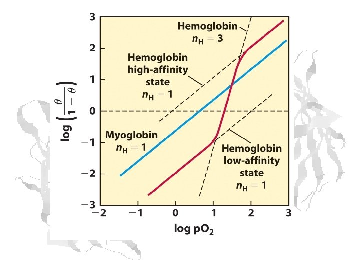
 (6) (7) Hill Equation (8) (9) (10) Myoglobin (11) Hemoglobin")
(5) (6) (7) Hill Equation (8) (9) (10) Myoglobin (11) Hemoglobin
 (1965) Only T and R conformations")
Cooperativity Models: Concerted Monod, Wyman, and Changeaux (MWC) (1965) Only T and R conformations exist The two states are in equilibrium T R transition involves shift in equilibrium constant
 (1966) There are intermediate conformations between")
Cooperativity Models: Sequential Koshland, Nemethy, and Filmer (KNF) (1966) There are intermediate conformations between T and R Intermediate conformations have intermediate binding affinities Change involves gradual conformational shift from more Tlike states to more R-like states
")
Hemoglobin T and R States Hb is more MWC-like than KNF-like T (Low Affinity) R (High Affinity)

Shift from T to R – another view
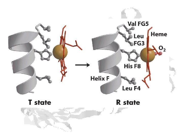

Structural Basis • O 2 Bound conformation does not permit several intersubunit bonds

Histidine “Ratchet” locks T and R states • Histidine at H 97 of 1 fits into socket between T 41 and P 44 in a 2 in the T state • In the R state, the valine side chain locks between T 38 and T 41.

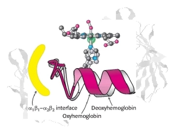
In the chains, the C teminal His makes a salt link with Asp FG 1 This holds the F helix in a position that keeps the Fe+2 out of the plain of the heme ring That in turn lowers the O 2 affinity Shift to the R state by the adjacent chain breaks salt link to C-terminal His, which moves it out of position to bind Asp FG 1 Relaxation of F helix allows heme Fe+2 to assume high-affinity position

Bohr Effect • The O 2 affinity of hemoglobin decreases with decreasing p. H • Improves delivery of oxygen to the tissues

Bohr Effect • C-terminal Histidine of the subunits is protonated at low p. H • His 146 can then form a salt link with Asp 94 in the deoxy (T) conformation • This stabilizes the T state of the protein.

Carbamate Formation Covalent binding at the N-terminus of each subunit • CO 2 transport is improved since some CO 2 is now being carried back to the lungs directly by hemoglobin • The release of H+ decreases p. H and increases the Bohr effect • Negatively charged carbamylated N-termini form salt link to the positive charge on Arginine 141. This salt link stabilizes the deoxy (T) form of the molecule and favors O 2 release.

2, 3 -Bisphoglycerate Binding
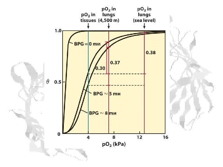

Combined Effects CO 2 , BPG and p. H are allosteric effectors of hemoglobin.

Fetal Hb • • Fetal hemoglobin has 2 α and 2 g chains The g chain is 72% identical to the chain. A His involved in binding to 2, 3 -BPG is replaced with Ser. Thus, fetal Hb has two less + charges than adult Hb. The binding affinity of fetal hemoglobin for 2, 3 -BPG is significantly lower than that of adult hemoglobin Thus, the O 2 saturation capacity of fetal hemoglobin is greater than that of adult hemoglobin This allows for the transfer of maternal O 2 to the developing fetus

Fetal Hb Binding Curve is Always to the Left of the Maternal Hb Binding Curve

Disease From a Hemoglobin Mutation

Sickle Cell • Misshapen cells cause vascular occlusion • Chronic anemia • Periodic episodes of pain • Autosplenectomy after infarct • Complications • Infection • Stroke • Renal Failure • Retinopathy • Life expectancy much improved since 60’s, but still shortened: 42 ♂ 48 ♀

Sickle Cell Complications Above: dactylitis Below: swollen, scarred spleen
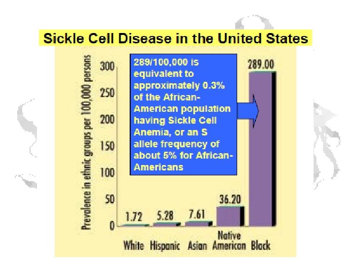

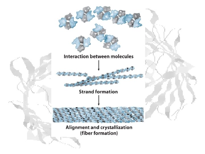
Sickle Cell • Cause: Glu 6 changed to Valine by gene mutation • Hydrophobic residue binds to pocket on adjacent chain of deoxygenated form • ~5% of American blacks carry gene • This is not a neutral mutation

Geographic Distribution of Hb. S

Malaria Belt

Heterozygote Advantage • Heterozygous individuals in Nigeria had a 29% higher likelihood of surviving to adulthood than homozygous normals. • The gene is maintained in the population by selection against both homozygotes.
")
Other O 2 Binding Proteins (w/o Heme)

Other O 2 Binding Proteins • Hemocyanins – Molluscs and some arthropods – Copper acts as binding metal – Cu(I) (colorless) Cu(II) (blue) – 75 k. Dal monomers (arthropods) • Each monomer has 2 Cu, binds 1 O 2 – Form dimers or hexamers – Polymers form very large complexes
 B. Single subunit from Limulus")
Hemocyanin structures A. 24 mer from Eurypelma (a tarantula) B. Single subunit from Limulus (horseshoe crab) C. 20 x 8 mer from Haliotis (Abalone) (each individual polypeptide is an 8 -fold repeat) d. C-terminal subunit from Octopus.

Other O 2 Binding Proteins • Hemerythrins – Sipunculids, brachiopods, priapulids, bacteria – Binuclear iron center – Fe(II) Fe(III) – 13 -14 k. Dal monomers • Each monomer has 2 Fe, binds 1 O 2 – Form (most often) octamers – Not cooperative Fe O-H + O=O / Fe A (deoxy) Fe-O-O : O··H / Fe B Fe-O-OH O / Fe C (oxy)

Sipunculid Priapulid Brachiopod

Hemerythrin

O 2 Binding Sites

Another Heme Protein That Doesn’t Bind O 2

The Disease - Chagas
 • Chronic Phase (10 -20 years post-infection)")
Symptoms • Acute Phase (weeks to months) • Chronic Phase (10 -20 years post-infection) • • • Swelling at the infection site Fever Fatigue Rash Body aches Headache Loss of appetite Nausea, diarrhea or vomiting Swollen glands Enlargement of your liver or spleen • • Irregular heartbeat Inflamed, enlarged heart (cardiomyopathy) Congestive heart failure Sudden cardiac arrest Difficulty swallowing due to enlarged esophagus Abdominal pain or constipation due to enlarged colon

Chagas Complications Acute Stage – swelling at bite location Chronic Stage megacolon Chronic Stage – cardiomyopathy, congestive heart failure

The Agent: Trypanosoma cruzi
 Triatoma gerstaeckeri (Local)")
The Vector – Cone Nosed Bugs Rhodnius prolixus (Tropical) Triatoma gerstaeckeri (Local)

The Protein - Nitrophorin • dimer of 20 kdal monomers • Heme contains Fe+3 • salivary glands of Triatomid bugs • bind nitrous oxide (NO)

Nitrophorin Action – slightly dramatized • Ravenous insect climbs onto face of peacefully sleeping human victim • Inserts hideous proboscis into helpless victim’s flesh • Nitrophorin, with NO bound, is injected into the bite wound • In the alkaline environment of the ghastly wound, NO is released • NO acts as vasodilator, prevents platelet accumulation • Empty binding site on nitrophorin binds histamine • Antihistamine effect prevents irritation to wake hapless blood donor.

Lest you think this is all theoretical… Potential future distribution in Texas From Emerging Infectious Diseases (2003) 9(1): 103 -105
- Slides: 73