Protein concentration determination inhwanspin yonsei ac kr s


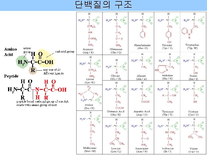


가 (lys, arg), (trp, tyr, his, phe)에 결합시의")


 로 standard 만든다. BSA 0 5 7.")
- Slides: 16
Protein concentration determination 담당교수 : 이승택 교수님 조교 : 이인환/이영훈 inhwan@spin. yonsei. ac. kr s 403호
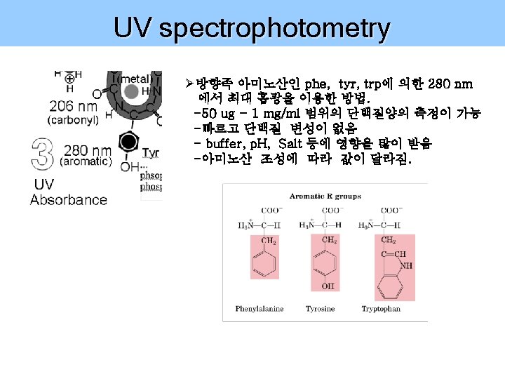
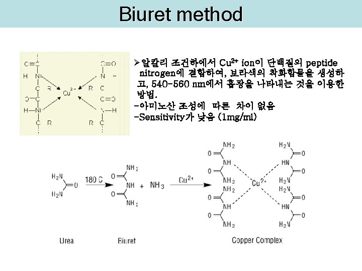
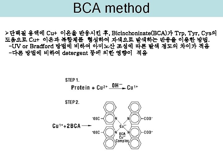
단백질 정량 방법 Ø UV spectrophotometry Ø Biuret method Ø BCA method Ø Lowry method Ø Bradford method
Lowry method 1 단계 : Biuret reaction : 알칼리 용액에서 Cu 2+와 protein peptide bond 간에 complex를 형성시켜서 Cu 2+가 Cu+로 환원된다. 2 단계 : Folin-Ciocalteu reaction : Cu+와 tryptophan, tyrosine, cysteine의 radical group이 Folin-Ciocalteu reagent (phosphomolybdate and phosphotungstate complex (노란색)를 환원시켜서 진한 청색으로 변한다. (yellow deep blue) 노란색 protein 500~
Bradford method Ø Coomassie Brilliant Blue G-250(CBBG)가 (lys, arg), (trp, tyr, his, phe)에 결합시의 흡광도의 변화를 측정 CBBG가 free dye의 형태로 있을 때 최대 흡광은 465 nm (red) 이지만, amino acid와 결합하여 anionic form일 때의 최대 흡광은 595 nm (blue) 인 특징을 이용. free dye(cationic) bound dye(anionic) Red <ㅡ> Green <ㅡ> Blue <ㅡ> Blue-Protein (470 nm) (650 nm) (590 nm) (595 nm) H+ http: //images. google. co. kr/imgres? imgurl=http: //www. medicine. mcgill. ca/physio/vlab/Other_exps/endo/images/coomassie. gif&imgrefurl=http: //www. medicine. mcgill. ca/physio/v lab/Other_exps/endo/spectro. htm&usg=__o. Xkz. OZQq. WGGM_Kk. W 3 r. EZ 8_v. RKc=&h=280&w=472&sz=31&hl=en&start=1&um=1&itbs=1&tbnid=1 N 1 qx. G 3 gt 1 plo. M: &tbnh=77&tbnw=129&prev=/images%3 Fq%3 Ddouble%2 Bprotona ted%2 Bcoomasie%2 Bbriliant%2 Bblue%2 Bbradford%26 um%3 D 1%26 hl%3 Den%26 newwindow%3 D 1%26 tbs%3 Disch: 1
단백질 정량분석법 UV, Bradford > Lowry, BCA > Biuret BCA 5
실험방법- Lowry ► Reagent A : 2% Na 2 CO 3 in 0. 1 N Na. OH Reagent B : 1 g Cu. SO 4. 5 H 2 O in 100 ml distilled water Reagent C : 2 g Potassuim Tartarate in 100 ml distilled water ► 위의 reagent A: 9. 8 ml 에 B : 0. 1 ml , C : 0. 1 ml을 차례로 15 ml tube에 넣고 섞어준다. ► BSA로 standard 를 만든다. stock : BSA 1 mg/ml BSA 0 5 7. 5 10 12. 5 15 sample 10 15 DW 200 195 192. 5 190 187. 5 185 DW 190 185 total 200 ul ► 각 standard 와 sample 에 위에서 만든 solution 1 ml 씩 넣고 vortexing 한다. ► room temp. 에서 15분간 방치한다. ► folin-phenol reagent를 D. W와 1: 1 dilution 하여 (500 ul : 500 ul) 하여 1 ml을 만든다. ► 각 standard 와 sample 에 folin-phenol reagent를 100 ul 를 가하고 바로 vortexing 한다. ► room temp. 에서 30분간 방치한다. ► spectrophotometer로 600 nm에서 흡광도를 잰다. ► standard curve를 그리고, 단백질량을 계산한다.
실험방법 - Bradford ► BSA (1 mg/ml) 로 standard 만든다. BSA 0 5 7. 5 10 12. 5 15 sample 10 15 DW 800 795 792. 5 790 787. 5 785 DW 790 785 total 800 ul ►각 microtube에 Bradford solution(5 X)를 200 ul 씩 넣어 final 1 X가 되도록 한 후, 바로 vortexing 한다. ► 2분간 반응시킨 후, 595 nm에서 흡광도를 잰다. ► standard curve를 그리고, 단백질량을 계산한다.