
Protein concentration determination hoonlimyonsei ac kr S 338

- Slides: 16
Protein concentration determination 담당교수 : 이승택 교수님 조교 : 임훈/ 남윤성 hoonlim@yonsei. ac. kr S 338호
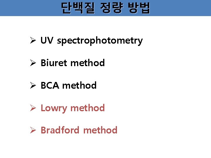
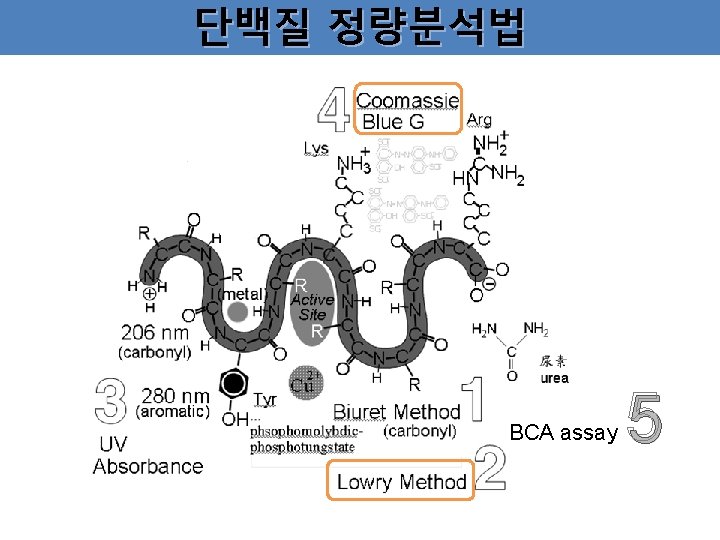
단백질 정량 방법 Ø UV spectrophotometry Ø Biuret method Ø BCA method Ø Lowry method Ø Bradford method
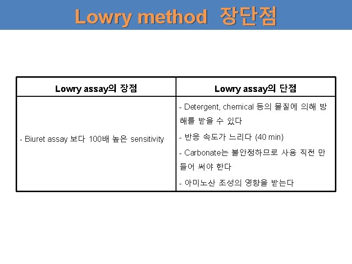
Lowry method Ø 구리이온이 peptide bond에 결합하는 Biuret method에 기초를 둠. Ø Cu 2+ 가 Folin reagent와 Folin-Ciocalteu반응을 하는 원리를 이용함. 1 단계 : Biuret reaction : 알칼리 용액에서 Cu 2+와 protein peptide bond 간에 complex를 형성시켜서 Cu 2+가 Cu+로 환원된다. 2 단계 : Folin-Ciocalteu reaction : Cu+와 Trp, Tyr의 radical group이 Folin-Ciocalteu reagent (phosphomolybdate and phosphotungstate complex (노란색)를 환원시켜서 진한 청색으로 변한다. (yellow deep blue) blue 500 ~ 750 nm
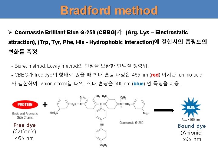
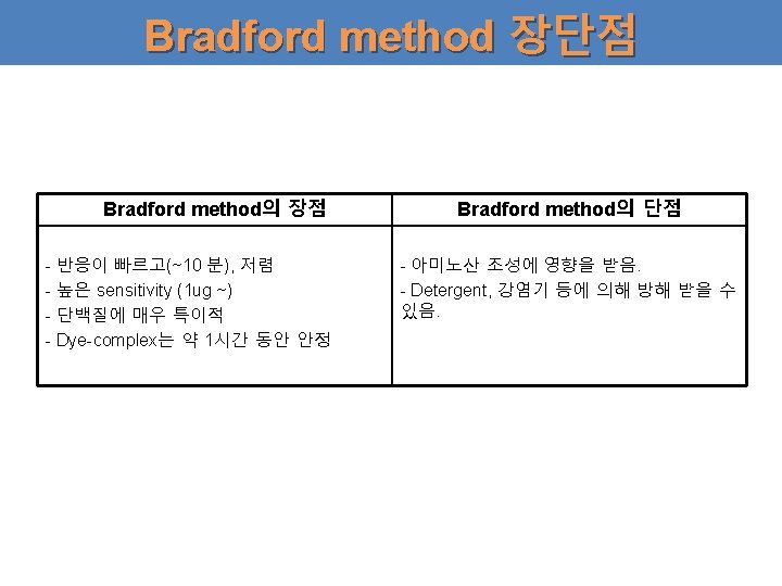
Bradford method Ø Coomassie Brilliant Blue G-250 (CBBG)가 (Arg, Lys – Electrostatic attraction), (Trp, Tyr, Phe, His - Hydrophobic interaction)에 결합시의 흡광도의 변화를 측정 - Biuret method, Lowry method의 단점을 보완한 단백질 정량법. - CBBG가 free dye의 형태로 있을 때 최대 흡광 파장은 465 nm (red) red 이지만, amino acid 와 결합하여 anionic form일 때의 최대 흡광은 595 nm (blue) blue 인 특징을 이용. Free dye (Cationic) 465 nm Bound dye (Anionic) 595 nm
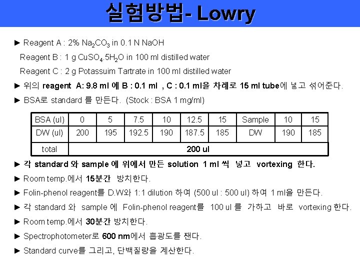
실험방법- Lowry ► Reagent A : 2% Na 2 CO 3 in 0. 1 N Na. OH Reagent B : 1 g Cu. SO 4. 5 H 2 O in 100 ml distilled water Reagent C : 2 g Potassuim Tartrate in 100 ml distilled water ► 위의 reagent A: 9. 8 ml 에 B : 0. 1 ml , C : 0. 1 ml을 차례로 15 ml tube에 넣고 섞어준다. ► BSA로 standard 를 만든다. (Stock : BSA 1 mg/ml) BSA (ul) 0 5 7. 5 10 12. 5 15 Sample 10 15 DW (ul) 200 195 192. 5 190 187. 5 185 DW 190 185 total 200 ul ► 각 standard 와 sample 에 위에서 만든 solution 1 ml 씩 넣고 vortexing 한다. ► Room temp. 에서 15분간 방치한다. ► Folin-phenol reagent를 D. W와 1: 1 dilution 하여 (500 ul : 500 ul) 하여 1 ml을 만든다. ► 각 standard 와 sample 에 Folin-phenol reagent를 100 ul 를 가하고 바로 vortexing 한다. ► Room temp. 에서 30분간 방치한다. ► Spectrophotometer로 600 nm에서 흡광도를 잰다. ► Standard curve를 그리고, 단백질량을 계산한다.
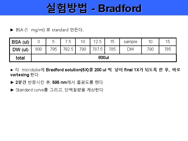
실험방법 - Bradford ► BSA (1 mg/ml) 로 standard 만든다. BSA (ul) 0 5 7. 5 10 12. 5 15 sample 10 15 DW (ul) 800 795 792. 5 790 787. 5 785 DW 790 785 total 800 ul ► 각 microtube에 Bradford solution(5 X)를 200 ul 씩 넣어 final 1 X가 되도록 한 후, 바로 vortexing 한다. ► 2분간 반응시킨 후, 595 nm에서 흡광도를 잰다. ► Standard curve를 그리고, 단백질량을 계산한다.