Prostate probe with SPECT technique NSS MIC 2010

Prostate probe with SPECT technique NSS – MIC 2010 - November 5 - Knoxville F. Garibaldi- INFN – Roma 1 – gr. Coll. ISS Ø the medical problem Ø the proposal Ø Layout Ø Multimodality Ø Si. PM/electronics Ø summary and outlook 1

Radionuclides imaging techniques 1. Collimator Only gammas that are perpendicular to imaging plane reach the detector Patient injected with radioactive drug. Drug localizes according 2. Scintillator Converts gammas to to its metabolic visible light properties. Gamma rays, emitted by 3. Photodetector radioactive decay, that exit the patient are Convert light to imaged. electrical signal 4. Readout Electronics Amplify electrical signal and interface to computer 5. Computer decoding procedure Elaborate signal and gives image output

PET mechanical collimation Compton Camera Multi pinhole
 - cheape(r) - extending the radiotracers available -")
Single photon techniques pros - simple(r) - cheape(r) - extending the radiotracers available - dual tracer looking at two different biological processes cons - efficiency - spatial resolution

Compton Prostate Imaging Probe Internal Compton Probe External Compton Probe

Predicted Internal Probe Performance 141 ke. V 171 ke. V 245 ke. V 511 ke. V 364 ke. V 4 mm Point-to-Point, 1 cm from probe (Monte Carlo simulation + ML reconstruction) Comparison with SPECT for In-111 Imaging Distance Compton Probe High-Sensitivity Collimator High-Resolution Collimator 10 cm Efficiency 1. 8 e-3 1. 11 e-4 4. 00 e-5 Resolution 2. 47 mm 15. 9 mm 10. 5 mm

Relative Uptake of In-111 Prostascint Organ Relative Uptakes Prostate 1. 0 Liver 2. 0 Blood 1. 5 Bone 0. 7 Kidney 1. 0 Spleen 1. 0 Bladder 0. 6 Rectum 0. 4 Testes 0. 6 Averaged from three In-111 Prostascint SPECT scans

Conventional SPECT Reconstructions 5: 1 10: 1 15: 1 20: 1 w / tumor Prostate bkgd 171 and 245 ke. V, 8. 8 M events / 40 slices Spatial resolution ~15 mm FWHM

Compton Prostate Probe Reconstructions 245 ke. V only, 1. 2 million events, 8 mm lesion Prostate 5: 1 10: 1 15: 1 20: 1 w / tumor bkgd Spatial resolution ~2 mm FWHM

Internal Detector Details 10– 12 layers of 1 mm thick Si detectors + position and orientation sensor Exploded View Assembled Unit

Compton Probe Promising but Challenging • First detector – Energy resolution – largely addressed – Timing resolution – still an issue – Packaging – solvable • Second detector – Countrate capability – solvable – Cost – always an issue • System – Image reconstruction – solvable
 (Very thin kapton tape with aluminum traces)")
Detector Packaging Use Tape Automated Bonding (TAB) (Very thin kapton tape with aluminum traces) Kapton microcables “Raw” energy spectrum Detector VATA ASIC Unfolded energy spectrum Kapton “hybrid” board

Timing • Desired time resolution <10 ns FWHM • Poor timing from Si is evident • Slower signal generation from events near backplane • Large range of pulse-height coupled with leading-edge trigger is a big issue time-walk • BGO-Silicon timing spectrum for 511 ke. V source Signal generation depends on 3 D interaction position and recoil electron direction time-jitter Signal generation at two biases for three depths

How Challenges Affect Performance • Consider anticipated countrate with In-111 Prostascint (from Monte Carlo simulations): – ~4 Mcps on second detector – ~40 kcps on scattering detector – 50 ns time window for present Si detectors (may need to be even larger) • Crandom= 2 x 4 x 106 x 4 x 104 x 50 x 10 -9 = 16, 000 cps ! • Ctrue was only ~10 kcps (or less) • Performance dominated by randoms! • Energy sum window can be used to reject randoms but only if the second detector also has good energy resolution
")
Single photon Compton camera ( N. Clinthorne. Michigan )

External Multipinhole Alternative External probes will have small FOV and limited-angle tomography but… • SPECT/CT can identify prostate region • Probe can be computer-steered to image desired FOV • Conventional SPECT can be used to “complete” probe data

Endorectal Multipinhole? 30 mm ~15 mm • Some tomographic capability • Requires high detector resolution (0. 5– 1 mm + depth-of-interaction) • High enough efficiency and resolution?

W. Moses – Rome workshop 2005

Radionuclides Single photon 111 In-Prosta. Scint is not a good radiotracer but a new one proposed by M. Pomper looks promising. The single photon endorectal probe provides 2 D imaging. We have to try to have 3 D images ( using multipinhole collimation and/or adding up a SPECT tomograph (spatial resolution would be dominated by the small probe (see later, the PET case))

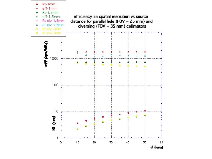
our proposal -insert scintillator pixels into square holes of the collimator better performances (spatial resolution (? ) and sensitivity (thicker scintillator)) -using diverging collimator better performances (reducing scan time) -using multipinhole collimation better performances (increasing sensitivity, tomographic recinstruction)
 Radiotracers available for SPECT and")
New radiotracers coming soon (M. Pomper , Johns Hopkins) Radiotracers available for SPECT and PET (from “New agents and Techniques for Imaging prostate cancer” A. Zahreer, S. Y. Cho, M. Pomper”, to be published on JNM ) SPECT: Prostascint, Bombesyn, 99 m. Technetium nanocolloid (limphonodes), other coming soon… PET C— 11 Choline, F-18 -Choline, Ga-68 Dotabomb (Hofmann (Rome workshop)) many others coming… (collaboration with Johns Hopkins for testing in ISS (mice models for prostate available) and/or at JHU) 22
- Slides: 22