Prokaryotic Cell Structure Bacteria shape and size Bacteria

Prokaryotic Cell Structure

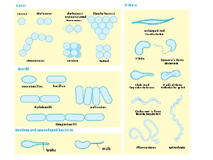
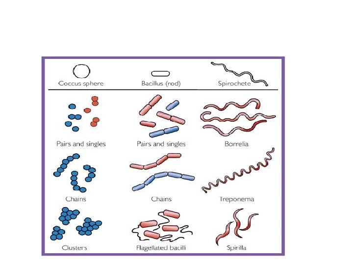
Bacteria – shape and size • Bacteria are believed to be the first cell to evolve – have no clear membrane bound nucleus or organelles • Bacteria vary in size and shape • Coccus • In pairs diplococcus. eg. Neisseria. sp. • Long chains – Streptococcus sp. • Irregular grape like clumps Staphylococcus sp. • Tetrads eg. Micrococcus sp.

Epulopiscium fishelsoni grows as large as 600 µm by 800 µm, a little smaller than a printed hyphen. Exceptionally large bacteris

• Bacillus: rod shape eg. Bacillus spp. – Coccobacilli – The shape of the rod’s end often varies – Some bacteria form long multinucleate filaments – eg. Actinomycetes • Sprillum: long rods twisted into spirals or helix – spirilla (rigid) spirochetes (if flexible) • Size: – Mycoplasma are only 100 200 nm in diameter – E. coli is 1. 1 to 1. 5 µm wide by 2. 0 to 6. 0 µm. long, – Spirochetes size reaches 500 µm in length

Structure and function of prokaryotes • Membrane systems • prokaryotic and eukaryotic membranes are similar in structure • Membranes of eukaryotic microorganisms serve to compartmentalize cell contents into organelles • Prokaryotic organisms contain only a single membranous structure, cytoplasmic mebrane or plasma membrane • measures 4 – 5 nm thick

• permeability barrier of the cell • involve in complex biochemical processes respiration • membranes are formed of a lipid bilayer, made of phospholipids • fatty acid portion hydrophobic, glycerol phosphate part hydrophilic • hydrophilic parts are exposed to the aqueous external environment

• the inner and outer sides of the cytoplasmic membrane have different properties • property of ‘sidedness’ is of great importance • overall structure of a membrane is maintained by hydrogen bonds and hydrophobic interactions • (Mg 2+, Ca 2+) help to stabilize the structure • Eucaryotic membranes differentiated from those of prokaryotes with sterols • Mycoplasmas, contain sterols

Plasma Membrane • Layer of phospho-lipids and proteins that separates cytoplasm from external environment. • Regulates flow of material in and out of cell.

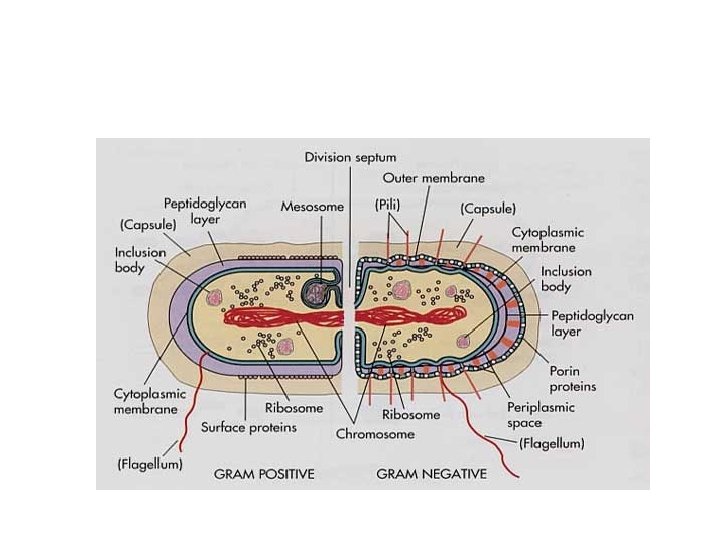
Cell walls • Bacterial cell wall is unique two broad categories, Gram positive and gram negative • Gram positive bacteria have a thick, single layered wall. Gram negative complex multilayered wall thin • Peptidoglycan layer is present in the cell walls • In Gram positive bacteria, bulk of the wall is peptidoglycan Gram negative it accounts for only the innermost layer
 and N acetylgucosamine (NAG) linked by")
• Peptidoglycan consists N acetylmuramic acid (NAM) and N acetylgucosamine (NAG) linked by bonds described as β 1 4 linkages • Gram positive cell walls contain another polymer called teichoic acid • Mycobacterium, Corynebacterium contain waxy esters of mycolic acids

Cell Wall • Rigid peptidoglycan polysaccharide coat that gives the cell shape and surround the cytoplasmic mem-brane. Offers protection from environment.
 • Some bacteria possess additional hair like structures")
Bacterial cell surface (Fimbriae and Pili) • Some bacteria possess additional hair like structures called fimbriae • shorter than flagella but numerous • to stick to a surface • Pili – specialised pili conjugation process • Glycocalyx (Slime / capsule) • Glycoclyx consists of polysaccharides, with glycoprotein • Hinders the engulfing (phagocytosis) • Also prevents desiccation.

PILI • • Short protein appendages Smaller than flagella Adhere bacteria to surfaces Used in conjugation for Exchange of genetic information • Aid Flotation by increasing buoyancy

Nucleoid • Region of the cytoplasm where chromosomal DNA is located. Usually a singular, circular chromosome. • Smaller circles of DNA called plasmids (extra chromosomal DNA) are also located in cytoplasm.

Nucleoid • • • Prokaryotic DNA is in circular form lack a nuclear envelope Bacterial DNA not associated with proteins The DNA is highly coiled Plasmids extrachromosdmal circular DNA

Ribosomes • Translate the genetic code into proteins. • Free-standing and distributed throughout the cytoplasm. • Bacterial ribosomes have two sub units 50 S and 30 S
 rotate")
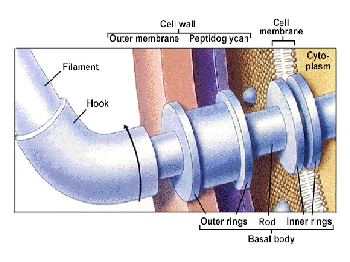
Flagella • • hair like structures called flagella (14 – 20 nm diameter) rotate like a ship’s propeller protein called Flagellin, flagellar subunits basal body rotates the flagellum to cause movement of the cell Arrangement of flagella Monotrichous: Eg. Vibrio cholerae Amphitrichous : Eg. Spirillum volutans Lophotrichous: Eg. Alcalegenes faecalis Peritrichous: Eg. E. coli

Monotrichous Lophotrichous Amphitrichous Peritrichous

Chemotaxis and Motility • Chemotaxis is the movement of an organism towards or away from a chemical • Positive chemotoxis movement towards a chemical (attractant); negative chemotaxis movement away from a chemical (repellent) • Bacterial movement is characterized by runs and tumbles • when an attractant present it is marked by larger runs and less frequent tumbles

Mesosome • Infolding of cell membrane. • Possible role in cell division. • Increases surface area. • Photosynthetic pigments or respira-tory chains here. • Http: //www. med. sc. edu: 85/fox/protobact. jpg

Other structures • Inclusion bodies for storage of materials • Poly β – hydroxybutyric acid (PHB), Granules of polyphosphate, volutin granules matachromatic granules generation of ATP and other cell costitutions

Bacterial endospores • Bacterial endospore is not a reproductive structure • Resistant to harsh environmental conditions • Bacillus and Clostridium produce endospores • Endospore is more complex than the vegetative cell • Dipicolinic acid (DPA) • Sporulation occurs due to environmental stress

Spore formation • The sporulation process successive stages • Preparatory stage • Forespore stage • Stage of cell wall formation • Maturation stage occurs in four
 • aerobic, high G C percentage gram")
Other Prokaryotes • Actinomycetes (The Filamentous Bacteria) • aerobic, high G C percentage gram positive bacteria form branching filaments or hyphae and asexual spores • closely resemble fungi in overall morphology • aerial hyphae, substrate hyphae • Septa • aerial hyphae reproduce asexually • Most actinomycetes are non motile • they break down hard organic materials like newspaper
 2. Aerial Hyphae")
Growth of Actinomycetes on agar plate 1. Chain of Conidiospores (Conidia) 2. Aerial Hyphae 3. Agar Surface 4. Substrate Hyphae
 cells. • Chemoheterotrophic lengths")
Spirochaetes • Gram negative bacteria, long, helically coiled (spiral shaped) cells. • Chemoheterotrophic lengths between 5 and 250 µm diameters around 0. 1 0. 6 µm • Flagella called axial filaments, cell membrane and outer membrane • cause a twisting motion which spirochaete will undergo asexual transverse binary fission • Most spirochaetes are free living and anaerobic
, • Disease causing members of this")
• Classification three families (Brachyspiraceae, Leptospiraceae, Spirochaetaceae), • Disease causing members of this phylum Leptospira species, Borrelia burgdorferi, Borrelia recurrentis, Treponema pallidum

Spirochaetes Treponema pallidum spirochetes

Cyanobacteria • Cyanobacteria , blue-green algae, blue-green bacteria obtain their energy through photosynthesis • significant component of the marine nitrogen cycle and an important primary producer

• The cyanobacteria were classified into five sections, I V. • Chlorococcales, Pleurocapsales, Oscillatoriales, Nostocales and Stigonematales

Mycoplasma • Mycoplasmas lack a cell wall • unaffected by many common antibiotics such as penicillin beta lactam antibiotics • parasitic or saprotrophic • pathogenic in humans, M. pneumoniae, • Mycoplasma is by definition restricted to vertebrate hosts • Cholesterol is required for the growth

Cell wall structure • M. pneumoniae cells are of small size and pleomorphic • Mycoplasmas are unusual among bacteria – possess sterols for the stability of their cytoplasmic membrane • low GC content

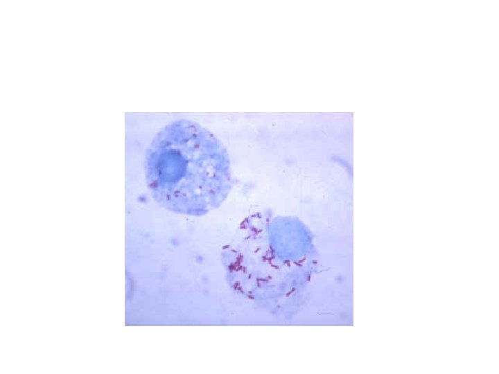
Rickettsiae • Rickettsia is a genus of motile, Gram negative, pleomorphic bacteria present as cocci, rods thread like • Obligate intracellular parasites, survival depends on entry, growth, and replication within the cytoplasm of eukaryotic host cells • cannot live in artificial nutrient environments are grown either in tissue or embryo cultures • Rickettsia carried as parasites by cause diseases typhus, rickettsialpox, Boutonneuse fever, African Tick Bite Fever, Rocky Mountain spotted fever, Australian Tick Typhus
 • Archaea although look like bacteria are not closely related to them")
Archaebacteria (Archaea) • Archaea although look like bacteria are not closely related to them • divided into two evolutionary lineages based on r. RNA sequences, crenarchaeotae, Euryarcheotae • Crenarchaeotae grow at high temperatures and metabolize elemental sulfur • Euryarchaetoes are methanogens some grow aerobically very high concentrations of salt • Archaea possess membrane lipids of branched chain hydrocarbons bound to one or two glycerol molecules by ether bonds

ARCHAEA Halobacteria sp
- Slides: 40