Programmed Cell death Saeb Aliwaini April2013 Introduction Human

Programmed Cell death Saeb Aliwaini April/2013

Introduction • Human Body makes 10 billion cells every day. • Cell death makes balance : • There are various forms of cell death. Amongst these, two well-known pathways are necrosis and apoptosis. Other less described cell death pathways are mitotic catastrophe , autophagy and necroptosis.
 • Apoptosis")
Apoptosis • Apo = from , ptosis = falling ( dropping off) • Apoptosis is characterized by specific biochemical and morphological features that culminate in shrinkage of the cell to apoptotic bodies that are engulfed by neighboring macrophages.

Major inducers of apoptosis A- Apoptosis occurs during normal cell turnover, tissue homeostasis, embryogenesis, induction and maintenance of immune tolerance. Other inducers - Irreparable DNA damage - Cell cycle perturbation - Death ligands, e. g. , Fas ligand - Growth factor withdrawal - Calcium influx -Free radicals - Radiation therapy - Chemotherapy drugs

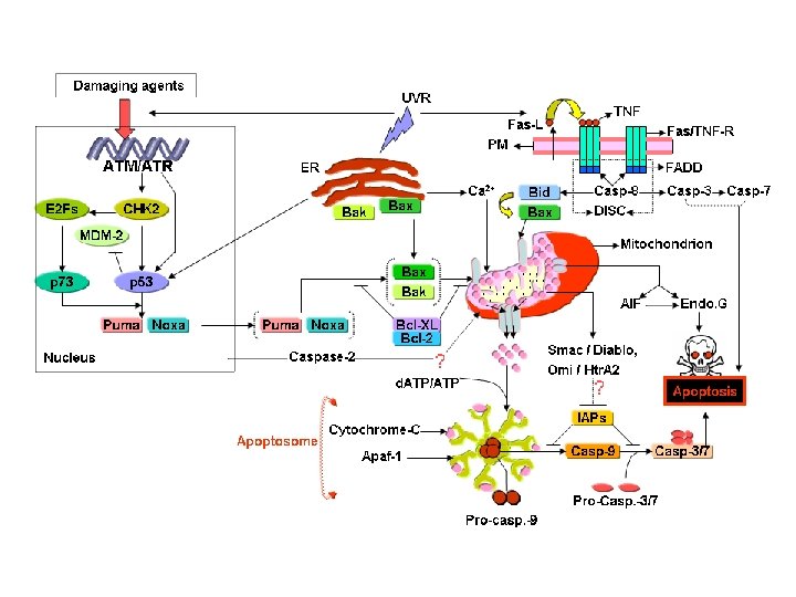
Mechanisms of Apoptosis • The molecular mechanisms of apoptosis were highly conserved through evolution

Mechanisms of Apoptosis

Caspases and Caspases dependent Apoptosis • Caspases: • The "c" of "caspase" refers to a cysteine protease, while the "aspase" refers to the enzyme's unique property to cleave after aspartic acid residues • Their proteolytic activity cleaves their substrate after Asp residues. • Caspases are synthesized in cells as catalytically inactive zymogens • Their activity is irreversible • Tow types of caspases in Apoptosis: Initiator and effector

Caspases and Caspases dependent Apoptosis - Is typically activated in the early stages of apoptosis. - It cleaves key cellular components: Such cytoskeleton proteins and nuclear proteins such as DNA repair enzymes. - Nuclear condensation and cell shrinkage. - The cells express (translocation of phosphatidyl serine from the inside of the cell to the outer surface).

A- Extrinsic pathway - Is mediated by death receptors Apoptosis via this mechanism is very rapid Induction of apoptosis by TNF receptor - Cell membrane receptors, which belong to tumor necrosis factor (TNF) family. - Including TNF-receptor 1, Fas, DR 3, DR 4, DR 5, and DR 6 - Specific ligands, such as TNF-α, lymphotoxin, Fas ligand - Conformational changes to form death inducing signaling complex. ( DISC) - Followed by recruitment of one of the caspases, typically caspase 8, to the DISC
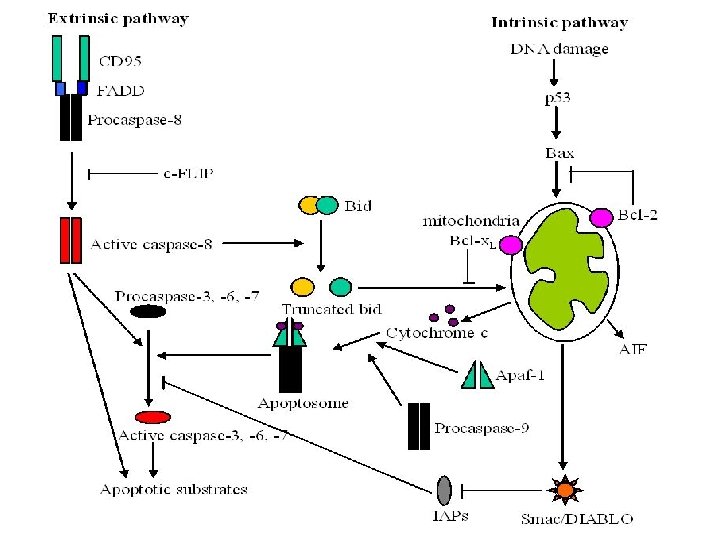

Caspases and Caspases dependent Apoptosis B- Intrinsic pathway - Also called mitochondrialmediated apoptosis. - Main inducer : DNA damage - The Bcl 2 families of proteins are the main mediators of this process. This family has up to 4 conserved domains, known as the Bcl 2 homology (BH) domains

• The Bcl 2 family contains both pro-apoptotic members and anti-apoptotic members. • The pro-apoptotic members : - Bax family that consist of Bax, Bok and Bak. - BH-3 only proteins, such as Bid, Bad, and Bim. - The anti-apoptotic members of the Bcl 2 family contain all 4 conserved domains, such as Bcl 2 and Bclxl. - Bcl 2 are found exclusively within intracellular membranes and within the cytosol.

How they work? • Pro apoptotic proteins : to release apoptotic-signaling factors from the mitochondria through mitochondrial outer membrane permeabilization (MOMP) via opening of the membrane permeability transition pore. • Anti-apoptotic: function by preventing apoptosis through heterodimerization with pro-apoptotic proteins and self over-expression.

- cytochrome c, Smac/DIABLO, and Htr. A 2/Omi are capsases dependent - The apoptosome formation and effector caspase activation that causes the token apoptotic events, such as chromatin condensation, plasma membrane asymmetry, and cellular blebbing

Caspase-independent apoptosis • Various factors are involved: 1 - Apoptosis-inducing factor: - The lysosomal protease cathepsin D trigger AIF. - AIF becomes an active cell killer when it is released to the cytosol. - With factor # 2 (endonuclease G) they induce peripheral chromatin condensation. - The lethal effects of AIF are controlled by the anti-apoptotic protein heat shock protein 70 that interacts with AIF and protects against its apoptogenic effects.
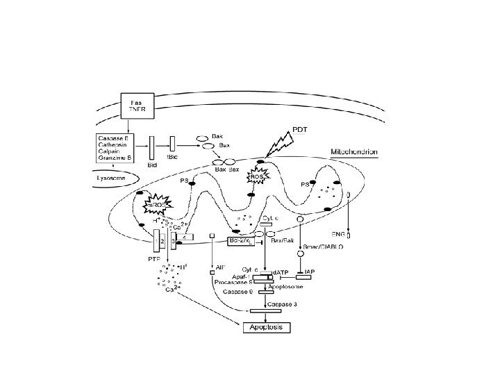

3 - Endoplasmic reticulum • Calcium release and activation of caspase 12

Inhibitor of apoptosis proteins: - The IAP proteins function as endogenous caspase inhibitors - NAIP, c-IAP 1, c-IAP 2, XIAP, and Survivin - Survivin prevent caspase-9 activation within a functional apoptosome - Control of mitosis- Survivin can enhance microtubule stability in metaphase spindle formation.

Signals involved in Apoptosis Stimulus

Cisplatin mechanism of action Tamoxifin, Doxorubicin, 5 FU and other chemotherapeutics Necrosis Necroptosis Autophagy Mitotic catastrophe

Apoptosis assays • 1. Cytomorphological alterations • 2. DNA fragmentation • 3. Detection of caspases, cleaved substrates, regulators andinhibitors • 4. Membrane alterations • 5. Mitochondrial assay
 • Detection of Caspases, Cleaved Substrates. (PARP, H")
• Transmission electron microscopy (TEM) • Detection of Caspases, Cleaved Substrates. (PARP, H 2 AX, . . ) • Apoptosis PCR microarray is a relatively new methodology that uses real-time PCR to profile the expression of at least 112 genes involved in apoptosis • Membrane Alterations externalization of phosphatidylserine residues on the outer plasma membrane of apoptotic cells allows detection via Annexin V • Mitochondrial Assays mitochondrial assays and cytochrome c release allow the detection of changes in the early phase of the intrinsic pathway.

Cytomorphological alterations
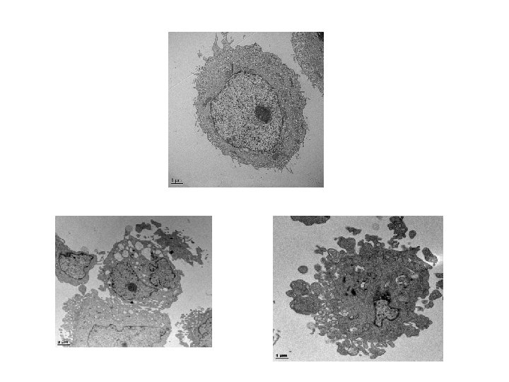

1. Cytomorphological alterations and 2 - DNA fragmentation

2. DNA fragmentation

2 -DNA fragmentation

A- Cell cycle analysis MDA-MB-231 cells 2 n Plate 3 X 10⁵ cells/well in 6 well plates M G 1 2 n Typical Cell cycle profile G 2 S G 0 -G 1 4 n 48 hours settle Treat with 0. 2 µm AJ-5 For 24 and 48 hours Trypsinize and fix with cold ethanol for at least 30 min FACS process and analysis Sub. G 1 S G 2 -M

MCF 7 MDA-MB-231 A- Cell cycle analysis AJ-5 induces a G 1 arrest and apoptosis (sub-G 1 peak) in breast cancer cells

3 - Membrane alterations Annexin V staining MDA-MB-231 cells Necrosis Late apoptosis Plate 3 X 10 5 cells/well in 6 well plates 48 hours settle Treat cells with 0. 2 µm AJ-5 for 24 and 48 hours Viable cells Early apoptosis Trypsinize and stain cells with (Annexin V and PI) FACS analysis 33

AJ-5 induces apoptosis in breast cancer cells in accordance with the cell cycle analysis.

Detection of caspases, cleaved substrates, regulators andinhibitors MCF 7 0. 0 µM AJ-5 0. 2 µM AJ-5 MDA-MB-231 0. 0 µM AJ-5 0. 2 µM AJ-5 - Nuclear fragmentation after treatment with 0. 2 µM AJ-5 for 24 h followed by Hoescht staining. AJ-5 treatment leads to an increase in PARP cleavage level and nuclear fragmentation confirming that it induces apoptosis.

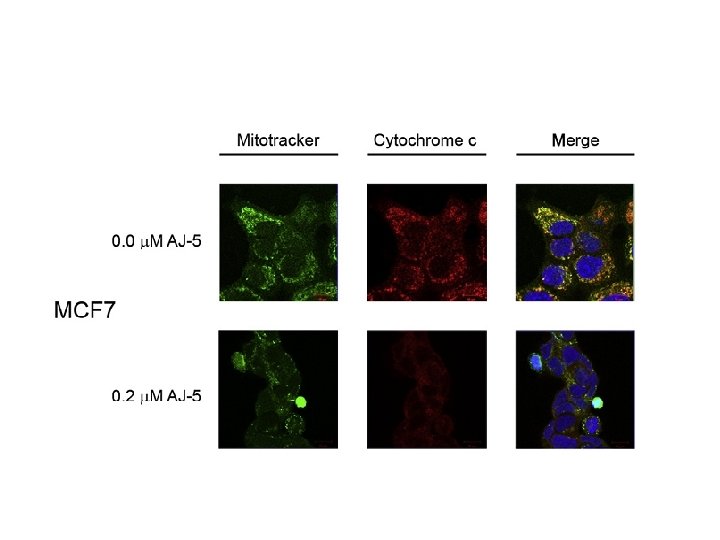
Methodologies: Apoptosis Assays • 5. Mitochondria assays o Cytochrome C release Gang et. al (2010)

Apoptosis Extrinsic pathway Caspase 8 MAPK Intrinsic pathway BH 3 proteins Bid, Bad, Puma, Noxa p 53 p 21 Cell cycle arrest Cyto C Effector Caspases 3, 7 and 6 Which apoptosis pathway is induced by AJ-5 ? 38

apoptosis pathway Markers? Check for intrinsic and extrinsic apoptosis pathways markers MCF 7 MDA-MB-231 AJ-5 activates both intrinsic and extrinsic as early as 1 h of the treatment
- Slides: 39