PRINCIPLES OF INNATE IMMUNITY THE INNATE IMMUNE SYSTEM


 *")

 * Rolling adhesion * Slowing down leukocytes (margination)")

 * Family of small soluble molecules that stimulate activation and migration")


 CELLS * Large granular lymphocytes that circulate in blood * Functions")
 CELLS * Activated NK cells release IFN-gamma which activates * Macrophages")
- Slides: 51
PRINCIPLES OF INNATE IMMUNITY
THE INNATE IMMUNE SYSTEM * First line of defense against pathogens * Components * * * * Complement system Macrophages and neutrophils Defensins Coagulation system Cytokines and inflammatory cytokines Inflammatory response Natural killer cells
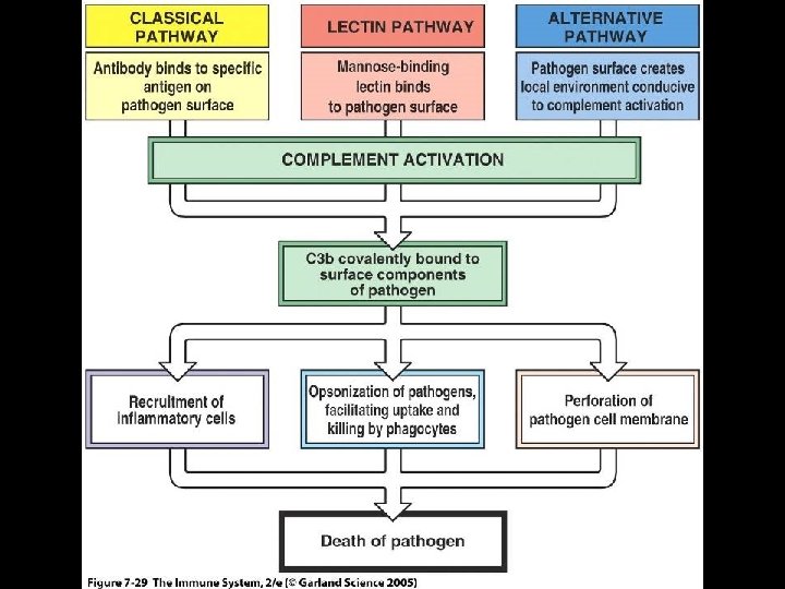
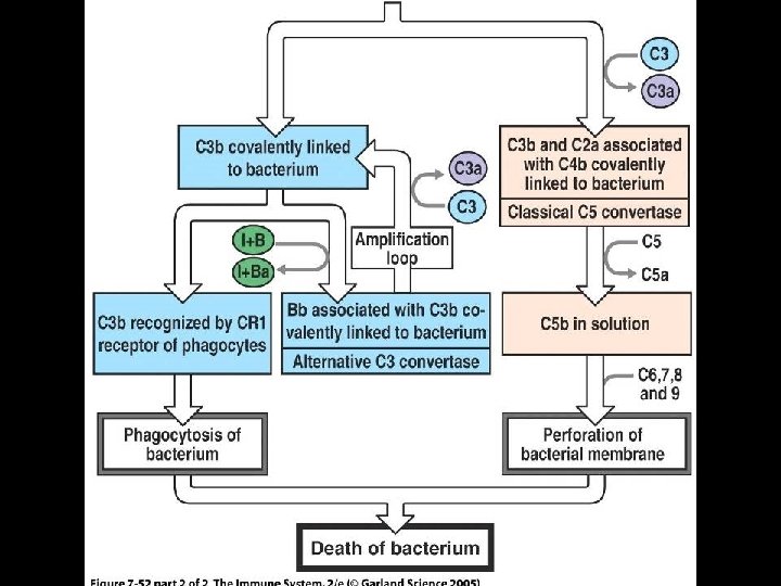
THE COMPLEMENT SYSTEM * A set of proteins widely distributed throughout body fluids and tissues * Proteins act in a cascade of reactions to attack extracellular forms of pathogens * Complement activation results in * Inflammatory response * Pathogens coated with complement * Complement coating of pathogens * Enhanced engulfment and destruction by phagocytes * Direct killing of pathogens
PATHWAYS OF COMPLEMENT ACTIVATION * Classic pathway * Activated by antibody * First discovered * Alternative pathway * Activated by some bacterial cell surfaces * Antibody not involved * Lectin pathway * Activated by mannose binding lectin * Antibody not involved
THE COMPLEMENT SYSTEM * Nomenclature has developed haphazardly * Proteins of classic pathway named with capital “C” followed by a numeral (C 1, C 2, C 3…. . C 9) * Cleavage fragments named as parent followed by lower case letter * “a” for smaller fragment (C 3 a) * “b” for larger fragment (C 3 b) * Some classic components participate in other 2 pathways
IMMUNOGLOBULINS IN ACTION *Complement fixation
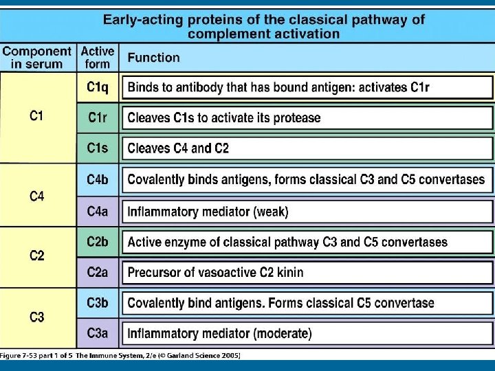
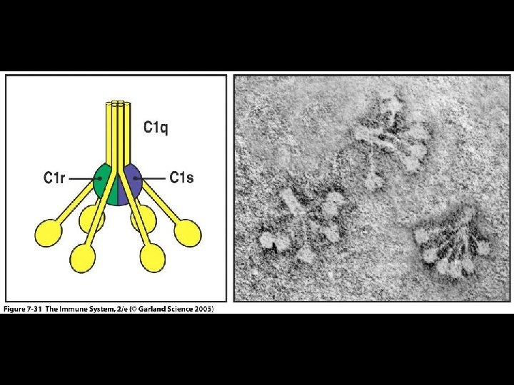
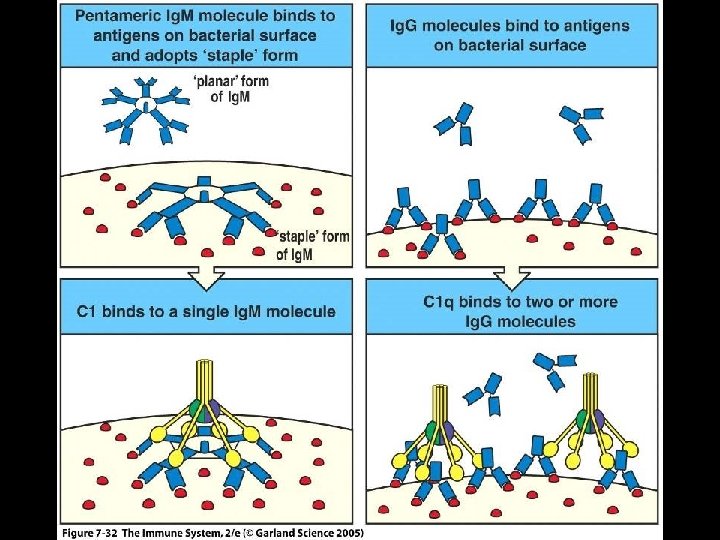
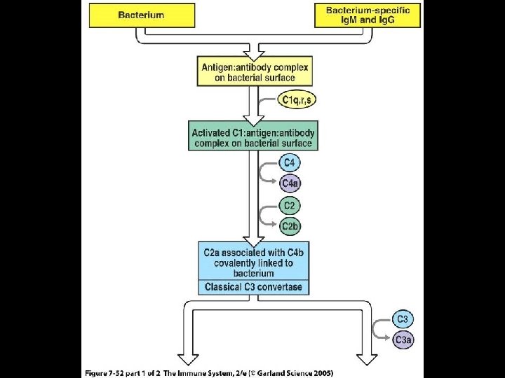
CLASSIC PATHWAY OF COMPLEMENT ACTIVATION * C 1 binds to Fc region of antibody part of Ab/Ag complex * C 1 is complex of 3 proteins * C 1 q is binding protein * C 1 r and C 1 s are proteases * C 1 q binds to Fc region of antibody which activates C 1 r which activates C 1 s * Most efficient at activating complement * Ig. M, Ig. G 1 and Ig. G 3
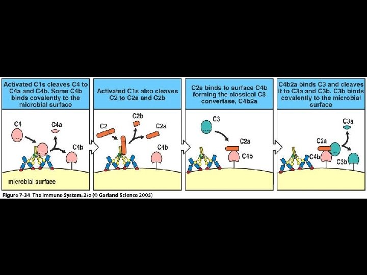
CLASSIC PATHWAY OF COMPLEMENT ACTIVATION * Activated C 1 s cleaves C 4 to * C 4 a and C 4 b * Activated C 1 s cleaves C 2 to * C 2 a and C 2 b * C 4 b and C 2 b form complex covalently bonded to pathogen surface * C 4 b/C 2 b complex (C 3 convertase) cleaves C 3 to * C 3 a and C 3 b
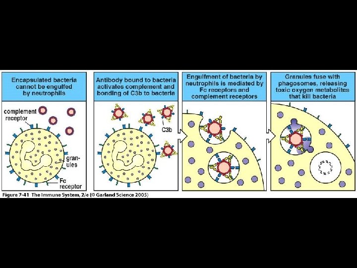
ANTIBODY AND COMPLEMENT ENHANCE PHAGOCYTOSIS * Enhanced phagocytosis especially important * Streptococcus pneumoniae * Haemophilus influenzae * Cryptococcus neoformans * Macrophages and neutrophils have receptors for * Antibody * Fc-gamma for Fc region * Complement receptor 1 (CR 1) for C 3 b
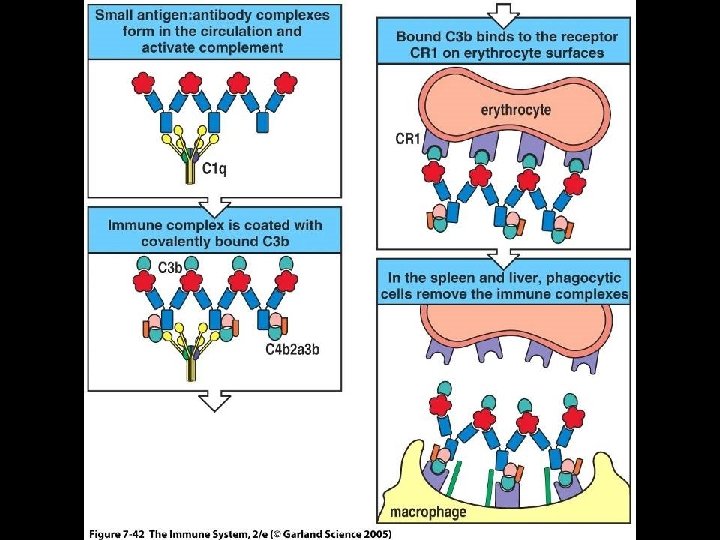
COMPLEMENT RECEPTORS REMOVE IMMUNE COMPLEXES * Immune complexes * Soluble antibody/antigen complexes * Form after immune response to most infections * IC must be removed to prevent precipitation and deposition on endothelial membranes * Kidneys * Removal of IC * Complement binds to IC * Erythrocytes bind to complement by CR 1
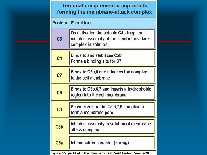
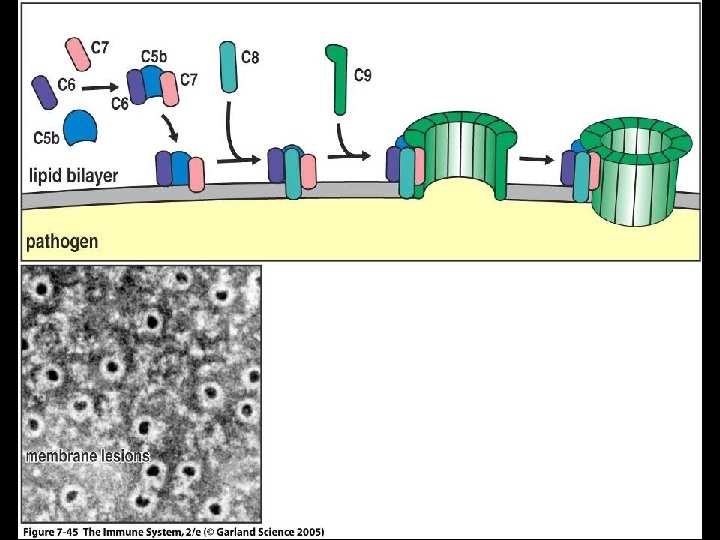
DIRECT KILLING OF PATHOGENS BY COMPLEMENT SYSTEM * Terminal complement proteins form “membrane attack complex” * Mechanism of attack by classic pathway * C 3 b binds to C 3 convertase (C 4 b, 2 b) / (C 4 b, 2 a) results in * C 5 convertase (C 4 b, 2 b, 3 b) / (C 4 b, 2 a, 3 b) * C 5 binds C 3 b of C 5 convertase * C 5 cleaved to * C 5 a and C 5 b * C 5 b initiates assembly of attack membrane components * C 6 – C 9 * Deficiency increases susceptibility to Neisseria meningitidis and Neisseria gonorrhoeae
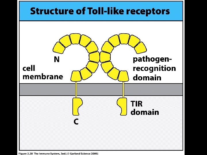
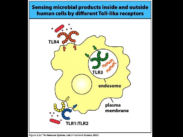
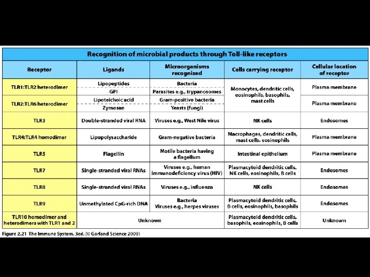
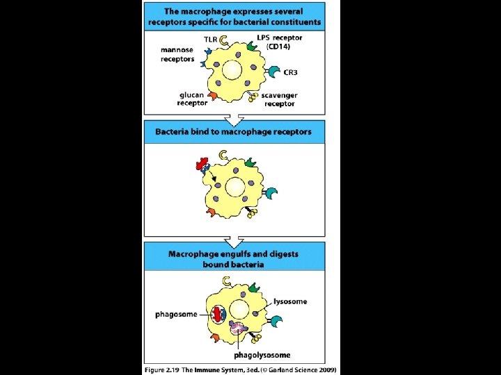
RECOGNITION OF PATHOGENS FOR PHAGOCYTOSIS * Mechanism of recognition * Toll-like receptors (innate immune receptors) * Toll-like receptors * Named for ‘Toll’ receptor in fruitfly * Polypeptides with horseshoe-shaped structure * Recognition by macrophages initiates activation * Phagocytosis * Secretion of cytokines
ACTIVATION OF MACROPHAGES * Activated macrophages secret * Cytokines * Chemokines (chemoattractant cytokines) * Inflammatory mediators * Cytokines and chemokines * Interleukin-1 (IL-1), IL-6, IL-8, IL-12 and TNF-alpha * Inflammatory mediators * Prostaglandins, leukotrienes, plasminogen activator, plateletactivating factor (PAF)
Figure 8 -15
MIGRATION OF NEUTROPHILS INTO TISSUE (EXTRAVASATION) * Rolling adhesion * Slowing down leukocytes (margination) * Weibel-Palade bodies in vascular endothelial cells secreting P and E selectins * Tight binding * Interaction between LFA-1 and ICAM-1 * Diapedesis * Passage between vascular endothelial cells * Migration to infection site
Figure 8 -19
Chemokines (Chemoattractant Cytokines) * Family of small soluble molecules that stimulate activation and migration of cells * Group classification * CC * Two adjacent cysteine amino acids * Chromosome 4 * CXC * Two separated cysteine amino acids * Chromosome 17
Figure 8 -16 part 1 of 3
Figure 8 -16 part 2 of 3
Figure 8 -16 part 3 of 3
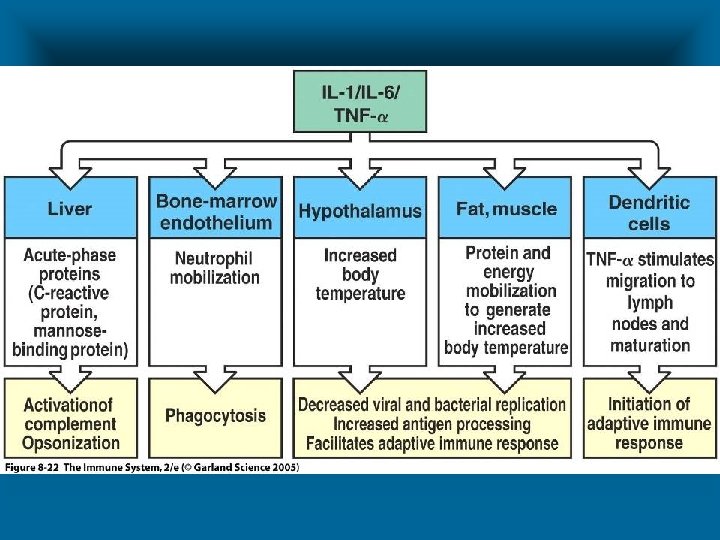
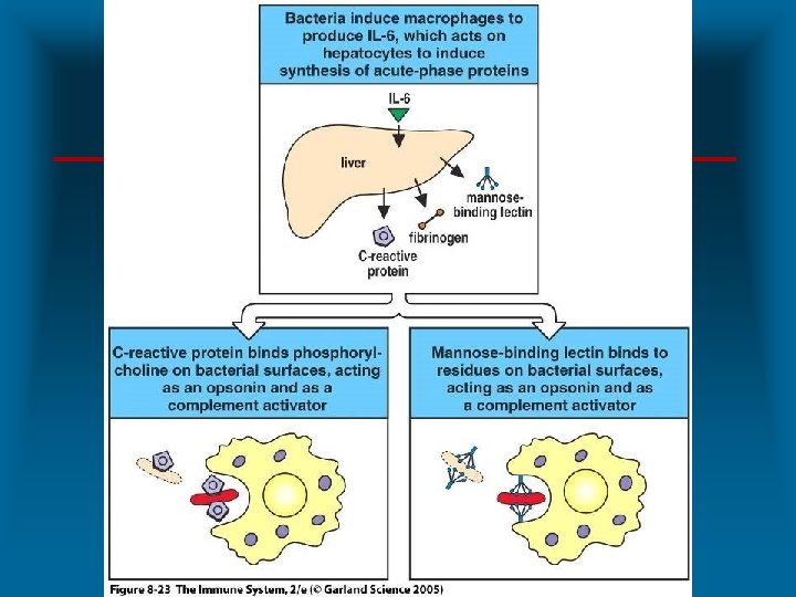
BIOLOGICAL ACTIVITY OF IL-1, IL 6 AND TNF-ALPHA * Induce hepatocytes to produce acute-phase proteins * C-reactive protein (CRP) * Mannose binding lectin (MBL) * Induce bone marrow to release neutrophils * Induce hypothalamus to raise temperature * Induce fat and muscle cells to generate heat
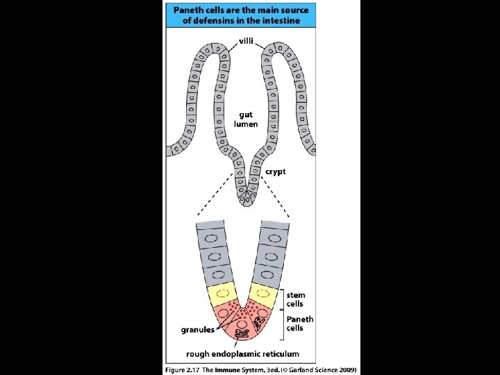
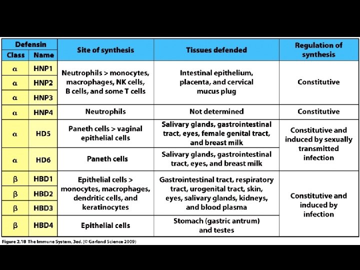
DEFENSINS * Family of amphipathic antimicrobial peptides * 35 to 40 amino acids * Mechanism of action * Disruption of cell membranes * Classification * Alpha * Neutrophils and Paneth cells * Beta * Epithelial cells of skin, respiratory tract and UG tract
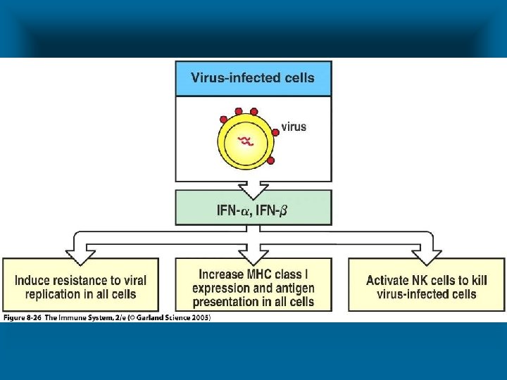
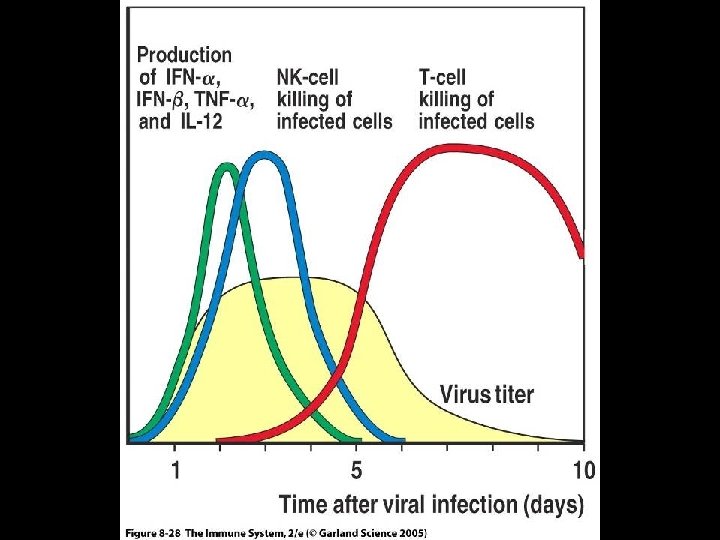
THE INNATE RESPONSE TO VIRAL PATHOGENS * Virus infected healthy cells produce * Interferon-alpha (IFN-alpha) * Interferon-beta (IFN-beta) * IFN-alpha and IFN-beta are type 1 interferons * Type 1 interferons * Inhibit virus replication * Activate natural killer (NK) cells * Increases expression of MHC-1 molecules
Figure 8 -25
NATURAL KILLER (NK) CELLS * Large granular lymphocytes that circulate in blood * Functions * Killing infected cells (cytotoxic) * Secretion of cytokines * Activation by * Type 1 interferons * Infected cells * Stimulates cytotoxic function * IL-12 and TNF-alpha * Macrophages * Stimulates cytokine secretion
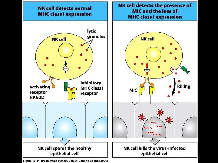
NATURAL KILLER (NK) CELLS * Activated NK cells release IFN-gamma which activates * Macrophages * Release IL-12 * Positive feedback system for NK and macrophages * Differentiate infected from uninfected cells * NK cells express receptors for MHC class I molecules * Binding of NK cells to MHC class I molecules turn off NK cells * NK cells provide innate immunity to intracellular pathogens