Principles of Diagnosis Dr Mohammad Arif Abid Dermatologist

Principles of Diagnosis Dr. Mohammad Arif Abid Dermatologist

SKIN ANATOMY

• What could be easier than the diagnosis of skin disease? The pathology is before your eyes! Why then do nondermatologists have such difficulty interpreting what they see? >3000 skin disease • hundreds of cutaneous diseases • single entity can vary in its appearance • skin diseases are dynamic and change in morphology What at first glance appeared to be simple to diagnose may later appear to be simply impossible.

• Dermatology is a morphologically oriented specialty. • As in other specialties, the medical history is important; however, the ability to interpret what is observed is even more important. • The diagnosis of skin disease must be approached in an orderly and logical manner.

A methodical approach History. brief history, noting duration, rate of onset, location, symptoms, family history, allergies, occupation, and previous treatment.

A methodical approach…… Primary lesion. Determine the primary lesion. Examine the lesions carefully; a hand lens is a valuable aid for studying skin lesions. Determine the nature of any secondary or special lesions. Distribution. Determine the extent of the eruption by having the patient disrobe completely

A methodical approach…… Differential diagnosis. Formulate a differential diagnosis. Tests. biopsy laboratory tests, potassium hydroxide, skin scrapings for scabies, Gram stain, cultures, patch tests, dark field examination, blood tests.

Examination technique • DISTRIBUTION. • The skin should be examined methodically. • An eye scan over wide areas is inefficient. • divide the skin surface into several sections and carefully study each section.

Examination technique…. . • DISTRIBUTION…. • patients may show small areas of their skin • Patients with rashes should receive a complete skin examination to determine: • distribution and confirm the diagnosis. • quantities of medication

PRIMARY LESIONS AND SURFACE CHARACTERISTICS. • Often the primary lesion is identified and the diagnosis is confirmed at this step • The physician should learn the surface characteristics of all the common entities and gain experience by examining known entities • flesh-colored papule might be a wart, sebaceous hyperplasia, or a basal cell carcinoma.

Approach to treatment • Successfully treatment belong to correct diagnosis • If a diagnosis has not been established, medications should not be prescribed ( topical steroids)


Primary lesions • Most skin diseases begin with a basic lesion that is referred to as a primary lesion. • Identification of the primary lesion is the key to accurate interpretation and description of cutaneous disease. • Its presence provides the initial orientation and allows the formulation of a differential diagnosis.

Secondary lesions • Secondary lesions develop during the evolutionary process of skin disease or are created by scratching or infection. • They may be the only type of lesion present, in which case the primary disease process must be inferred

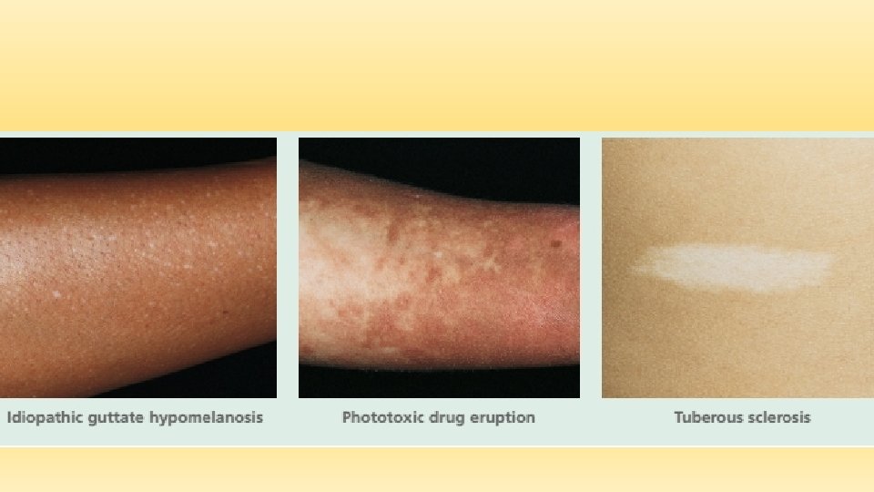
Macule • A circumscribed, flat discoloration that may be brown, blue, red, or hypopigmented • Hypopigmented( tinea versicolor) • Depigmented ( vitiligo) • Brown( melisma lentigo ) • Blue ( mangolian spot) • Red ( viral exanthema)


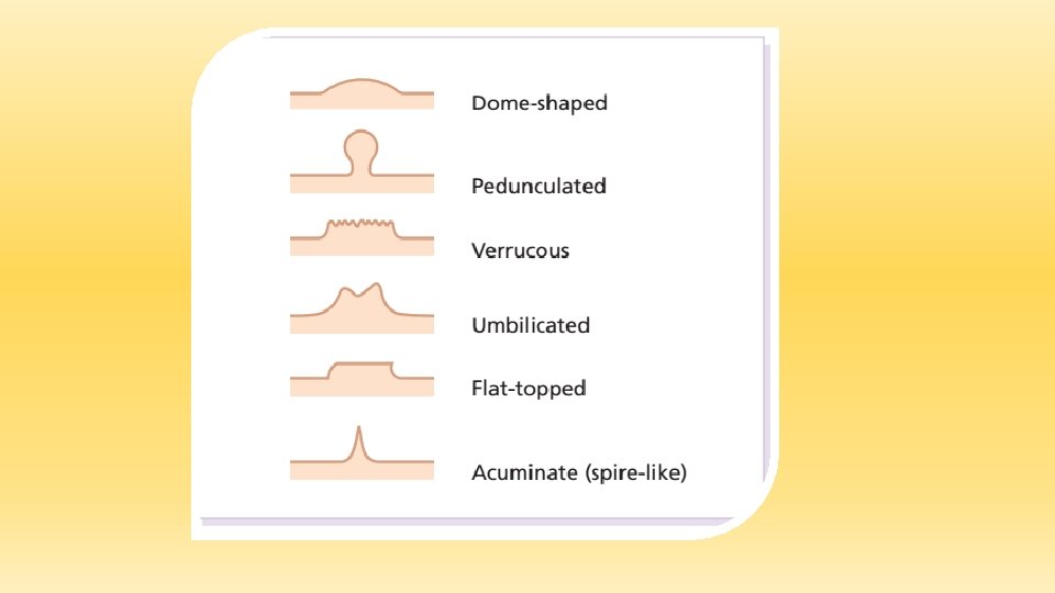
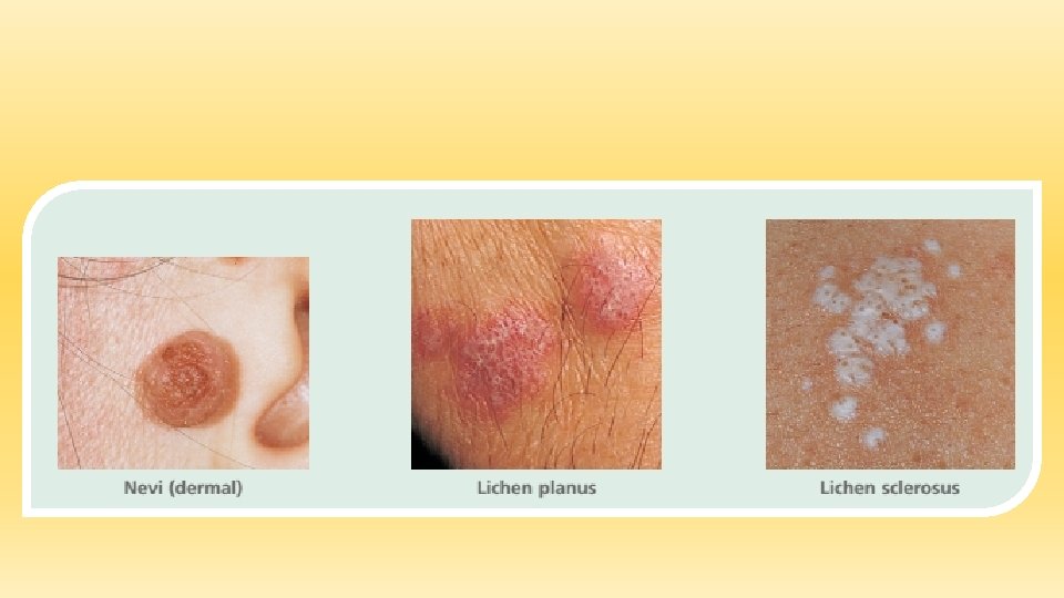
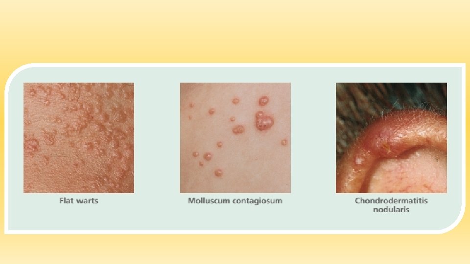
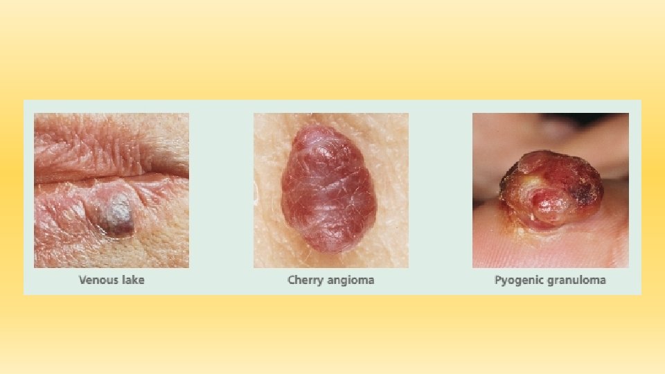
Papule • Papule An elevated solid lesion up to 0. 5 cm in diameter; color varies; papules may become confluent and form plaques • Flesh colored, yellow, or white • Achrochordon (skin tag • Brown • Nevi • Red • Acne • Blue or violaceous • Blue nevus

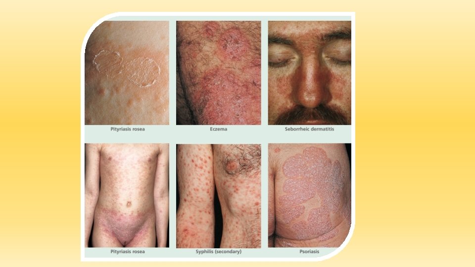
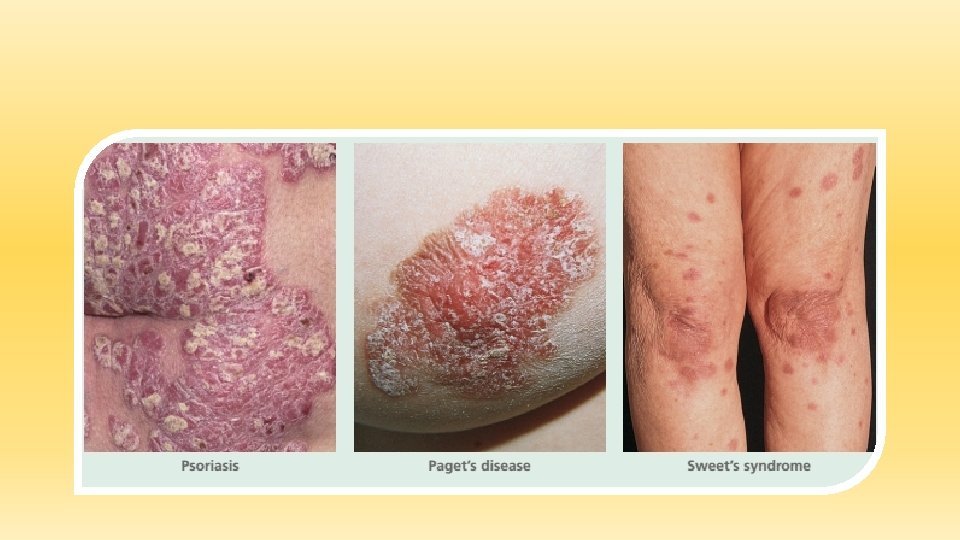
Plaque • A circumscribed, elevated, superficial, solid lesion more than 0. 5 cm in diameter, often formed by the confluence of papules • Chronic cutaneous (discoid) lupus erythematosus • Pityriasis rosea • Tinea corporis • Psoriasis

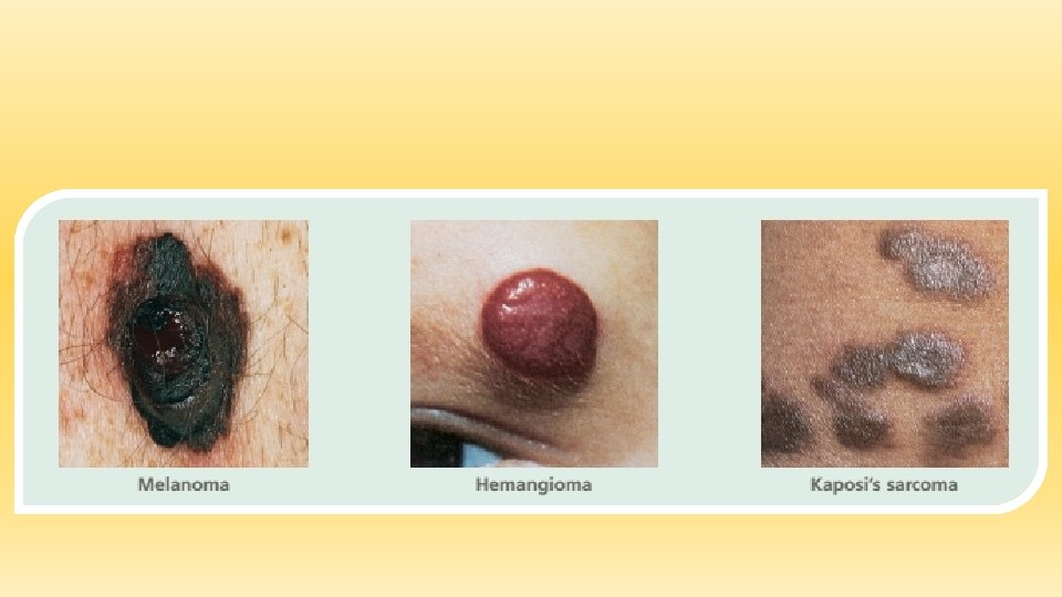
Nodule • Nodule A circumscribed, elevated, solid lesion more than 0. 5 cm in diameter; a large nodule is referred to as a tumor • Basal cell carcinoma • Cutaneous T-cell lymphoma • Erythema nodosum • Melanoma • Metastatic carcinoma Neurofibromatosis

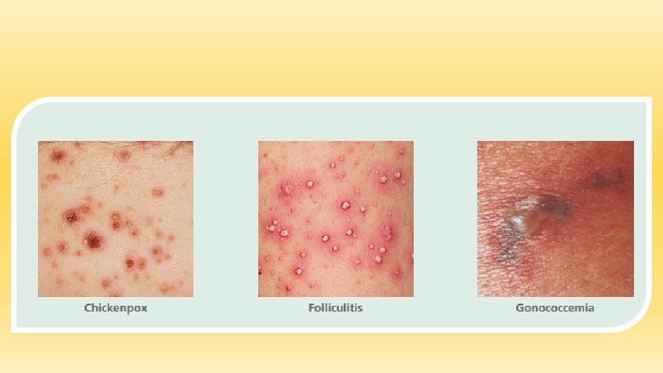
Pustule • A circumscribed collection of leukocytes and free fluid that varies in size • Acne • Candidiasis • Herpes zoster • Impetigo
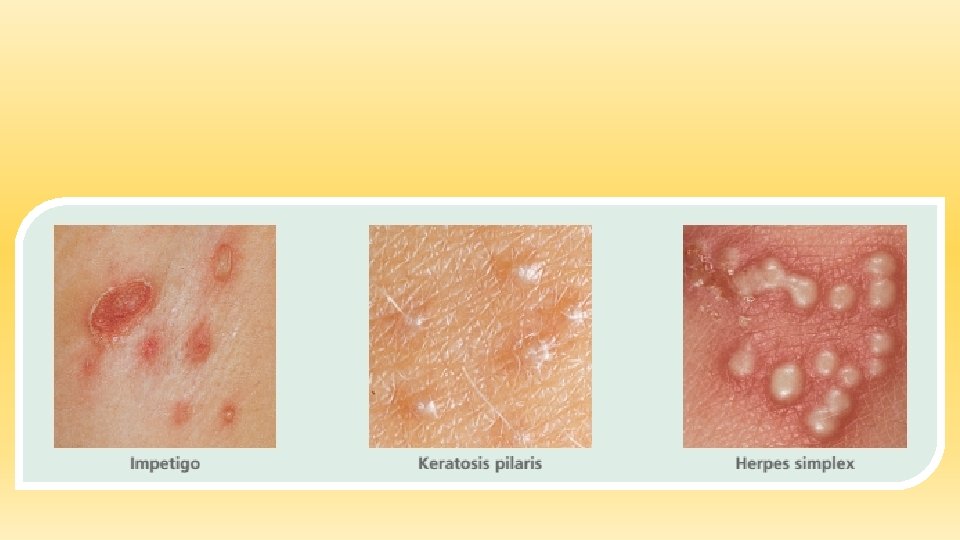

Vesicle • A circumscribed collection of free fluid up to 0. 5 cm in diameter • Benign familial chronic pemphigus • Cat-scratch disease • Herpes zoster • Impetigo
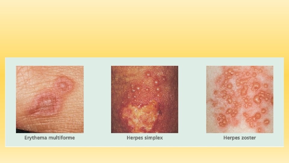

Bulla • A circumscribed collection of free fluid more than 0. 5 cm in diameter • Bullae in diabetics • Bullous pemphigoid • Fixed drug eruption • Herpes gestationis
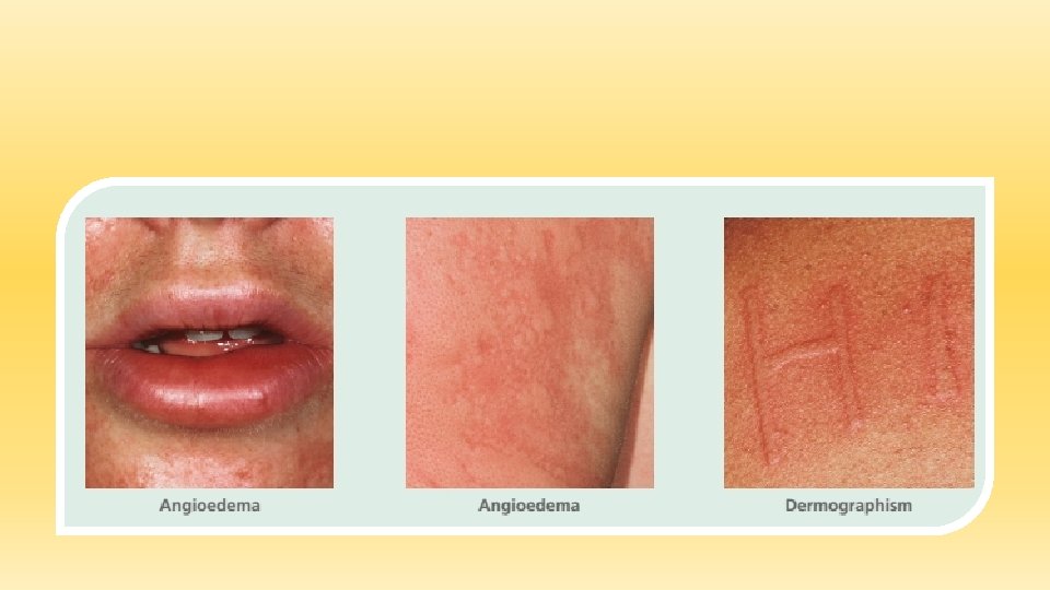
 • A firm, edematous plaque resulting from infiltration of the dermis with")
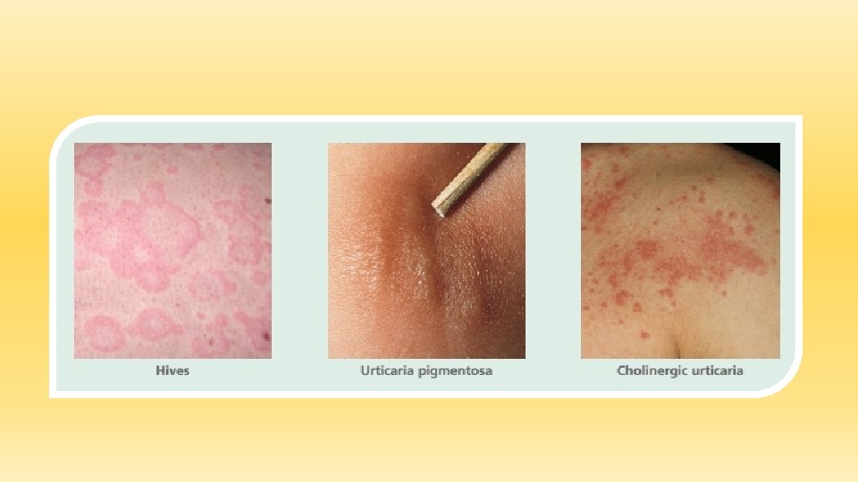
Wheal (hive) • A firm, edematous plaque resulting from infiltration of the dermis with fluid; wheals are transient and may last only a few hours • Angioedema • Bullous pemphigoid • Cholinergic urticarial • Urticaria

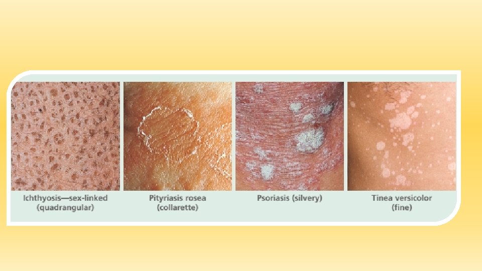
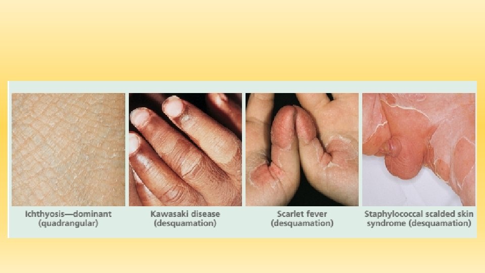
SECONDARY SKIN LESIONS—SCALES • Excess dead epidermal cells that are produced by abnormal keratinization and shedding Fine to stratified Erythema craquelé Ichthyosis—dominant • Scaling in sheets (desquamation) Kawasaki disease Scarlet fever

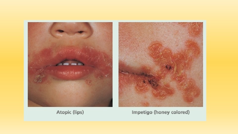
SECONDARY SKIN LESIONS—CRUSTS • A collection of dried serum and cellular debris and blood ; a scab • Acute eczematous inflammation • Atopic (face) • Impetigo (honey colored) • Pemphigus foliaceus

SECONDARY SKIN LESIONS—EROSIONS AND ULCERS • A focal loss of epidermis; erosions do not penetrate below the dermoepidermal junction and therefore heal without scarring • Candidiasis • Dermatophyte infection • Perlèche • Sun-damaged skin


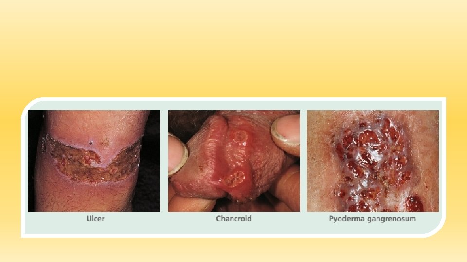
SECONDARY SKIN LESIONS—ULCERS • A focal loss of epidermis and dermis; ulcers heal with scarring • Aphthae • Chancroid • Decubitus • Neoplasms • Syphilis (chancre) • Stasis ulcers

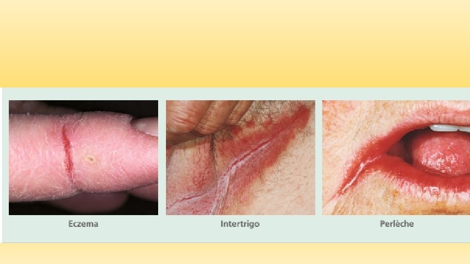
SECONDARY SKIN LESIONS—FISSURES • A linear loss of epidermis and dermis with sharply defined, nearly vertical walls • Chapping (hands, feet) • Eczema (fingertip) • Intertrigo Perlèche

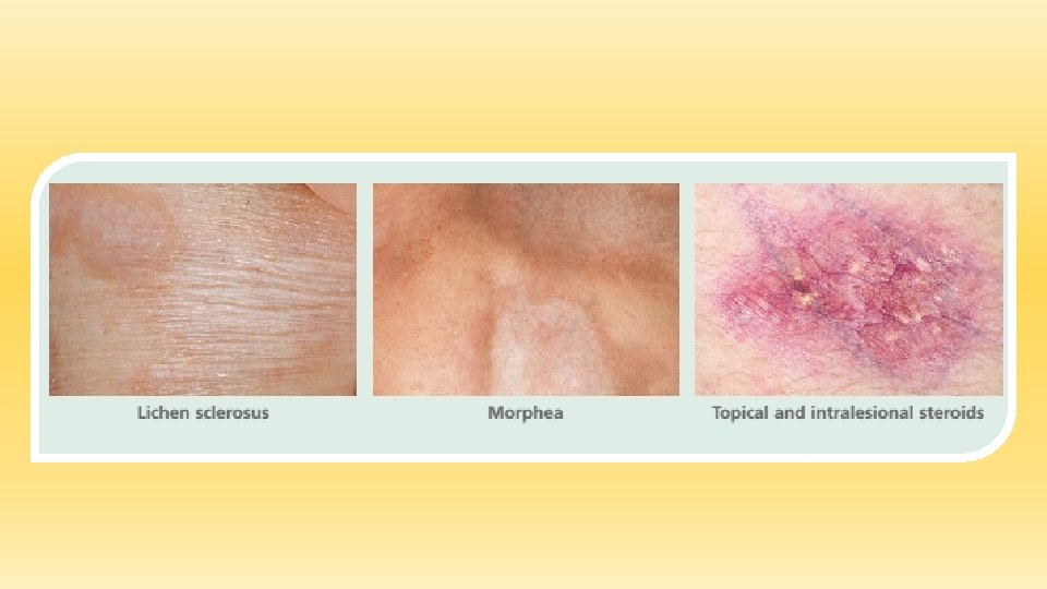
SECONDARY SKIN LESIONS—ATROPHY • A depression in the skin resulting from thinning of the epidermis or dermis • Chronic cutaneous (discoid) lupus erythematosus • Radiodermatitis • Striae

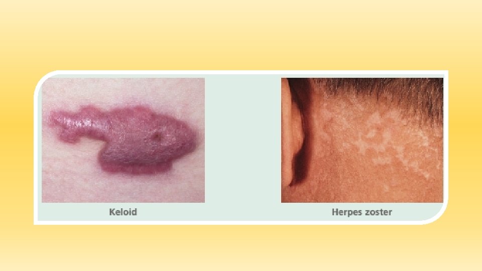
SECONDARY SKIN LESIONS—SCARS • Scar An abnormal formation of connective tissue implying dermal damage; after injury or surgery scars are initially thick and pink but with time become white and atrophic • Acne • Bullous pemphigoid • Hidradenitis suppurativa • Keloid


SPECIAL SKIN LESIONS • Excoriation An erosion caused by scratching; excoriations are often linear

SPECIAL SKIN LESIONS…… • Comedone A plug of sebaceous and keratinous material lodged in the opening of a hair follicle; the follicular orifice may be dilated (blackhead) or nar rowed (whitehead or closed come done)

SPECIAL SKIN LESIONS…… • Milia A small, superficial keratin cyst with no visible opening

SPECIAL SKIN LESIONS…… • Cyst • A circumscribed lesion with a wall and a lumen; the lumen may contain fluid or solid matter

SPECIAL SKIN LESIONS…… • Petechiae A circumscribed deposit of blood less than 0. 5 cm in diameter

SPECIAL SKIN LESIONS…… • Purpura A circumscribed deposit of blood greater than 0. 5 cm in diameter

SPECIAL SKIN LESIONS…… • Burrow A narrow, elevated, tortuous channel produced by a parasite

SPECIAL SKIN LESIONS…… • Lichenification An area of thickened epidermis induced by scratching; skin lines are accentuated so the surface looks like a washboard

SPECIAL SKIN LESIONS…… • Telangiectasia Dilated superficial blood vessels

SPECIAL SKIN LESIONS……

Any Question?

Thanks
- Slides: 65