Principle Skeletal Muscles 1 Muscles of Facial Expression


- Slides: 110
Principle Skeletal Muscles 1 Muscles of Facial Expression, Muscles that Move the Mandible and Muscles that Move the Eyeballs
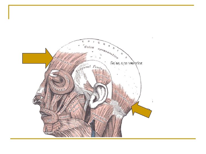
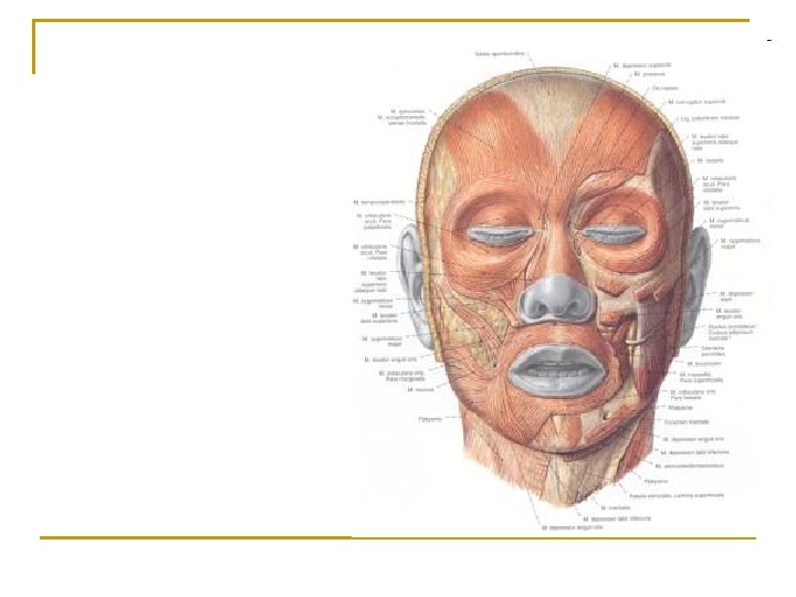
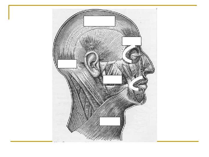
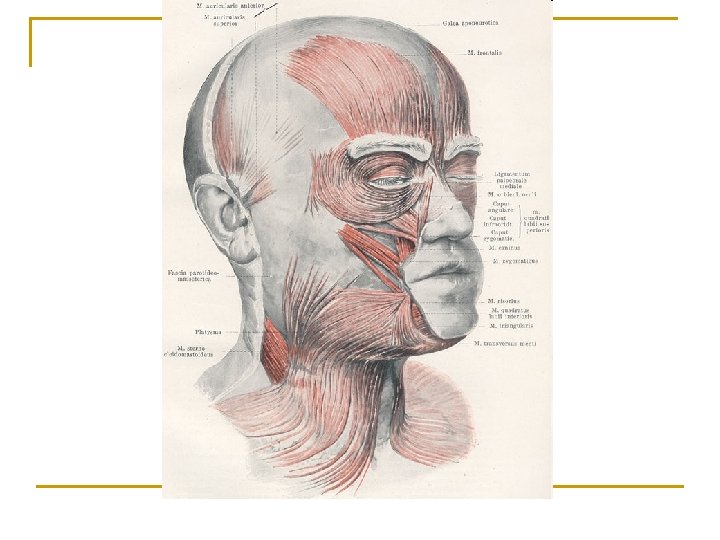
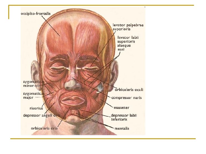
Muscles of Facial Expression n The muscles of facial expression provide humans with the ability to express a wide variety of emotions. The muscles themselves lie within the layers of superficial fascia. The origins are generally in the fascia or bones of the skull and insertions into the skin of the face
Occipitofrontalis Front and Occipital Bellies n Front Belly: q q q n Origin – Epicranial Aponeurosis Insertion – Skin superior to orbit Action – Draws scalp forward, raises eyebrows and wrinkles skin of forehead horizontally Occipital Belly q q q Origin – Occipital and Temporal Bones Insertion – Epicranial Aponeurosis Action – Draws scalp backwards
Orbicularis Oris n n n Origin – muscle fibers surrounding opening of mouth Insertion – Skin at corner of mouth Action – Closes and protrudes lips, compresses lips against teeth and shapes lips during speech
Zygomaticus Major n n n Origin – zygomatic bone Insertion – Skin at angle of mouth and orbicularis oris Action – Draws corners of mouth outward and upward as in smiling
Buccinator n n n Origin – Maxilla and Mandible Insertion – Orbicularis Oris Action – presses cheeks against teeth and lips, as in whistling; draws corner of mouth laterally, assists in chewing be keeping food between teeth
Platysma n n n Origin – Fascia over deltoid and pectoralis major muscles Insertion – Mandible, muscles around mouth and skin of lower face Action – Draws outer part of lower lip downward and backward as in pouting; depresses mandible
Orbicularis Oculi n n n Origin – Medial wall of orbit Insertion – Circular path around orbit Action – Closes eye; wrinkles forehead vertically
Levator Palpebrae Superioris n n n Origin – Roof of Orbit Insertion – Skin of upper eyelid Action - Opens Eye
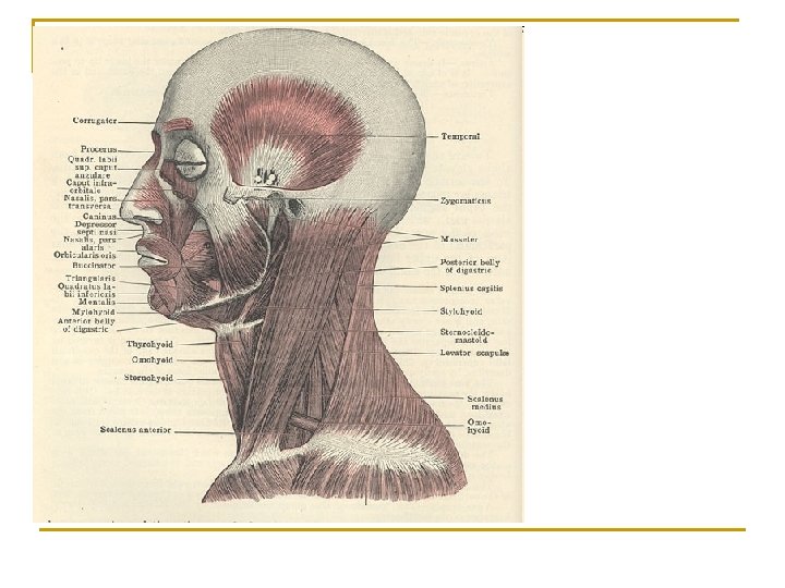
Muscles that Move the Mandible n Also known as muscles of mastication because they are used for biting and chewing. These muscles also assist in speech.
Masseter n n n Origin – Maxilla and Zygomatic Arch Insertion – Mandible Action – Elevates and retracts mandible
Temporalis n n n Origin – Temporal Bone Insertion – Mandible Action – Elevates and retracts mandible
Medial Pterigoid n n n Origin – Sphenoid bone and maxilla Insertion – Mandible Action – elevates and protracts mandible and moves mandible from side to side
Lateral Pterygoid n n n Origin – Sphenoid Bone Insertion – TMJ Action – Protracts mandible, depresses mandible and moves mandible from side to side
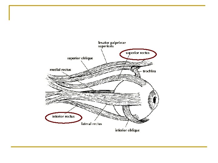
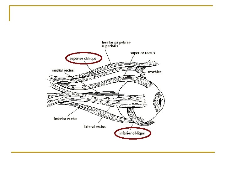
Muscles that Move the Eyeballs n Movement of the eyeballs are controlled by three pairs of extrinsic muscles. These are among the fastest contracting and most precisely controlled skeletal muscles of the body.
Superior Rectus/Inferior Rectus n Superior: q q q n Origin – Tendinous ring attached to bony orbit around the optic foramen Insertion – Superior and central part of the eyeball Action – Moves eyeball upward and medially and rotates its medially Inferior: q q q Origin - Tendinous ring attached to bony orbit around the optic foramen Insertion – Inferior and central part of the eyeball Action – Moves eyeball downward and medially and rotates it laterally
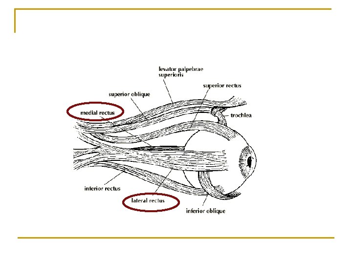
Lateral/Medial Rectus n Lateral: q q q n Origin – Tendinous ring attached to bony orbit around the optic foramen Insertion – Lateral Side of Eyeball Action – Moves eyeball laterally Medial: q q q Origin – Tendinous ring attached to bony orbit around the optic foramen Insertion – Medial Side of Eyeball Action – Moves eyeball medially
Superior/Inferior Oblique n Superior: q q q n Origin – Tendinous ring attached to bony orbit around the optic foramen Insertion – Eyeball between superior and lateral recti Action – moves eyeball downward, laterally and rotates medially Inferior: q q q Origin – Maxilla Insertion – eyeball between inferior and lateral recti Action – moves eyeball upward and laterally and rotates it laterally
Cadaver – face dissection
Principle Skeletal Muscles 2 Muscles that act on the abdominal wall, muscles used in breathing and muscles that move the pectoral girdle
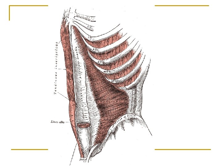
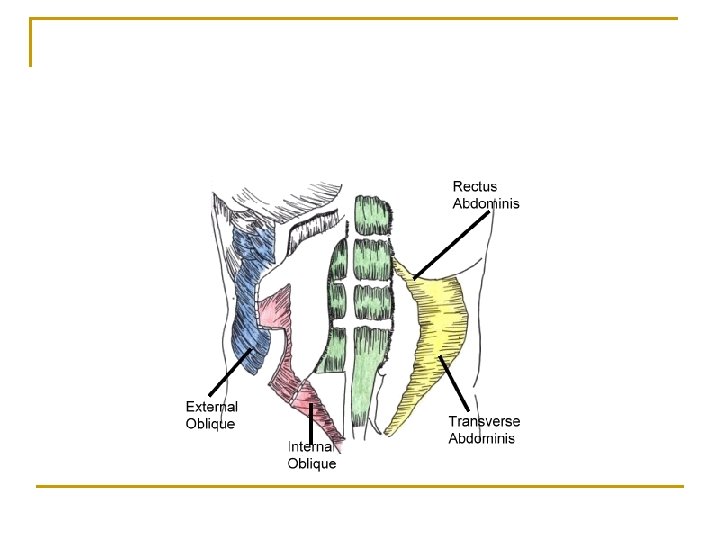
MUSCLES THAT ACT ON THE ANTERIOR ABDOMINAL WALL n The anterior abdominal wall is composed of skin, fascia and 4 pairs of muscles. q q Tendinous Intersections – bands of connective tissue that divides the rectus abdominis Linea Alba – tough fibrous band extending from xiphoid process to pubic symphysis
Rectus Abdominis n n n Origin – Pubis and Pubic Symphysis Insertion – Costal Cartilage and Xiphoid Process Action – Flexes vertebral column and compresses abdomen
Linea Alba Tendinous Intersections
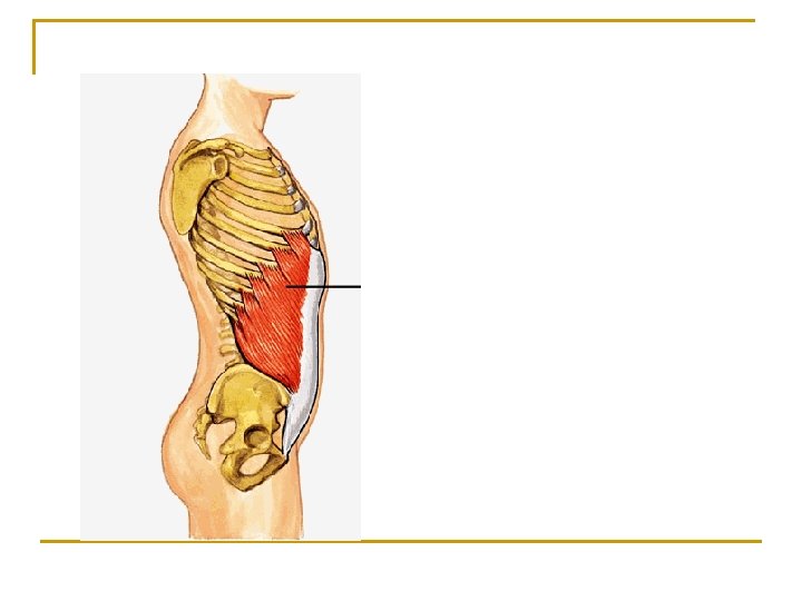
External Oblique n n n Origin – Lower 8 ribs Insertion – Crest of Ilium and Linea Alba Action – Compresses abdomen, flexes vertebral column. Singularly rotates vertebral column
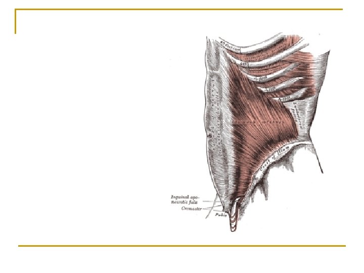
Internal Oblique n n n Origin – Ilium, inguinal ligament Insertion – Costal Cartilage and linea alba Action - Compresses abdomen, flexes vertebral column. Singularly rotates vertebral column
Transverse Abdominis n n n Origin – Ilium, inguinal ligament, lumbar fascia, and costal cartilage Insertion – Xiphoid Process, linea alba and pubis Action – Compress Abdomen
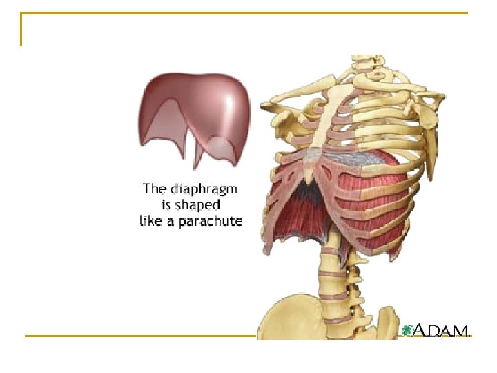
Muscles Used in Breathing n These muscles alter the size of the thoracic cavity so that breathing can occur. Inhalation occurs when the thoracic cavity increases in size and exhalation occurs when the thoracic cavity decreases in size
Diaphragm n n n Origin – xiphoid process, costal cartilage and lumbar vertebrae Insertion – central tendon Action – increases the vertical dimension of the thoracic cavity resulting in inhalation.
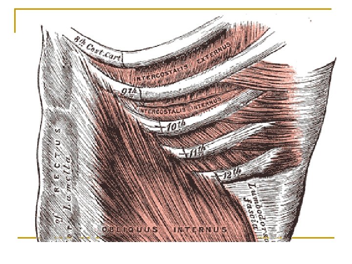
External and Internal Intercostals n n n Origin – ribs Insertion – ribs Action: q q External – increases the anteroposterior and lateral dimensions of thoracic cage resulting in inhalation Internal – decreases the antroposterior and lateral dimensions resulting in forceful exhalation
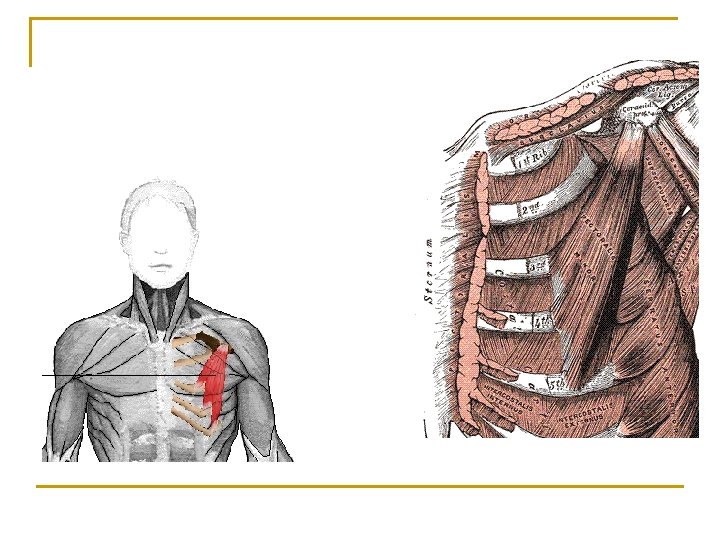
Muscles that Move the Pectoral Girdle n n These muscles are divided into anterior (pectoralis minor and serratus anterior) and posterior (trapezius, levator scapulae and rhomboid major) thoracic muscles based on their location. The main action of the muscles is to hold the scapula in place so that is can function as a stable origin for the muscles that move the humerus
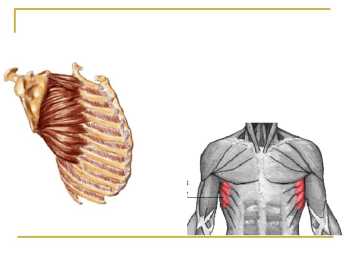
Pectoralis minor n n n Origin – Ribs 3 -5 Insertion – Scapula Action – depresses scapula, moves it laterally and forward
Serratus Anterior n n n Origin – Upper 8 or 9 ribs Insertion – Scapula Action – Moves scapula laterally and forward. AKA: “the boxer’s muscle” because it is important in horizontal arm movements like punching.
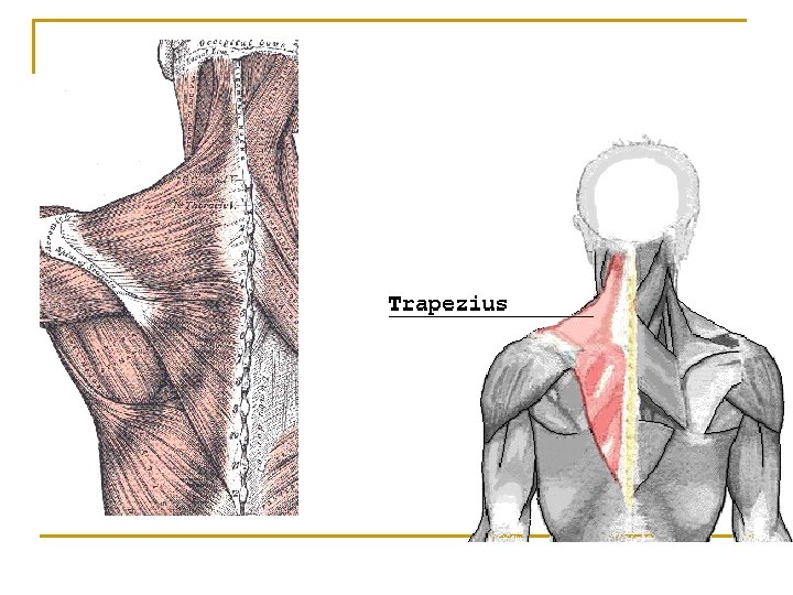
Trapezius n n n Origin – occipital bone, spines of C 7 and thoracic vertebrae Insertion – clavicle and scapula Action – Elevates clavicle, moves scapula medially
Levator Scapulae n n n Origin – C 1 – C 5 Insertion – Scapula Action – elevates scapula
Rhomboid Major n n n Origin – Spines of T 2 -T 5 Insertion – Scapula Action – Elevates scapula, moves it medially
Principle Skeletal Muscles 3 Muscles that move the Vertebral Column, Muscles that move the femur, tibia, fibula foot and toes
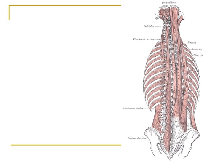
Erector Spinae n n n Origin – All ribs, cervical, thoracic and lumbar vertebrae Insertion – Occipital Bone, ribs and vertebrae Action – Extends head; extends and laterally flexes vertebral column
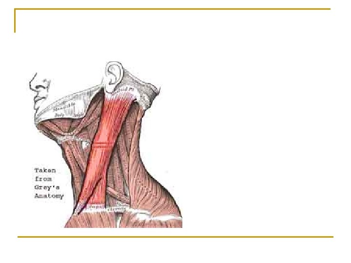
STERNOCLEIDOMASTOID!!! n n n Origin – sternum and clavicle Insertion – Mastoid process of temporal bone Action – flex cervical spine or rotate head
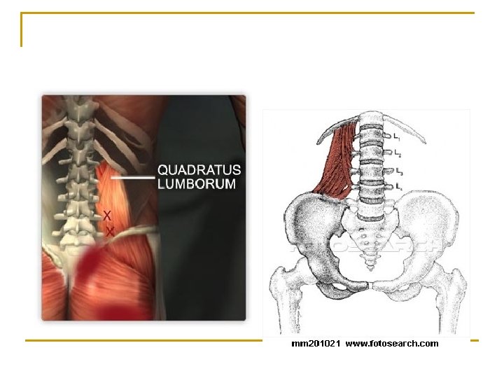
Quadratus Lumborum n n n Origin – Ilium Insertion – 12 th rib and upper 4 lumbar vertebrae Action – Extend lumbar spine when both are contracted. Flexes lumbar spine when one is contracted
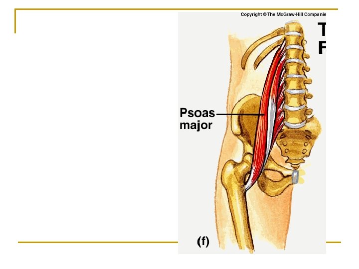
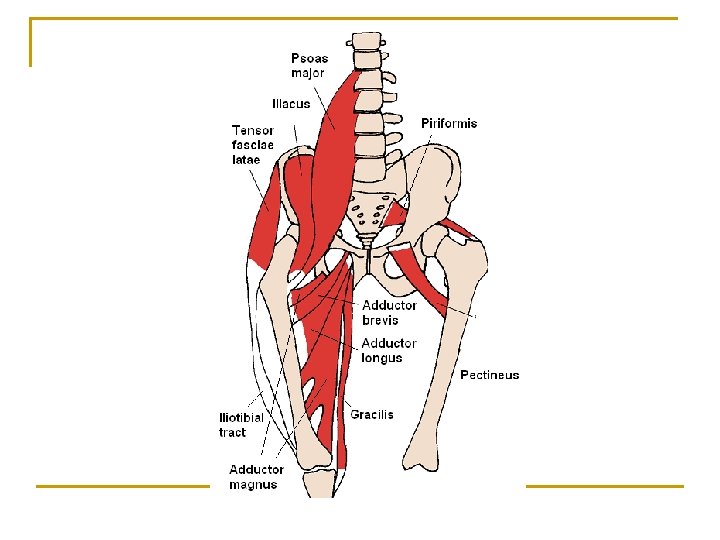
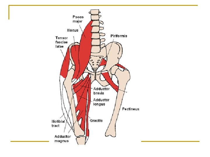
Psoas Major n n n Origin – Lumbar Vertebrae Insertion – Femur Action – Flexes and rotates thigh laterally at the hip
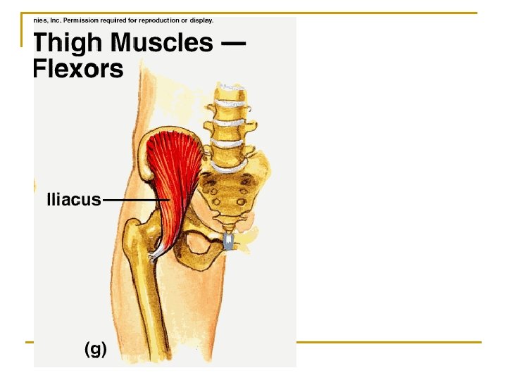
Iliacus n n n Origin – Ilium Insertion – Femur Action – Flexes and rotates thigh laterally at hip
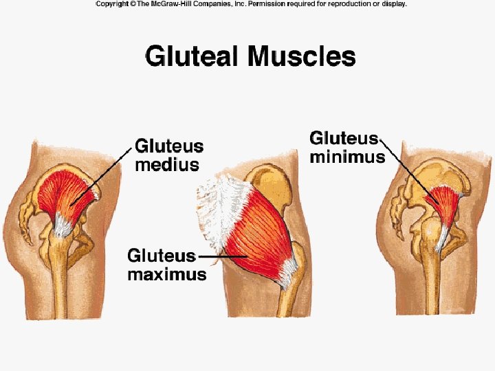
Gluteal Muscles n Gluteus Maximus, Minimus and Medius q q q Origin – Ilium Insertion – Femur Actions – n n Maximus: Extends and rotates thigh laterally at hip Medius and Minimus – Abducts and rotates thigh medially at hip
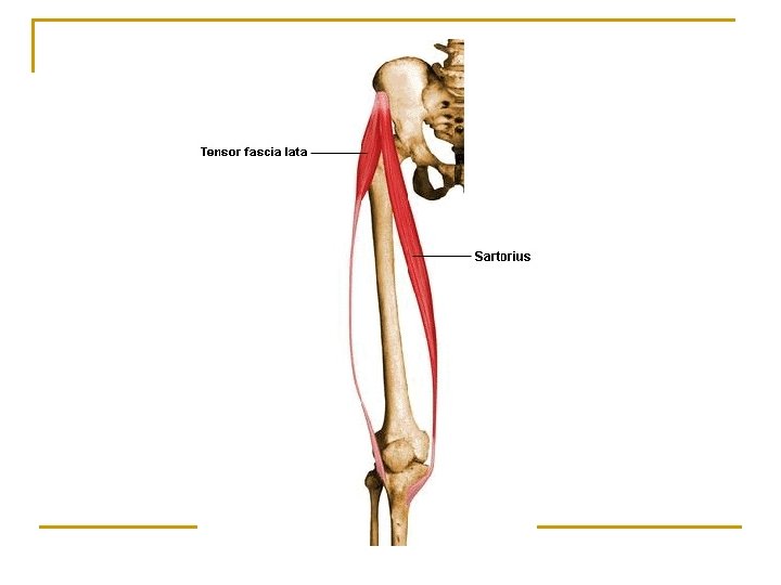
Tensor Fasciae Latae n n n Origin – Ilium Insertion – Tibia Action – Flexes and abducts the thigh at the hip
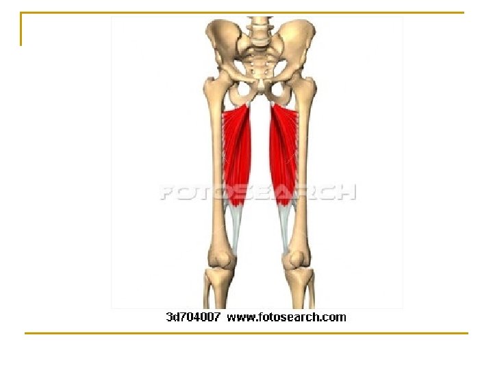
Adductor Longus n n n Origin – Pubis and Pubic Symphysis Insertion – Femur Action – Adducts, medially rotates and flexes thigh at hip
Adductor Magnus n n n Origin - Pubis and Ischium Insertion – Femur Action – Adducts, flexes, medially rotates and extends thigh
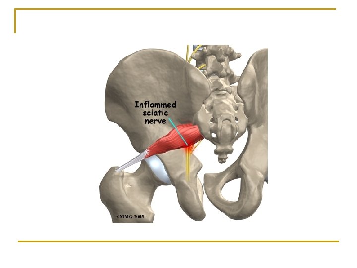
Piriformus n n n Origin – Sacrum Insertion – Femur Action – Rotates thigh laterally
Gracilis n n n Origin – Pubic Symphysis Insertion – Tibia Action – Adducts and medially rotates thigh at hip and flexes knee
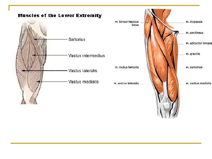
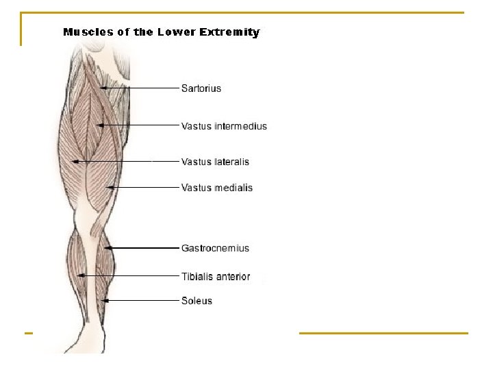
Quadriceps Femoris n Composed of 4 parts: q q n Rectus Femorus Vastus Lateralis Vastus Medialis Vastus Intermedius Action – Extend leg at knee joint
Sartorius n n n Origin – Ilium Insertion – Tibia Action – Flexes leg at knee, abducts and laterally rotates thigh at hip joint (like corssing legs)
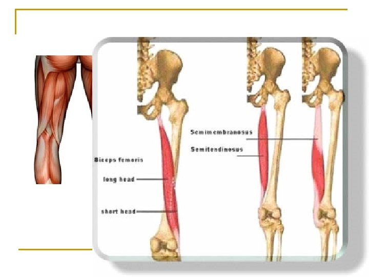
Hamstrings n Group of 3 muscles: q q q n Biceps Femoris Semitendinosus Semimembranosus Action – flexes leg at knee, extends thigh at hip
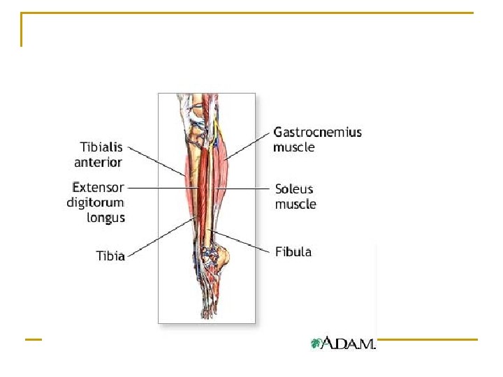
Tibialis Anterior n n n Origin – Tibia Insertion – 1 st metatarsal and 1 st cuniform (tarsal) Action – dorsiflex and invert foot
Peroneus Longus n n n Origin – Fibula and Tibia Insertion – 1 st metatarsal and 1 st cuniform Action – Plantar flexes and everts foot
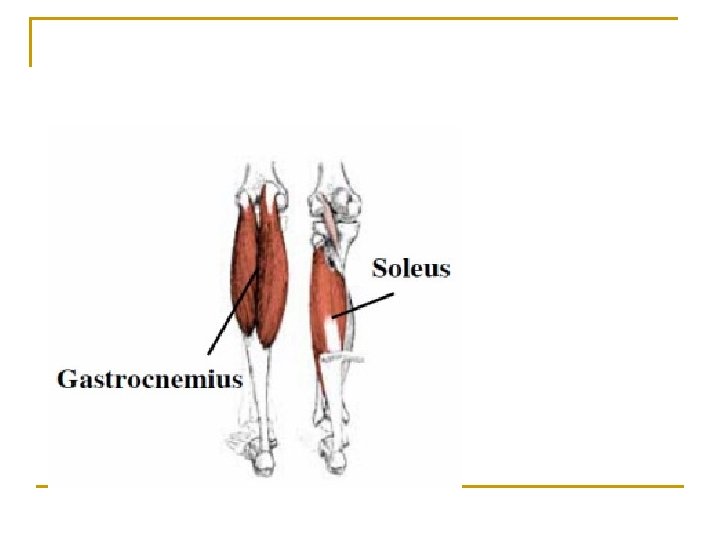
Gastrocnemius n n n Origin – Femur Insertion – Calcaneus Action – Plantar Flexion; flexes leg at knee
Soleus n n n Origin – Fibula and Tibia Insertion – Calcaneus Action – Plantar Flexion
Flexor Digitorum Longus n n n Origin – Tibia Insertion – Distal Phalanges Action – Flexes toes
Principle Skeletal Muscles Upper Extremity
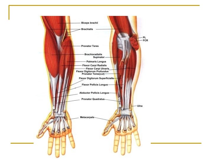
Biceps brachii n Origin – Scapula n Insertion – Radius n Action – Flexes and supinates forearm at elbow
Brachialis n Origin – Humerus n Insertion – Ulna n Action – Flexes forearm at elbow joint
Brachioradialis Origin – Humerus Insertion – Radius Action – Flexes forearm at elbow joint
Triceps Brachii Origin – Scapula and Humerus Insertion – Ulna Action – Extends forearm at elbow joint
Supinator Origin – Humerus and Ulna Insertion – Radius Action – Supinates Forearm
Pronator teres Origin – Humerus and Ulna Insertion – Radius Action – Pronates Forearm
Pronator Quadratus Origin –Ulna Insertion – Radius Action – Pronates Forearm
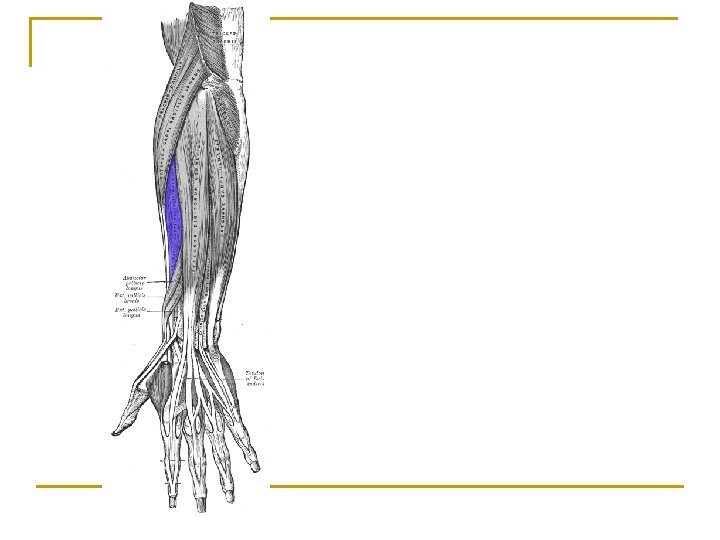
Flexors n n n Origins – Humerus or Humerus and Ulna Insertions – Carpals, metacarpals and phalanges Actions – Flexes wrist, hands and fingers
Extensors n n n Origins – Humerus or Humerus and Ulna Insertions – Metacarpals and phalanges Actions – Extends, adducts and abducts wrist, hands and fingers
Pectoralis Major Origin – Clavicle, sternum and 6 th & 7 th ribs Insertion – Humerus Action – Adducts and rotates arm medially
Pectoralis Minor Origin – 3 rd – 5 th ribs Insertion – scapula Action – depresses scapula
Latissimus Dorsi Origin – Spines of lower vertebrae Insertion – Humerus Action – Extends, adducts and rotates arm at shoulder
Deltoid Origin – Clavicle and scapula Insertion – Humerus Action – Abducts, flexes, extends and rotates arm at shoulder
Subscapularis Origin –Scapula Insertion – Humerus Action – Rotates arm medially
Coracobrachialis Origin –Scapula Insertion – Humerus Action – flexes and adducts arm at shoulder
Serratus Anterior Origin – Upper 8 ribs Insertion – Scapula Action – Moves scapula laterally and forward (horizontal arm movement)
Trapezius Origin – Occipital Bone and spines of thoracic vertebrae Insertion – Clavicle and Scapula Action – Elevates clavicle, moves scapula medially, extends head
Levator Scapulae Origin – Upper 4 or 5 cervical vertebrae Insertion – Scapula Action – elevates scapula