PRICE 0 45 Ovarian Tumors DR Omar AbuAzzam

| PRICE: 0. 45| Ovarian Tumors DR Omar Abu-Azzam
 may be : 1) Physiological cysts 2) Neoplastic")
Ovarian cyst ( or mass ) may be : 1) Physiological cysts 2) Neoplastic : benign, borderline, malignant and secondaries 3) Tuboovarian abscess 4) Ectopic pregnancy 5) Endometrioma 6) Polycystic ovary syndrome 7) Theca leutein cysts 8) Luteoma
 ovarian cysts 1) Follicular cyst 2) Corpus luteum cyst ● In the process")
Physiological(functional) ovarian cysts 1) Follicular cyst 2) Corpus luteum cyst ● In the process of normal ovulation, a follicle develops to maturity and then ruptures to release an ovum; this is followed by formation and subsequent involution of the corpus luteum. - Follicular cysts arise when rupture does not occur and the follicle continues to grow; When it becomes larger than 3 cm, it is called a follicular cyst. - Corpus luteum cysts occur when the corpus luteum fails to involute and continues to enlarge after ovulation (the corpus luteum enlarges for the first six weeks of pregnancy and doubles its prepregnancy size). ● They are a small cysts, usually less than 10 cm in size. ● They are generally asymptomatic , unless cyst accidents occur ( Rupture , Bleeding , Ovarian torsion ). ● Most spontaneously resolve within a few weeks, but some persist for several months.

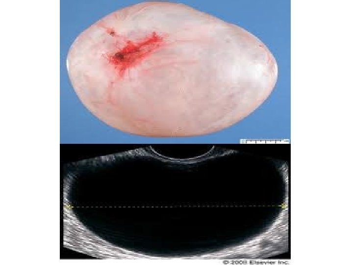
On ultrasound examination : Follicular cysts appear as simple unilocular, anechoic cysts with a thin, smooth wall. There should be no enhancing nodules or other solid components, no enhancing septations, and no more than physiologic ascites.

Corpus luteum cysts appear usually as a small complex ovarian cyst with wall vascularity on power Doppler analysis. The characteristic circular Doppler appearance is called the 'ring of fire'. - May appear as simple cyst.
. ●")
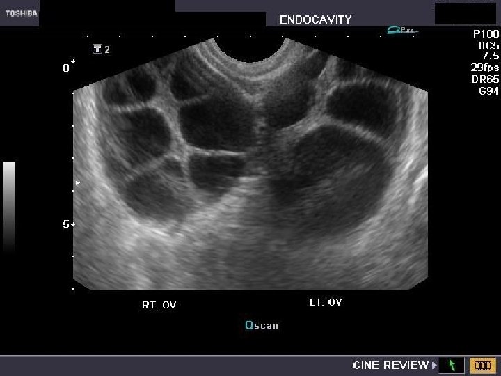
Theca lutein cysts ● Theca lutein cysts (also called lutein cysts, hyperreactio luteinalis). ● They are luteinized follicle cysts that form as a result of overstimulation from high h. CG levels or hypersensitivity to h. CG. ● Bilateral multiseptated cystic adnexal masses. ● Occur in a woman with gestational trophoblastic disease, multiple gestation, ovarian hyperstimulation, or a pregnancy complicated by fetal hydrops. ● They can also occur in a normal pregnancy due to hypersensitivity to normal levels of h. CG. ● Most are asymptomatic, but maternal virilization, hyperemesis gravidarum, preeclampsia, or thyroid dysfunction may occur. ● The cysts gradually resolve weeks to months after the source of h. CG is eliminated.

Luteoma ● Luteoma is a rare nonneoplastic ovarian change associated with pregnancy that can simulate a neoplasm on clinical, gross, or microscopic examination. ● The size of the mass can range from 1 to 25 cm in diameter, but is usually 6 to 10 cm in diameter. ● Luteomas involute spontaneously after delivery or are adequately treated by a conservative surgical approach. ● It is associated with an increases of sex hormones, primarily progesterone and testosterone. so may cause masculinization (virilization) of the mother ( 36% of cases) and the possible masculinization of the female fetus. (Out of the 36% of women who show male characteristics from the luteoma, 75% of female fetuses will also show signs of masculinization).

- Signs of maternal musculinization : include acne, the growth of dark hair (especially on the face), deepening of the voice, temporal balding, and Clitoromegaly. → An increase in testosterone levels in the mother doesn't necessarily mean masculinization will occur. - Signs of female fetal musculinization : labioscrotal fusion and enlarged clitoris (Luteomas can cause the fetus to be born with an ambiguous sex necessitating a sex to be chosen for the fetus depending on how the parents want to raise the infant). ● Diagnosis : Luteomas are not often detected before delivery. Most luteomas are found during surgery if a caesarean section is performed or when some other surgery is performed. - The diagnosis should be suspected in the presence of a solid adnexal mass and maternal hirsutism or virilization. ( during pregnancy ) ● Risk factors : 1) PCOS 2) Past history of luteoma 3) Multiple pregnancy 4) Advanced maternal age

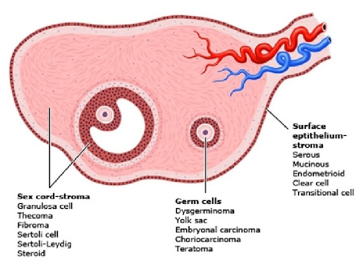
Ovarian Neoplasms ● Ovarian neoplasms arise from the surface epithelium, germ cells, and sex-cordstromal tissue and may be benign, borderline or malignant. These neoplasms persist unless excised. ● Epithelial tumors account for 60 -65% of all ovarian tumors, germ cell tumors account for 30% and sex-cord stromal tumors account for 8%.

Benign ovarian neoplasms ● Predominantly they occur in premenopausal women, they are uncommon in premenarchal and postmenopausal women. - Among premenopausal patients, more than 90% of cases are benign, as opposed to just 60% in the postmenopausal population. ● Presentation : 1) Asymptomatic : incidental finding on examination or imaging. 2) Lower abdominal pain. (Mild pain) 3) Ovariaan cyst accidents : torsion, rupture or hemorrhage. - Present as acute abdomen. 4) Dyspareunia. 5) Pressure effects, eg on the bladder, causing urinary frequency, or on venous return, causing varicose veins and leg oedema. 6) Ascites ( e. g Meigs' syndrome). 7) Endocrine : hormone-secreting tumours may cause virilisation, menstrual irregularities or postmenopausal bleeding. This is uncommon though. ● Diagnosis : by histopathology. ● Treatment : ovarian cystectomy / oopherectomy.
 Epithelial ovarian tumors : 1) Serous cystadenoma 2) Mucinous cystadenoma")
Benign ovarian neoplasms A) Epithelial ovarian tumors : 1) Serous cystadenoma 2) Mucinous cystadenoma 3) Endometrioid cystadenoma 4) Brenner tumor B) Germ cell ovarian tumors : 1) Mature cystic teratoma (dermoid cyst) 2) Mature solid teratoma 3) Monodermal highly specialized teratomas (e. g. , struma ovarii, carcinoid) C) Sex-cord stromal cell ovarian tumors : 1) Fibroma 2) Thecoma

Serous cystadenoma ● The most common benign ovarian neoplasms. ● The peak incidence is at the 4 th to 5 th decades of life. ● Pathology : - Grossly : They are thin-walled, usually unilocular (or sometimes multilocular) , that contain clear, straw-coloured fluid and range in size from 5 to over 20 cm. Bilateral in 20 -25%. - Microscopically : The cyst lining consists of a simple epithelium with cilia that may be columnar or flat. As with other serous tumours, psammomatous calcification can be a feature.

Mucinous cystadenoma ● The second most common benign ovarian neoplasms. ● The peak incidence occurs between 30 -50 years of age. ● Grossly : They are thin-walled, usually multilocular (or sometimes unilocular) , that contain mucin, generally tend to be larger than serous cystadenomas at presentation (they can attain an enormous size), bilateral in 5%. ● Histologically : A single layer of tall columnar mucinous epithelial cells with basally placed nuclei and containing apical mucin, lines the cystic spaces. No ciliated cells are present, resemblance to endocervical type epithelium.

Endometrioid cystadenoma ● Histopathology : The glands resemble normal endometrium. Stroma is moderately cellular. No cytologic atypia, no architectural complexity. ● Many of these tumors arise from endometriosis. Brenner tumor ● They are rare ovarian tumors, The peak incidence is at the 4 th to 5 th decades of life. ● The majority of these tumours are benign. However, they can be malignant. ● Generally asymptomatic. ● On gross pathological examination : They are solid, sharply circumscribed and pale yellow-tan in colour. 90% are unilateral. The tumours can vary in size from less than 1 centimetre to 30 centimetres. ● Histologically : There are nests of transitional-type epithelial cells with longitudinal nuclear grooves (coffee bean nuclei) lying in abundant fibrous stroma.
 ● The most common germ cell tumor and the")
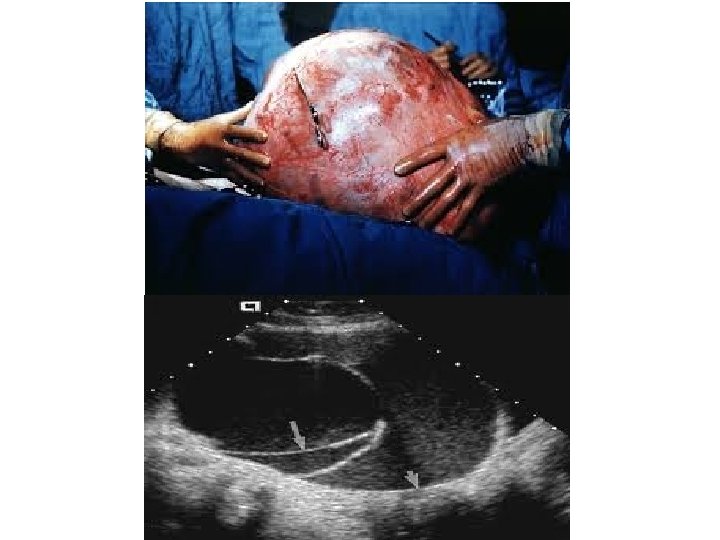
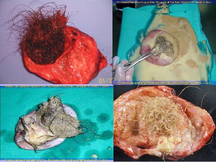
Mature cystic teratoma (Dermoid cyst) ● The most common germ cell tumor and the most common ovarian tumor in women the second and third decade of life. ● Histopathology : Mature cystic teratomas contain mature tissue of ectodermal (eg, skin, hair follicles, sebaceous glands), mesodermal (eg, muscle, urinary), and endodermal origin (eg, lung, gastrointestinal). ● Grossly : Most cysts are unilocular. The inner lining of every mature cystic teratoma contains single or multiple white shiny masses projecting from the wall toward the center of the cysts. When hair, other dermal appendages, bone and teeth are present, they usually arise from this protuberance. This protuberance is referred to as the Rokitansky protuberance. The Rokitansky protuberance is a common site of malignant transformation. ● They are bilateral in 10 -17 % of cases.

Clinical manifestations : ● Most women with dermoid cysts are asymptomatic. ● The most common complication associated with mature cystic teratomas is torsion. The reported rate of torsion ranges from 3. 2 -16%. Torsion is more common in tumors of intermediate size than in small or extremely large tumors. ● Rupture of dermoid cysts with spillage of sebaceous material into the abdominal cavity can occur, but is uncommon. Shock and hemorrhage are the immediate sequelae of rupture; a marked granulomatous reaction (chemical peritonitis) may subsequently develop and lead to formation of dense adhesions. - The rate of spontaneous rupture of mature cystic teratomas is low, ranging from 1. 23. 8%. This has been ascribed to the thick capsule surrounding this lesion. Diagnosis : These tumors have a characteristic ultrasound appearance, which allows reasonably accurate noninvasive diagnosis in many cases. The reported specificity is 98 -100 %. Definitive diagnosis is made at the time of surgical excision.

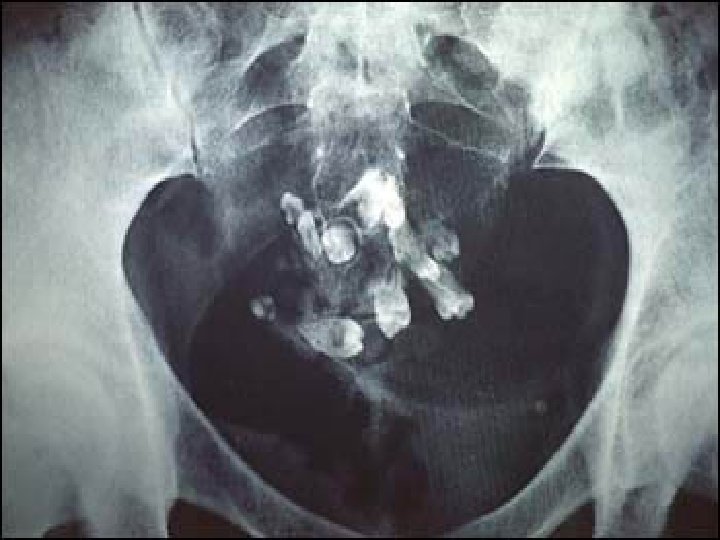
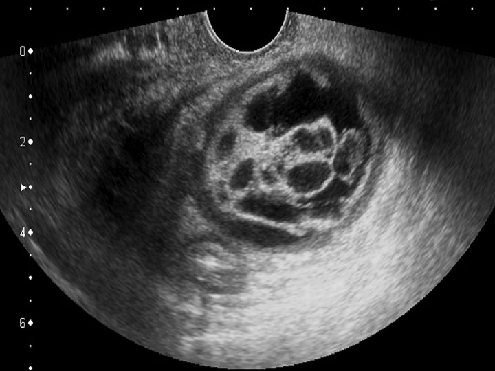
Imaging : ● Plain abdomino-pelvic X-ray : May show calcific and tooth components with the pelvis. ● Ultrasound : Typically an ovarian dermoid is seen as a complex cystic adnexal mass with some mural components. Most lesions are unilocular. The spectrum of sonographic features includes : 1 - Rokitansky nodule - dermoid plug. 2 - Diffusely or partially echogenic mass with posterior sound attenuation owing to Sebaceous material and hair within the cyst cavity ( echogenic interface at edge of mass that obscures deep structures) : the tip of the iceberg sign. 3 - Echogenic, shadowing calcific or dental (tooth) components. 4 - Presence of fluid-fluid levels. 5 - Multiple thin, echogenic bands caused by hair in the cyst cavity : the dot-dash pattern.
 and speckled debris within the cyst are characteristic of")
The echogenic focus (between markers) and speckled debris within the cyst are characteristic of a benign cystic teratoma

Treatment : Ovarian cystectomy, via either laparoscopy or laparotomy. With either approach, the abdomen should be copiously irrigated to avoid a chemical peritonitis from spillage of the sebaceous cyst fluid. Malignant transformation : Malignant transformation occurs in 0. 2 -2 % of mature cystic teratomas. Although any of the components of a mature cystic teratoma may undergo malignant degeneration, squamous cell carcinoma arising from the ectoderm is the most common secondary neoplasm. Risk factors for malignant neoplasm in a mature cystic teratoma include age over 45 years, tumor diameter greater than 10 cm, rapid growth, and findings on imaging (eg, low resistance intra-tumor flow on Doppler).

Monodermal highly specialized teratomas The specialized or monodermal teratomas are a rare and remarkable subset of teratomas that consist of a predominant mature histologic cell type, the most common of which are struma ovarii and carcinoid, a well-differentiated neuroendocrine neoplasm. They are usually unilateral, although a contralateral teratoma may be present. Struma ovarii : Is a teratoma predominantly composed of mature thyroid tissue. The secretion of thyroid hormones results in clinical hyperthyroidism in 25 -35 % of patients. Struma ovarii is uncommon, comprising approximately 2. 7 % of ovarian teratomas. It is often associated with a mature cystic teratoma and rarely with a cystadenoma. Most cases of struma ovarii are benign and can be managed by excision of the ovary or by unilateral salpingo-oophorectomy. However, malignant change may occur in struma ovarii, but it is exceedingly rare.

Carcinoid neoplasms : Ovarian carcinoid neoplasms are rare. Primary ovarian carcinoid neoplasms are usually unilateral, localized to the ovary, and indistinguishable histologically from metastasis. They have similar appearances to those that arise in any other site (eg, gastrointestinal or respiratory). They are comprised of nests and cords of relatively bland cells with endocrine features and a fine vascular network. Some carcinoid neoplasms secrete bioactive polypeptides and amines, producing a constellation of symptoms, predominantly flushing and diarrhea. Carcinoid syndrome develops in about one-third of cases, and it can develop without hepatic metastases due to direct venous drainage from the ovary into the systemic circulation. 5 -hydroxyindoleacetic acid, a metabolite of serotonin, is excreted in the urine and can be used to confirm the diagnosis of carcinoid syndrome, and as a marker of disease activity in patients with advanced disease or the carcinoid syndrome. Carcinoid tumors metastatic to the ovary are even more rare; they tend to be bilateral and arise from primary ileal carcinoid tumors. In such cases, disseminated abdominal disease is common.

Mature solid teratoma In rare instances, a teratoma is solid but is composed entirely of benign-appearing heterogeneous collections of tissue and organized structures derived from all three cell layers. Most mature solid teratomas are unilateral and benign, although peritoneal implants have been described. Grossly, it may be difficult/impossible to differentiate these neoplasms from malignant solid immature teratomas, which are almost always solid and they therefore require liberal sampling.

Fibroma ● Rare benign ovarian neoplasm. The most common sex cord ovarian tumour. ● They tend to occur mostly during perimenopause and postmenopause, the median age having been reported to be about 52 years. ● Presentation : 1) Asymptomatic. 2) Ascites : 40% of tumors > 6 cm are associated with ascites. 3) Right side pleural effusion. - Meigs syndrome = Ovarian fibroma + Ascites + Pleural effusion ( right side) → Fibromas are seen in 75% of patients with nevoid basal cell carcinoma syndrome. ● Grossly : Mean 6 cm, usually unilateral, solid, lobulated, firm, white and may have myxoid change. ● Histopahology : There are intersecting bundles of spindle cells producing collagen. May have hyaline bands and edema; no atypia.
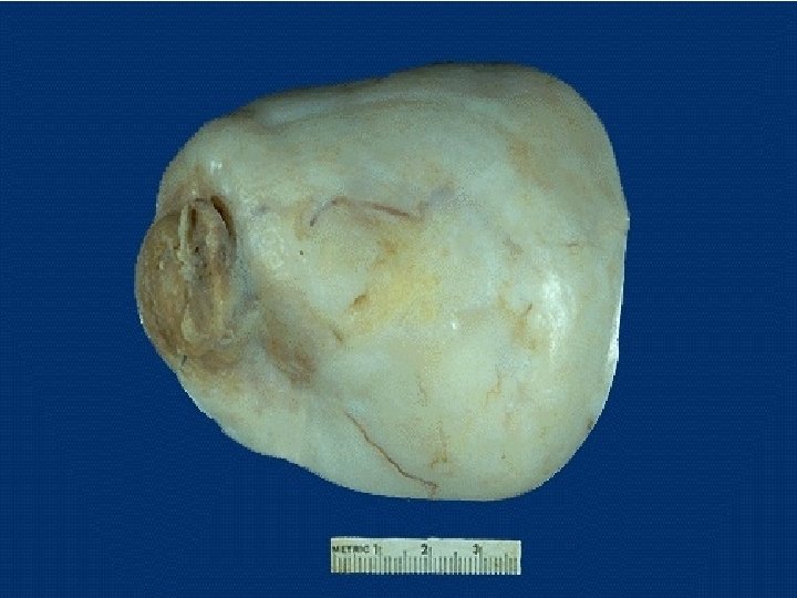

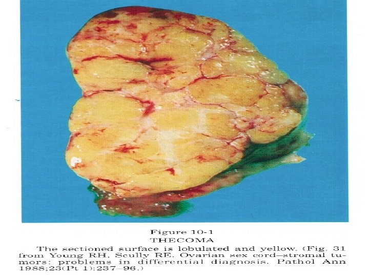
● Rare benign ovarian neoplasm. Thecoma ● Occur in old women (mean age 59 years; 84% after menopause). ● Grossly : Unilateral (90%), well-defined, firm, solid and covered by intact ovarian serosa, Variable size; usually yellow; often a mixture with fibroma (white). ● Histopathology : Spindle cells with moderate pale cytoplasm containing lipid droplets and central nuclei. Intervening stroma has collagen deposition and focal hyaline plaque formation. → Most thecomas appear as a mixture of fibroma and thecoma components, so-called ovarian fibrothecoma. ● Presentation : 1) They are typically estrogen-producing tumors ( 60% ) : abnormal uterine bleeding - 20% have concurrent endometrial carcinoma 2) Androgen : producing tumors ( 10% ). 3) Non fuctioning tumor.

Epithelial Ovarian cancer Epidemiology Risk factors Histopathology Pathogenesis Clinical features and diagnosis Staging and surgical treatment Adjuvant therapy Screening

Epidemiology ● The second most common gynecologic malignancy in developed countries. - Incidence of 9. 4 per 100, 000 women and a mortality rate of 5. 1 per 100, 000. ● The most common cause of gynecologic cancer death and the fifth leading cause of cancer death in women. ● The lifetime risk of developing ovarian cancer in US is. 1. 4%. ● The majority of ovarian malignancies (95 %) are derived from epithelial cells; the remainder arise from other ovarian cell types (germ cell tumors, sex cordstromal tumors). ● Serous carcinoma, the most common histologic subtype of epithelial ovarian carcinoma (75 percent of epithelial carcinomas). ● The majority of ovarian cancers are diagnosed at an advanced stage : confined to primary site (15 %); spread to regional lymph nodes (17 %); distant metastases (61%); and unstaged (7 %).
 Age : - The incidence increases with increasing age. - The")
Risk factors (1) Age : - The incidence increases with increasing age. - The average at diagnosis of ovarian cancer is 63 years old. - The incidence by age is : ●<20 years old – 0. 2 to 1. 4 per 100, 000 ● 20 to 29 – 1. 8 to 2. 2 per 100, 000 ● 20 to 39 – 3. 1 to 5. 1 per 100, 000 ● 40 to 49 – 9. 0 to 15. 2 per 100, 000 ● 50 to 59 – 21. 8 to 28. 3 per 100, 000 ● 60 to 69 – 36. 2 to 41. 5 per 100, 000 ●≥ 70 – 47. 6 to 56. 7 per 100, 000 → The age at diagnosis of ovarian cancer is younger among women with a hereditary ovarian cancer syndrome. The risk of ovarian cancer reaches 2 -3 % in women with a BRCA 1 gene mutation at age 35 and for those with a BRCA 2 mutation at age 50. The typical age at diagnosis of ovarian cancer in women with Lynch syndrome (hereditary nonpolyposis colon cancer) is 43 to 50 years old.
 Reproductive and hormonal factors : 1 - Early menarche ( < 12 years")
(2) Reproductive and hormonal factors : 1 - Early menarche ( < 12 years ). 2 - late menopause ( > 52 years ). 3 - Nulliparity. 4 - Infertility. - Ovulation induction for treatment of infertility does not appear to increase the risk. 5 - Endometriosis : - Increased risk of clear cell , endometrioid , and low grade serous EOC, but not high grade serous or mucinous EOC. - A literature review estimated that the risk of malignant transformation of ovarian endometriosis was 2. 5 %. - Endometriosis-associated EOC appears to develop in younger women and have a better prognosis than most cases of EOC. 6 - Polycystic ovarian syndrome. 7 - Postmenopausal hormone therapy : The absolute risk of ovarian cancer with postmenopausal hormone therapy appears to be small. 8 - Intrauterine device : Data from the NHS showed an association between use of an intrauterine device and increased risk of ovarian cancer; the type of intrauterine device was not specified.
 Genetic factors : ● Several ovarian cancer susceptibility genes have been identified, primarily")
(3) Genetic factors : ● Several ovarian cancer susceptibility genes have been identified, primarily BRCA 1 and 2 and the mismatch repair genes (associated with Lynch syndrome); other genes include RAD 51 C, RAD 51 D, and BRIP 1. - It is estimated that BRCA gene mutations and Lynch syndrome account for 10 to 15% of ovarian cancer cases. - The estimated lifetime risk of ovarian cancer is 35 -46 % for BRCA 1 mutation carriers and 13 -23 % for BRCA 2 mutation carriers. - Lynch syndrome : Lynch syndrome (hereditary nonpolyposis colorectal cancer; HNPCC) is associated with other cancers, in particular, endometrial, ovarian, urogenital, and other gastrointestinal primaries. Colorectal cancer is the hallmark disease for Lynch syndrome; endometrial cancer is the second most common malignancy in affected women (occurring in up to 70 %), but ovarian cancer is also increased in frequency. The lifetime risk of ovarian cancer in women with Lynch syndrome is 3 -14 % compared with 1. 5 % in the general population. Women with Lynch syndrome account for 1 % of ovarian cancers. (4) Family history of ovarian cancer : A personal or family history of breast cancer had been thought of as a risk factor for ovarian cancer; however, BRCA gene mutations appear to account for most of this increased risk.
 Environmental factors : 1 - Cigarette smoking : Current smoking or past smoking")
(5) Environmental factors : 1 - Cigarette smoking : Current smoking or past smoking appears to increase the risk of mucinous ovarian cancer, but not other types of EOC. 2 - Talc and asbestos : e. g, genital use of talcum powder (talc). 3 - Obesity : High body mass index (BMI) appears to increase ovarian cancer risk. - No association between alcohol intake and the risk of EOC. - There is no clear relationship between physical activity and ovarian cancer risk. - No association between vitamin D supplementation and ovarian cancer risk.

Protective factors The factors with the strongest association with a reduced risk of ovarian cancer include : 1) Oral contraceptives: - Larger reductions in ovarian cancer risk occurred with increasing duration of OC use (RR decreased by approximately 20 % for each five years of use; by 15 years, the risk of ovarian cancer was reduced by 50 %). Importantly, the protective effect persisted for 30 years after cessation of OCs. - There are no data regarding the use of non-oral estrogen-progestin contraceptives (ring, patch) for ovarian cancer prevention. 2) Multiparity : - The EOC risk appears to decrease with an increasing parity. - History of multiple gestation or later age (>35 years old) at last pregnancy are protective against EOC. 3) Gynecological surgeries (Salpingo-oophorectomy, Tubal ligation or Hysterectomy) - Hysterectomy without oophorectomy was associated with a reduction in the risk of ovarian cancer. 4) Breastfeeding. → Systematic reviews regarding a potentially protective effect against ovarian cancer of nonsteroidal antiinflammatory drugs (NSAIDs) have yielded conflicting results.
 that explains the link between many of the risk factors")
Pathogenesis The pathogenetic mechanism(s) that explains the link between many of the risk factors and development of epithelial ovarian carcinoma (EOC) have not been determined. Traditionally, two main hypotheses were proposed: ●Incessant ovulation : Repeated ovulation results in minor trauma to the ovarian epithelium, which in turn, can lead to malignant transformation. Support for this hypothesis is derived from the observation that women with periodic suppression of ovulation as a result of oral contraceptive use, pregnancy, or lactation have a lower incidence of epithelial ovarian carcinoma. ●Exposure to gonadotropins : Persistent ovarian exposure to gonadotropins and elevated estradiol concentrations may be carcinogenic. This hypothesis is supported by the observation that experimentally induced ovarian tumors contain gonadotropin receptors. In contrast, evidence shows that a history of a multiple gestation is associated with a decreased risk of EOC. According to the gonadotropic hypothesis, women with a history of multiple gestations should be at increased risk of EOC because they have higher levels of gonadotropins during their fertile years and they have a greater incidence of double ovulations per menstrual cycle. Furthermore, one case control study failed to demonstrate a relationship between serum levels of luteinizing hormone and the risk of ovarian cancer.

Histopathology Currently, based upon histopathology, immunohistochemistry, and molecular genetic analysis, the five main subtypes of epithelial ovarian, fallopian tubal, and peritoneal carcinomas and their relative proportions are : ●High-grade serous carcinoma (70 -80 %) ●Endometrioid carcinoma (10 %) ●Clear cell carcinomas (10 %) ●Mucinous carcinoma (3 %) ●Low-grade serous carcinoma (<5 %)

HIGH-GRADE SEROUS CARCINOMA ● Accounts for approximately 70 -80 % of all malignant ovarian neoplasms. ● The peak age range is 45 to 65 years with a mean of 57 years. ● Most HGSC is diagnosed at an advanced stage (stage III or IV) and has a poor overall prognosis. HGSC that is confined to the ovary at diagnosis is rare (<10 percent). ● Relatively chemosensitive. ● Gross pathology : HGSC can range in size from microscopic to greater than 20 cm in diameter. The external surfaces of the neoplasm may be smooth or have friable surface papillae. The mass is typically cystic and multilocular with serous or bloody fluid and soft friable papillary excrescences. Other areas may be solid and range from soft to firm, depending on the tumor stroma. Gross metastases are often found throughout the peritoneum and the omentum. These are typically firm nodules of various sizes that may coalesce into larger masses, often an omental cake. ● Microscopic pathology : Microscopically, HGSC may have a variety of architectural patterns. These include: complex papillary, glandular, microcystic, and solid patterns. Psammoma bodies may be present, but are rarely as numerous as in low grade serous carcinoma (LGSC). The mitotic rate is often very high, with the threshold for HGSC defined as ≥ 12 mitotic figures per 10 high powered fields. The key feature of HGSC, regardless of the overall architectural pattern, is the marked cytologic atypia with prominent mitotic activity. The atypical nuclei are hyperchromatic with a threefold or greater variation in nuclear size, and tumor giant cells are common.

LOW-GRADE SEROUS CARCINOMA ● LGSC is uncommon and accounts for fewer than 5 % of all cases of ovarian carcinoma. ● Like HGSC, LGSC is typically diagnosed at an advanced stage, and consequently, the long-term prognosis is poor. ● Relative insensitivity to platinum-based chemotherapy. ● Gross pathology : LGSC is often grossly indistinguishable from HGSC or serous borderline neoplasms. ● Microscopic pathology : presence of destructive stromal invasion. composed of small papillae lined by neoplastic cells that exhibit uniform nuclei with less than threefold variability in size. it has a much lower mitotic activity than that observed in HGSC, with <12 mitoses present per 10 high powered fields (HPF). Another distinct feature that may be found in LGSC is a hyalinized stroma with numerous psammoma bodies.

ENDOMETRIOID CARCINOMA ● Accounts for approximately 10 % of all ovarian carcinomas. ● Presents most frequently in women in their 40 s and 50 s, with a mean patient age of 56 years. ● Endometrioid carcinomas are most often identified at an early stage (unlike serous carcinomas), consequently these patients have a much better prognosis. ● Relatively chemosensitive. ● Ovarian endometrioid carcinoma is often associated with and believed to arise from endometriosis (up to 42 % of patients have evidence of ovarian or pelvic endometriosis). ● Endometrioid ovarian carcinoma is associated with carcinoma of the endometrium in 15 -20 % of cases.

● Gross pathology : Endometrioid carcinoma can have a variable appearance. It may be cystic or solid. Residual foci of endometriosis can often be identified with the typical "chocolate cyst" appearance of an endometrioma. The outer surfaces are generally smooth. It is usually confined to one ovary (bilaterality favors metastasis from endometrium). ● Microscopic pathology : Histologically, endometrioid carcinoma of the ovary resembles low-grade endometrioid endometrial carcinoma. The majority of ovarian endometrioid carcinomas have a complex glandular, cribriform, and/or villoglandular architectural pattern with back-to-back growth or elongated or round glands with smooth luminal contours. The glands are typically lined by stratified columnar cells with scant eosinophilic cytoplasm and low to intermediate grade nuclei.

CLEAR CELL CARCINOMA ● Accounts for approximately 5 - 10 % of all ovarian carcinomas. ● Presents most commonly in perimenopausal women in their late 40 s or 50 s. ● Often presents at an early stage (stage I or II) and has a relatively good prognosis due to the absence of distant metastases. ● Insensitive to platinum-based chemotherapy. ● Associated with an increased risk of vascular thrombotic events and paraneoplastic hypercalcemia. ● Similar to endometrioid carcinoma, clear cell ovarian carcinoma is often associated with, and likely arises from, endometriosis. ● Gross pathology : Ovarian clear cell carcinoma often presents as a large mass with an average size of 15 cm. The tumor usually consists of a thick-walled uni- or multilocular cyst with yellowish, fleshy nodules protruding into the lumen of the cyst(s), and watery or mucinous fluid within the cyst(s). However, the neoplasm may

● Microscopic pathology : Clear cell carcinomas display an array of different histologic patterns that often occur together within the same neoplasm. The most common patterns are solid, tubulocystic, and papillary. clear cell carcinomas often contain a prominent hyalinized stroma. The neoplastic cells often have distinct cell borders with nuclei of varying sizes and shapes.

MUCINOUS CARCINOMA ● Accounts for 3 - 4 % of primary ovarian cancers. ● Present in perimenopausal women in their late 40 s to early 50 s. ● Nearly all mucinous carcinomas of the ovary present with early stage disease, usually stage I. ● Gross pathology : Primary ovarian mucinous carcinoma range size from 8 to 20 cm, but can be much larger. It is typically cystic or solid, unilateral, and confined to the ovary. The external surface is usually smooth without surface involvement. Primary ovarian mucinous carcinomas do not present with gross pseudomyxoma peritonei. Although pseudomyxoma peritonei historically was believed to result from rupture of primary ovarian mucinous neoplasm, it is now accepted that pseudomyxoma peritonei almost always results from metastasis to the ovary, often from an appendiceal primary. The primary site should be sought in these cases. ● Microscopic pathology : The cells of ovarian mucinous carcinomas can resemble those of the intestine, endocervix, or gastric pylorus, although the vast majority of these neoplasms have gastrointestinal differentiation.
, fallopian tubal")
Clinical features and diagnosis The clinical presentation of epithelial ovarian carcinoma (EOC), fallopian tubal carcinoma, and peritoneal carcinoma may be either acute or subacute. - Women who present in an acute fashion are typically those with advanced disease who present with a condition that requires urgent care and evaluation (eg, pleural effusion, bowel obstruction and VTE). - EOC may present in a subacute fashion (eg, adnexal mass, pelvic or abdominal pain, gastrointestinal symptoms) in women with either early or advanced disease. These conditions are usually evaluated in an outpatient setting. Adnexal mass : may be discovered due to symptoms of pelvic pain or pressure or it may be found on a routine pelvic examination or an imaging study performed for another indication. Pelvic or abdominal symptoms : Bloating, Urinary urgency or frequency, Difficulty eating or feeling full quickly, Pelvic or abdominal pain. - The symptoms associated with ovarian cancer are nonspecific. - The type or severity of symptom does not reliably correspond to disease stage.

Other subacute presentations : 1 - Postmenopausal bleeding. 2 - Rectal bleeding. 3 - For fallopian tubal carcinoma, a classic triad of symptoms has been described: clear or blood-tinged vaginal discharge, pelvic pain, and a pelvic mass. The vaginal discharge, referred to as hydrops tubae profluens, has been regarded as pathognomonic for the disease. However, vaginal discharge is not present in most women with fallopian tubal cancer (only 2 of 12 in one series ). 4 - Atypical glandular cells on cervical cytology : Women with a finding of atypical glandular cells should be evaluated for cervical and endometrial carcinoma. 5 - Paraneoplastic syndromes : cerebellar degeneration, polyneuritis, dermatomyositis, hemolytic anemia, disseminated intravascular coagulation, acanthosis, or nephrotic syndrome. 6 - Palpable inguinal or cervical lymphadenopathy. 7 - Incidental operative finding.

→ Definitive diagnosis of ovarian cancer requires surgery. Once an adnexal mass has been identified, the evaluation to exclude malignancy includes a medical history, physical examination, imaging studies, and laboratory evaluation for tumor markers.
 Cancer antigen 125 ( CA 125 ) : ● The")
Tumor serum biomarkers (1) Cancer antigen 125 ( CA 125 ) : ● The CA 125 antigen is a large transmembrane glycoprotein derived from both coelomic (pericardium, pleura, peritoneum) and müllerian (fallopian tubal, endometrial, endocervical) epithelia. ● Normal value : CA 125: ≤ 35 U/Ml. ● Serum CA 125 is the most commonly used laboratory test for the evaluation of adnexal masses for epithelial ovarian cancer (EOC). ● CA 125 testing alone has a low sensitivity, particularly for early stage ovarian cancer. only raised in 50% of early stage disease. ● CA-125 being raised in numerous conditions including fibroids, endometriosis, adenomyosis , pelvic inflammatory disease, liver cirrhosis with or without ascites , Cancers of the endometrium, breast, lung, and pancreas.

● In postmenopausal women, the sensitivity for ovarian cancer was 69 -87 %, specificity was 81 -93 %. For premenopausal women, the sensitivity was 50 – 74%, specificity was 69 -78 %. ● CA 125 is not consistently produced by some histologic types of epithelial ovarian cancer, including: mucinous, clear cell, and mixed müllerian ovarian tumors. ● Serum CA 125 >200 U/m. L had been used as a criterion for referral to a gynecologic oncologist.
 human epididymis protein 4 (HE 4) : - The laboratory reference range of")
(2) human epididymis protein 4 (HE 4) : - The laboratory reference range of HE 4 is ≤ 150 p. M. (3) Carcinoembryonic antigen (CEA) : CEA may be elevated in malignancies that produce the protein, particularly mucinous cancers associated with the gastrointestinal tract or ovary. CEA may also be elevated in the following malignancies : Breast, Pancreas, Thyroid, Lung. - The typical upper limit of normal for CEA in non-smokers is 3. 8 micrograms per liter (mcg/L). For smokers, the upper limit of normal is 5. 5 mcg/L. (4) Cancer antigen 19 -9 (CA 19 -9) : Is a mucin protein that may be elevated in ovarian cancer.

Imaging ● Pelvic ultrasound is the single most effective way of evaluating an ovarian mass. With transvaginal ultrasonography being preferable due to its increased sensitivity over transabdominal ultrasound. ● Pelvic and abdominal imaging : Imaging studies can help to assess for the presence of ascites and the extent of disease in women with suspected intraabdominal spread of EOC. Abdominal and pelvic computerized tomography (CT) or magnetic resonance imaging (MRI) are the most commonly used modalities. ● Chest radiography is performed in most patients to evaluate for pleural effusion, pulmonary metastases, and mediastinal lymphadenopathy.
 was originally developed in")
Risk of Malignancy Index The risk of malignancy index (RMI) was originally developed in 1990 and is a multimodality approach that combines serum CA 125, pelvic ultrasound, and menopausal status into an index score to predict the risk of ovarian cancer in women with an adnexal mass. ● RMI I combines three presurgical features: serum CA-125 level (IU/ml); menopausal status (M); and ultrasound score (U). RMI = U x M x CA-125 - The ultrasound result is scored 1 point for each of the following characteristics: multilocular cysts, solid areas, metastases, ascites and bilateral lesions. U = 0 (for an ultrasound score of 0), U = 1 (for an ultrasound score of 1), U = 3 (for an ultrasound score of 2– 5). - The menopausal status is scored as 1 = premenopausal and 3 = postmenopausal. ( Postmenopausal can be defined as women who have had no period for more than one year or women over the age of 50 who have had a hysterectomy) - Serum CA-125 is measured in IU/ml and can vary between zero to hundreds or even thousands of units. ● RMI I score of >200 = high risk ( sensitivity 78%, specificity 87% for ovarian cancer ) RMI I score < 25 = low risk ( risk of cancer < 3%)

→ Cancer of the ovary, fallopian tube, or peritoneum often requires surgery for : 1 - Diagnosis 2 - Staging 3 - Management

STAGING SYSTEM Cancer of the ovary is surgically staged according to the 2014 International Federation of Gynecology and Obstetrics (FIGO) : STAGE I (Tumor confined to ovaries) IA : Tumor limited to 1 ovary, capsule intact, no tumor on surface, negative washings IB : Tumor involves both ovaries , otherwise like IA IC : Tumor limited to 1 or both ovaries IC 1 : Surgical spill IC 2 : Capsule rupture before surgery or tumor on ovarian surface IC 3 : Malignant cells in the ascites or peritoneal washings STAGE II (Tumor involves 1 or both ovaries with pelvic extension (below the pelvic brim) or primary peritoneal cancer) IIA : Extension and/or implant on uterus and/or Fallopian tubes IIB : Extension to other pelvic intraperitoneal tissues

STAGE III (Tumor involves 1 or both ovaries with cytologically or histologically confirmed spread to the peritoneum outside the pelvis and/or metastasis to the retroperitoneal lymph nodes) IIIA : Positive retroperitoneal lymph nodes and/or microscopic metastasis beyond the pelvis IIIA 1 : Positive retroperitoneal lymph nodes only IIIA 1(i) : Metastasis ≤ 10 mm IIIA 1(ii) : Metastasis > 10 mm IIIA 2 : Microscopic, extrapelvic (above the brim) peritoneal involvement ± positive retroperitoneal lymph nodes IIIB : Macroscopic, extrapelvic, peritoneal metastasis ≤ 2 cm ± positive retroperitoneal lymph nodes. Includes extension to capsule of liver/spleen IIIC : Macroscopic, extrapelvic, peritoneal metastasis > 2 cm ± positive retroperitoneal lymph nodes. Includes extension to capsule of liver/spleen STAGE IV (Distant metastasis excluding peritoneal metastasis) IVA : Pleural effusion with positive cytology IVB : Hepatic and/or splenic parenchymal metastasis, metastasis to extra-abdominal organs (including inguinal lymph nodes and lymph nodes outside of the abdominal cavity)
 Histologic type including grading should be designated")
Other major recommendations are as follows: 1) Histologic type including grading should be designated at staging. 2) Primary site (ovary, Fallopian tube or peritoneum) should be designated where possible. 3) Tumors that may otherwise qualify for stage I but involved with dense adhesions justify upgrading to stage II if tumor cells are histologically proven to be present in the adhesions.
 Total hysterectomy 2) Bilateral salpingo-oophorectomy 3) Pelvic and paraaortic lymph")
STAGING PROCEDURE : 1) Total hysterectomy 2) Bilateral salpingo-oophorectomy 3) Pelvic and paraaortic lymph node dissection 4) Peritoneal cytology 5) Omentectomy 6) Cytology of the diaphragm 7) Biopsy of any areas where metastases are suspected - Laparotomy is generally used, but laparoscopic or robot-assisted approaches are used by some surgeons for selected patients. → Frozen section : The diagnosis of ovarian, tubal, or peritoneal cancer is usually made at time of surgery.

Cytoreductive surgery is the cornerstone of therapy for ovarian, tubal, and peritoneal carcinoma. There are several potential benefits of aggressive primary surgical management in women with epithelial ovarian carcinoma (EOC), particularly for those with advanced disease : 1) Optimal response to postoperative systemic chemotherapy is achieved in the setting of minimal disease burden. 2) Disease-related symptoms (eg, abdominal pain, increased abdominal girth, dyspnea, early satiety) are related to tumor burden. Removal of bulky disease rapidly improves symptoms and quality of life. 3) Ovarian neoplasms produce multiple cytokines, at least some of which are immunosuppressive (eg, interleukin-10, vascular endothelial growth factor). Removal of tumor bulk may improve or restore host immune competence. 4) Cytoreduction is associated with increased survival. The volume of residual disease remaining after cytoreductive surgery correlates inversely with survival.

The terminology regarding extent of cytoreduction is: ●Complete cytoreduction – Cytoreduction to no grossly visible disease. ●Optimal cytoreduction – The Gynecologic Oncology Group (GOG) defines optimal cytoreduction as residual disease that is ≤ 1 cm in maximum tumor diameter. ●Suboptimal cytoreduction – Cytoreduction with tumor nodules >1 cm remaining. → Stage IA : In young women, conservative surgery (unilateral salpingooophorectomy) for fertility preservation is being considered. full surgical staging, including washings, omentectomy, appendectomy, and node biopsies, should be done and should be negative.

Adjuvant therapy Women with EOC should undergo adjuvant treatment with the exception of those with grade 1, stage IA/IB EOC. The preferred regimen is a platinum-taxane combination regimen. ● For women with early-stage disease, carboplatin and paclitaxel (as intravenously [IV] infusions every three weeks for three to six cycles) is usually administered. ● For women with advanced EOC, the options depend on the surgical outcomes : A) For women with optimally cytoreduced EOC (ie, <1 cm residual disease at the completion of surgery), there are multiple options for treatment, all of which are acceptable. These include : 1 - Standard chemotherapy (ie, carboplatin and paclitaxel administered IV every three weeks for six cycles) OR 2 - Dose-dense chemotherapy (ie, IV carboplatin on day 1 plus IV paclitaxel on days 1, 8, 15 every three weeks for six cycles) OR 3 - IV plus intraperitoneal (IP) chemotherapy (ie, IP cisplatin on day 1, IV paclitaxel on day 2, and IP paclitaxel on day 8 every three weeks for six cycles). B) Women with suboptimally cytoreduced EOC (ie, ≥ 1 cm residual disease) are not candidates for IP administration of chemotherapy. These patients usually receive standard or dose-dense chemotherapy for six cycles.
 refers to the administration of systemic chemotherapy")
→ Neoadjuvant therapy — Neoadjuvant chemotherapy (NACT) refers to the administration of systemic chemotherapy before definitive surgery. The goal of NACT is to reduce perioperative morbidity and mortality and increase the likelihood of a complete resection of disease at the time of cytoreductive surgery. In most cases of women treated with NACT, further chemotherapy is administered following surgery (ie, adjuvant chemotherapy).

SPECIFIC LONG TERM EFFECTS IN OVARIAN CANCER SURVIVORS In general, ovarian cancer survivorship can be characterized as issues involving one of the four domains of quality of life (QOL) : the physical, emotional (psychological), spiritual, and social. 1) Neurotoxicity : Muscle cramps, Ringing in ears or trouble hearing, Discomfort in feet, Trouble walking, Numbness and weakness in the hands. 2) Cognitive dysfunction 3) Fatigue 4) Gastrointestinal toxicity 5) Surgical complications : e. g, bowel obstruction, which can be caused by intraabdominal adhesions secondary to prior surgery. 6) Loss of fertility 7) Sexual dysfunction 8) Menopause : Menopausal hormonal therapy does not appear to increase the risk of either recurrence or mortality. 9) Psychological effects (Depression, Anxiety , Guilt , Poor body image).

PROGNOSIS Overall five-year survival in women with ovarian cancer is less than 45 %.
 younger age :")
The major prognostic factors associated with improved outcome were : 1) younger age : Younger patients are more likely to have a favorable prognosis because they are more likely to have tumors of less aggressive histology and lower grade, and better baseline performance status. 2) Low volume of residual disease. 3) Good performance status. 4) Serous histology.
 Office visits (including physical and pelvic exams) every three to six")
POSTTREATMENT SURVEILLANCE 1) Office visits (including physical and pelvic exams) every three to six months up to five years posttreatment, then annually. 2) CA-125 or other tumor markers (eg, HE 4) every visit if initially elevated, if indicated 3) Other testing (ie, chemistry profile or complete blood count, CT scan, or transvaginal ultrasound in women who underwent fertility-sparing surgery) only as clinically indicated. → Standard imaging techniques, including ultrasound, computerized tomography (CT), magnetic resonance imaging (MRI), and positron emission tomography (PET), have limited sensitivity to detect recurrent EOC.

Screening for ovarian cancer ● Screening for high risk women : There is a consensus that women at average risk for ovarian cancer should NOT undergo screening. ● Screening tests : Measurement of CA 125 and transvaginal pelvic ultrasound.

Ovarian germ cell neoplasms ● Derived from primordial germ cells of the ovary. ● They may be benign or malignant. ● Comprise approximately 20 -25 % of ovarian neoplasms overall, but account for only about 5 % of all malignant ovarian neoplasms. ● Arise primarily in young women between 10 and 30 years of age and represent 70% of ovarian neoplasms in this age group. ● Histological types : 1) Teratoma : - Mature teratoma : - Mature cystic teratoma ( Dermoid cyst ) - Mature solid teratoma - Immature teratoma 2) Dysgerminomas 3) Endodermal sinus (yolk sac) tumors 4) Rare OGCNs : Pure embryonal carcinomas, nongestational choriocarcinomas, and pure polyembryoma 5) Mixed germ cell tumors

● OGCNs grow rapidly, unlike the more common epithelial ovarian neoplasms, yet most patients present with stage IA disease (limited to one ovary). ● The majority are present as unilateral ovarian mass. ● Immature teratoma : account for 35. 6 % of all malignant OGCNs Dysgerminomas : account for 32. 8 % of malignant OGCNs Endodermal sinus tumors : account for 14 to 20 percent of all malignant OGCNs Embryonal carcinoma : accounts for 4 % of malignant OGCNs Mixed germ cell neoplasms : account for 5. 3 % of all malignant OGCNs Non-gestational choriocarcinoma : 2. 1 % of all malignant OGCNs (the estimated incidence of a primary ovarian choriocarcinoma is 1 in 369, 000).

Clinical manifestations - OGCNs often produce hormones, particularly the beta subunit of human chorionic gonadotropin (h. CG). - Patients typically present with one or more of the following signs and symptoms: 1) Abdominal enlargement — from the mass itself, ascites, or both. 2) Abdominal pain — from rupture or torsion. 3) Precocious puberty, abnormal vaginal bleeding — presumably from h. CG production. 4) Symptoms of pregnancy — from h. CG production. ● 85% of women with an OGCN have both abdominal pain and an abdominal mass. ● Fever or vaginal bleeding occurs in 10 %. ● OGCNs tend to be large (median size 16 cm). ● Ascites, rupture (pre- or intraoperative), and torsion are reported in 20, and 5 % of cases, respectively.

● The diagnosis is made by histology at time of surgical excision. The diagnosis is strongly suggested preoperatively by the presence of an adnexal mass on pelvic imaging and an elevated level of an associated tumor marker (eg, h. CG, alpha fetoprotein [AFP]). Tumor markers OGCNs are often associated with hormonal or enzymatic activity. 1) h. CG —embryonal cell carcinomas, and ovarian choriocarcinomas, mixed germ cell tumors, and some dysgerminomas. 2) AFP — endodermal sinus tumors, embryonal cell carcinomas and polyembryoma carcinomas, mixed germ cell tumors, and some immature teratomas ; most dysgerminomas are associated with a normal AFP. 3) Lactate dehydrogenase (LDH) — dysgerminomas. ● Staging : as EOC staging.
 Surgery : unilateral salpingo-oopherectomy / cytoreduction. 2) Chemotherapy : usually very sensitive")
Treatment 1) Surgery : unilateral salpingo-oopherectomy / cytoreduction. 2) Chemotherapy : usually very sensitive to chemotherapy. 3) Radiation therapy.

sex cord-stromal neoplasms Are a heterogeneous group of benign or malignant neoplasms that develop from the dividing cell population that would normally give rise to cells surrounding the oocytes, including the cells that produce ovarian hormones (the nongerm cell and nonepithelial components of the gonads). ● Malignant ovarian sex cord-stromal neoplasms are rare, comprising only 1. 2 % of all primary ovarian cancers (malignant neoplasms). ● In contrast with epithelial ovarian cancer, most patients with malignant sex cordstromal neoplasms are diagnosed with early-stage disease; even malignant neoplasms are generally considered to be low-grade. ● Lymph node metastases are rare from these neoplasms. ● The age distribution of sex cord-stromal neoplasms is somewhat younger than that of epithelial neoplasms, The average at diagnosis of sex cord-stromal neoplasms was 50 years (compared with 61 years for epithelial cancer). ● Histological types : fibromas, fibro-thecomas, granulosa cell neoplasms, Sertoli or Sertoli-Leydig cell neoplasms, and gynandroblastoma.

Clinical presentation Women with sex cord-stromal neoplasms generally present in the same manner as women with epithelial ovarian neoplasms (eg, abdominal or pelvic symptoms, finding of adnexal mass on examination or imaging). In addition, sex cord-stromal neoplasms that secrete estrogens or androgens result in endocrine-dependent clinical manifestations. The diagnosis should therefore be suspected in patients who present with signs of estrogen excess (eg, precocious puberty in a child, abnormal uterine bleeding, endometrial hyperplasia or carcinoma) or androgen excess (virilization), especially if an adnexal mass is present Tumor markers Some sex cord-stromal neoplasms secrete proteins (eg, inhibin, estrogen, androgens), which can serve as diagnostic markers. → For women with sex cord-stromal neoplasm, endometrial sampling should be performed. MANAGEMENT : The majority of ovarian sex cord-stromal tumors are benign and are managed surgically. Malignant sex cord-stromal tumors are often detected at an early stage and managed with surgery alone. Lymphadenectomy is not typically required. Adjuvant chemotherapy is an option in selected patients.

Ovarian tumors of low malignant potential Tumors of low malignant potential (also called borderline tumors) are a heterogeneous group of lesions defined histologically by atypical epithelial proliferation without stromal invasion. ● Account for 10 -20 % of ovarian epithelial tumors. ● The average at diagnosis is 40 to 60 years old, but the highest frequency relative to invasive ovarian cancer of these tumors occurs in the 15 to 29 year-old age group. Thus, the disease frequently affects women with a desire to preserve childbearing potential. HISTOLOGIC TYPES Serous borderline tumors, Mucinous borderline tumors, Endometrioid, clear-cell, and transitional cell (Brenner) borderline tumors. - The majority of borderline tumors are serous, Approximately 75 % of patients are diagnosed with stage I disease; 25 -50 % of tumors are bilateral. - Mucinous tumors are the other common cellular type; 90 % are stage I and fewer than 10 % are bilateral.

● Borderline tumors are staged using the same criteria as other ovarian tumors. Most women present with stage I disease (approximately 70 percent); stage II and III disease are relatively infrequent. TREATMENT (1) Fertility preserving surgery — Borderline tumors generally have an excellent prognosis and can be treated conservatively in women who wish to preserve their fertility or are pregnant at the time of diagnosis. Unilateral salpingo-oophorectomy, or, in some instances, ovarian cystectomy may be performed. - The overall risk of recurrence after conservative surgery ranges from 7 -30 %, and recurrences typically show borderline histology, not invasive cancer. (2) Hysterectomy and bilateral salpingo-oophorectomy — Total abdominal hysterectomy plus bilateral salpingo-oophorectomy (TAH-BSO) is recommended for women who are not planning pregnancy or have advanced stage disease. → The use of adjuvant chemotherapy is controversial : most physicians recommend chemotherapy after aggressive surgical debulking only if invasive implants are identified.

PROGNOSIS The prognosis depends upon the stage and histologic features of the tumor, but is generally good. 5 years survival rate : ●Stage I – 99% ●Stage II – 98% ●Stage III – 96% ●Stage IV – 77%
- Slides: 82