
Presented By Dr Saurav Singh PDCC Neuroanaesthesia Moderator
Presented By: Dr. Saurav Singh (PDCC Neuroanaesthesia) Moderator: Dr. Sanjay Agarwal 1
OVERVIEW • Introduction. • Role of Dexmedetomidine in clinical neurosurgical anaesthesia – ü Cerebral Vascular Disease. ü Intracranial Tumor Surgery. ü Traumatic Brain Injury (TBI). ü Spinal Cord Injury. ü Intraoperative Neurophysiology Monitoring. ü Sedation in Paediatric Patients With Neurosurgical Diseases. ü Benefits of Analgesia in Neurosurgery. ü The Neurocritical Care Unit. ü Awake Craniotomy. ü Epilepsy Surgeries Requiring Intraoperative Mapping. ü Stereotactic Neurosurgery/Deep Brain Stimulation. • Summary (PDCC Neuroanaesthesia) 2
INTRODUCTION (PDCC Neuroanaesthesia) 3
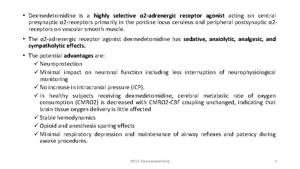
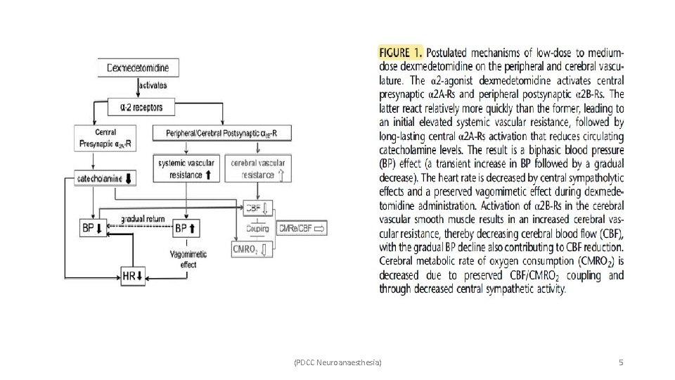
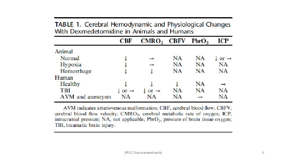
• Dexmedetomidine is a highly selective α 2 -adrenergic receptor agonist acting on central presynaptic α 2 -receptors primarily in the pontine locus ceruleus and peripheral postsynaptic α 2 receptors on vascular smooth muscle. • The α 2 -adrenergic receptor agonist dexmedetomidine has sedative, anxiolytic, analgesic, and sympatholytic effects. • The potential advantages are: ü Neuroprotection ü Minimal impact on neuronal function including less interruption of neurophysiological monitoring ü No increase in intracranial pressure (ICP). ü In healthy subjects receiving dexmedetomidine, cerebral metabolic rate of oxygen consumption (CMRO 2) is decreased with CMRO 2 -CBF coupling unchanged, indicating that brain tissue oxygen delivery is little affected ü Stable hemodynamics ü Opioid anesthesia sparing effects ü Minimal respiratory depression and maintenance of airway reflexes and patency during awake procedures. (PDCC Neuroanaesthesia) 4
(PDCC Neuroanaesthesia) 5
(PDCC Neuroanaesthesia) 6
(PDCC Neuroanaesthesia) 7
ROLE OF DEXMEDETOMIDINE IN CLINICAL NEUROSURGICAL ANAESTHESIA (PDCC Neuroanaesthesia) 8
CEREBRAL VASCULAR DISEASE • Carotid endarterectomy (CEA): ü Patients frequently have concomitant peripheral vascular disease and other related comorbidities, and are prone to hemodynamic fluctuations caused by surgical stimulation or perioperative medications. ü Intraoperative hypotension and the reduction in CBF caused by dexmedetomidine can potentially risk inadequate oxygen delivery that may necessitate the use of intracarotid shunts during awake CEA (although a prospective case series showed that dexmedetomidine as the primary sedative did not increase the incidence of shunts as compared with historical controls. ü Hypotensive episodes in the post anesthesia care unit after CEA, however, are more frequent with dexmedetomidine, and patients may require more hemodynamic interventions. ü There is no direct clinical evidence that dexmedetomidine exacerbates impaired dynamic cerebral vascular autoregulation in carotid artery stenosis. ü Similarly, there is no human evidence that dexmedetomidine worsens or causes cerebral ischemia, although high-dose dexmedetomidine was found to be associated with ischemic brain injury exacerbation in animals. (PDCC Neuroanaesthesia) 9
• Intracranial aneurysm/ SAH/ AVM/ ICH: ü The relationship between CBF and CMRO 2 is preserved when using dexmedetomidine in patients with unruptured intracranial aneurysms or arterial venous malformation. ü In a small retrospective study, sedation after unruptured cerebral aneurysm clipping with dexmedetomidine infusion (0. 4 to 0. 9 μg/kg/h) showed comparable blood pressure to propofol (0. 5 to 5. 0 mg/kg/h), but with a lower heart rate. ü Sixty-six percent of dexmedetomidine sedated patients, however, required additional propofol boluses to prevent agitation while intubated. ü In a recent case series of 12 patients, coil embolization of intracranial aneurysms was performed successfully under monitored anesthesia care (MAC) with dexmedetomidine without adverse hemodynamic or respiratory events. ü Nevertheless, these case series are retrospective and underpowered, and prospective randomized trials are needed to elucidate the effectiveness and safety of adding dexmedetomidine during intracranial aneurysm surgery, which requires systemic and cerebral hemodynamic stability. (PDCC Neuroanaesthesia) 10
ü Animal studies of SAH with cerebral vasospasm have found that dexmedetomidine attenuated brain edema, reduced vasospasm, and ameliorated neurological deficits. ü However, clinical neuronal protection related to dexmedetomidine use in patients with SAH has not been confirmed. ü Animal studies have implied that after focal cerebral infarction, dexmedetomidine infusion improved microregional oxygen supply/consumption balance, thereby decreasing cortical infarction size with more cell survival compared with saline. This neuronal protective effect of dexmedetomidine was stronger when combined with propofol or lidocaine. ü However, this neuroprotective effect in cerebral ischemia was not mediated by central αadrenoceptors and not related to the inhibition of presynaptic norepinephrine or glutamate release, but rather by inhibiting the stress hormone and inflammatory response, as well as through activation of a signaling pathway of cell growth, proliferation, and survival. ü In response to stroke, certain inflammatory mediators and stress hormones are activated, which can be inhibited by dexmedetomidine based on preclinical studies. (PDCC Neuroanaesthesia) 11
INTRACRANIAL TUMOR SURGERY • Dexmedetomidine prevents sudden increases in ICP and brain swelling. • As an anesthetic adjuvant results in less cardiovascular variability during operations and an early emergence. • In a prospective randomized controlled trial, infusing dexmedetomidine at 0. 7 μg/kg/h as an adjunct to propofol-fentanyl anesthesia in this setting improved hemodynamic stability and reduced fentanyl and antihypertensive agent consumption. • A systematic review of 254 patients from 5 randomized controlled trials involving functional endoscopic sinus surgery found that dexmedetomidine provided better surgical visibility than saline control or sevoflurane alone as its hypotensive action and hemodynamic stability reduced intraoperative bleeding, although the quality of the operative field was similar among dexmedetomidine, esmolol, and remifentanil groups. • In a recent randomized controlled trial, 4 commonly used anesthetic agents for neurosurgery with different mechanisms of action (midazolam, propofol, fentanyl, and dexmedetomidine) were titrated to an equivalent mild sedation in patients with supratentorial mass lesions before any surgical intervention. Dexmedetomidine compared with the other agents resulted in a much lower incidence of unmasked or exacerbated neurological deficits. (PDCC Neuroanaesthesia) 12
• A recent animal study found α 2 -adrenoceptor expression in C 6 glioma cells (a malignant glioma cell line), so that dexmedetomidine may have an interaction with the tumor through regulating multiple molecular signal pathways and directly activating α 2 -adrenoceptors in gliomas. • This seems to indicate that the “response” of tumors to anesthetics is strongly associated with the tumors’ histologic properties. • Therefore, to evaluate dexemedetomidine’s effects on neurological outcome in brain tumor patients, extensive disease-centered studies with carefully defined clinical phenotypes are needed. (PDCC Neuroanaesthesia) 13
TRAUMATIC BRAIN INJURY (TBI) • Sedation in TBI may have therapeutic significance besides facilitating airway control. • The purpose of sedation in TBI includes optimizing CMRO 2 and CBF, reducing elevated ICP, and preventing secondary brain injury. • In a retrospective case series, in which 85 severe TBI patients in the intensive care unit (ICU) received a median dose of dexmedetomidine of 0. 49 μg/kg/h for 32 hours (median infusion period) to maintain “cooperative sedation, ” midazolam and propofol requirements nearly disappeared indicating the effectiveness of dexmedetomidine as the sole agent for mild sedation for TBI patients. • Another prospective study observed 198 severe TBI patients who received dexmedetomidine and/or propofol sedation. Dexmedetomidine was associated with longer “calm to light sedation” targets compared with propofol alone in the first 7 days after infusion. • Although dexmedetomidine was associated with a higher degree of hypotension compared with propofol, there were no differences in adverse events between the groups. (PDCC Neuroanaesthesia) 14
• A case report of an alcohol-dependent TBI patient showed that dexmedetomidine as a continuous infusion (0. 5 to 1. 5 μg/kg/h for 8 d) facilitated sedation and controlled agitation when benzodiazepine treatment failed, without neurological or respiratory depression. • Systemic hypotension or hypertension can be disastrous for TBI patients with impaired cerebral autoregulation because it can decrease cerebral perfusion pressure, increase ICP, and lead to poor clinical outcomes. • Moderate to severe TBI patients may also suffer paroxysmal sympathetic hyperactivity (PSH) due to elevated ICP, which presents with elevated heart rate, blood pressure, respiratory rate, temperature, sweating, and posturing. • A case report showed that 0. 2 to 0. 7 μg/kg/h dexmedetomidine continuous infusion effectively controlled this syndrome when routine medication therapy failed. • Another retrospective study included 90 severe TBI patients who received dexmedetomidine or propofol/midazolam sedation in the neuro ICU for consecutive days, and found that the dexmedetomidine regimen group had a lower probability to be diagnosed with PSH. (PDCC Neuroanaesthesia) 15
SPINAL CORD INJURY • Recent animal studies have found that intravenous dexmedetomidine attenuated spinal ventral neuronal degeneration and preserved neurological function and neuronal viability after transient spinal cord ischemia or ischemia-reperfusion. • These beneficial effects were associated with improved cell survival and antiapoptotic factors, as well as with the attenuation of microglial activation, proinflammatory cytokine production, decreased interleukin-6, tumor necrosis factor-alpha, and reduced neutrophil infiltration, all of which indicate an anti-inflammatory effect. • Clinically, there is no direct evidence of improved functional outcome by virtue of dexmedetomidine’s theoretical spinal cord protective or anti-inflammatory effects. • In a single case of a focal inflammatory spinal cord disorder, namely transverse myelitis, hemodynamic instability was reported as the major concern response. In that case, dexmedetomidine sedation resulted in severe hypertension and bradycardia, which may have been due to an exaggerated peripheral vasoconstrictor response due to the lack of spinal reflexes. (PDCC Neuroanaesthesia) 16
• In terms of injury biomarkers, a prospective study found reduced levels of the stress hormone cortisol and the inflammatory response marker interleukin-10 after intraoperative dexmedetomidine infusion in cervical spine surgery. • However, those stress response biomarkers and cytokine concentrations could not be correlated with any postoperative functional recovery parameter. • Although dexmedetomidine does not attenuate the injury in patients with cervical cord lesions, its practical use lies perhaps in helping to prevent secondary injury by facilitating awake fiberoptic intubation. • A subsequent randomized controlled trial related to the performance of awake fiberoptic intubation in simulated cervical injury patients found that adding dexmedetomidine sedation (1 μg/kg bolus over 10 min followed by 0. 5 to 0. 7 μg/ kg/h) did not compromise hemodynamic instability and resulted in better patient tolerance and satisfaction compared with the use of midazolam alone. (PDCC Neuroanaesthesia) 17
INTRAOPERATIVE NEUROPHYSIOLOGY MONITORING • Intraoperative neurological injuries are usually detected as changes in the latencies or amplitudes of evoked potentials (EPs). • As such, intraoperative EP monitoring in spine surgeries should preferably be minimally affected by the anesthetic regimen. • Dexmedetomidine has been increasingly used to reduce the requirement of other anesthetics that may impair EP signal acquisition, and its effects on EPs have been observed and investigated thoroughly in spine surgery. • SSEPs are generally well maintained under dexmedetomidine (0. 2 to 0. 7 μg/kg/h) during spine surgeries, with retention of the ability to monitor consistent and reproducible potentials. • For motor evoked potentials (MEPs), most clinical studies found that clinically relevant doses of dexmedetomidine also do not affect the signal, but patients receiving a higher loading dose (1 μg/kg) or with a higher plasma concentration (0. 8 ng/m. L) do experience MEP amplitude reduction or even signal loss. (PDCC Neuroanaesthesia) 18
• Of note, all currently available evidence regarding the effects of dexmedetomidine on EPs stems from spine surgeries. • The impact of dexmedetomidine on EPs in the setting of different neuronal pathologic diseases may vary, especially for those with impaired cerebrospinal tracts. • As for intracranial surgeries, knowledge of how dexmedetomidine affects EP acquisition and the latencies and amplitudes of MEPs and SSEPs for intracranial tumor or cerebrovascular pathologies is lacking. (PDCC Neuroanaesthesia) 19
SEDATION IN PEDIATRIC PATIENTS WITH NEUROSURGICAL DISEASES • Sedation is usually required for imaging studies (eg, magnetic resonance imaging [MRI], computed tomography [CT]) in pediatric patients. • Separation, confinement, and an unfamiliar environment create agitation and anxiety for children, and in situations in which general anesthesia may not be preferred, effective sedation with minimal respiratory depression is highly beneficial. • A previous review extensively summarized early clinical studies of using dexmedetomidine as the primary or rescue sedative during MRI or CT examination in pediatric populations. • In this review, dexmedetomidine was superior to midazolam but inferior to propofol in providing effective sedation, and this may be because propofol led to fewer procedure interruptions and better parental satisfaction according to a comparison study of 1 - to 7 -year old children undergoing MRI with propofol or dexmedetomidine sedation. • Furthermore, the recovery time was slower in the dexmedetomidine cohort compared with those who received propofol. • However, the dosage of dexmedetomidine ranged widely, from 0. 3 to 2 μg/kg bolus over 10 to 15 minutes followed by 0. 5 to 1. 5 μg/kg/h infusion rate, and there was no optimal dosage identified. (PDCC Neuroanaesthesia) 20
• For outpatient EEG monitoring in children, intravenously administered dexmedetomidine provides effective sedation without agitation, hemodynamic fluctuation, or respiratory depression, and was superior to midazolam in terms of minimum “drug effect” on the quality of acquired EEG. • Sedation for intracranial radiotherapy for a 21 -month old child was reported to be safely achieved with dexmedetomidine sedation, providing a smooth sedation “induction” and fairly rapid recovery with airway protection. • Perioperative infusion of dexmedetomidine at 0. 2 μg/kg/h as an adjunct sedative reduced sevoflurane-related emergence delirium in children aged 1 to 10 years. • Clinical research defining the optimal dosing strategy and duration of dexmedetomidine infusion for sedation in pediatric TBI is lacking. • Notably, dexmedetomidine was reported to be associated with significant bradycardia during therapeutic hypothermia when combined with remifentanil for sedation in children with TBI. • Despite the few animal studies there is a lack of clinical evidence regarding the potential neuronal protective or toxic effects of dexmedetomidine in developing human brains following neurological injury, or how dexmedetomidine affects CBF, CMRO 2, and ICP in children. • Further translational studies and clinical evidence are needed. (PDCC Neuroanaesthesia) 21
BENEFITS OF ANALGESIA IN NEUROSURGERY • Postoperative pain following craniotomy or spinal surgery may be significant and can lead to postoperative agitation and hypertension, which should be properly managed to minimize potential intracerebral haemorrhage and/or vasogenic edema. • Opioids are effective in controlling pain but may cause respiratory depression. • Dexmedetomidine can maintain airway reflexes and patency in spontaneously breathing patients while providing analgesic effects. • A previous meta-analysis included eight small sample size randomized controlled trials and found that dexmedetomidine reduced intraoperative opioid consumption during intracranial procedures, although the administration time and dose were variable among the included studies. • Subsequent randomized controlled trials demonstrated that infusion of dexmedetomidine between 0. 2 and 0. 7 μg/kg/h during and after the operation reduced postoperative pain after craniotomy and spine surgeries. • It should be noted that Dexmedetomidine analgesic effect was not as potent as that of remifentanil, and those receiving dexmedetomidine had longer emergence times. (PDCC Neuroanaesthesia) 22
• To avoid such delayed emergence, a particular study made use of a 0. 4 or 0. 8 μg/kg bolus of dexmedetomidine for 10 minutes 1 h before the end of supratentorial craniotomy, with its secondary outcome showing less postsurgical pain compared with control during emergence and after transport to the ICU. • The analgesic efficacy of dexmedetomidine has also been shown in studies in spine surgery, mainly lumbar laminectomy, discectomy, or posterior lumbar interbody fusion. For spinal tumor surgery, no retrospective or prospective study of dexmedetomidine’s analgesic efficacy was identified. • Compared with oral clonidine premedication, dexmedetomidine possesses a noninferior opioid anesthetic-sparing effect, and equal postoperative recovery time, hemodynamic stability, and blood loss during spine surgery. • A randomized controlled trial study found that intraoperative dexmedetomidine infusion at 0. 5 μg/kg/h compared with saline improved the quality of recovery and reduced fatigue in the early postoperative period after major spine surgery, when continuously infusing 0. 2 μg/kg/h for another 24 hours postoperatively. • The effectiveness of dexmedetomidine as an analgesic was demonstrated both when given as an intravenous infusion and as an epidural injection. (PDCC Neuroanaesthesia) 23
• Neither for craniotomy nor for spinal surgery has a standard dexmedetomidine administration strategy been established with regards to dose, time of initiation, duration, and combination with other drugs. • Moreover, the intensity of pain varies with different craniotomy approaches, for example, supratentorial craniotomies are associated with less pain than infratentorial procedures. (PDCC Neuroanaesthesia) 24
THE NEUROCRITICAL CARE UNIT • It is exceptionally important in the neurosurgical population to control agitation and provide analgesia, so as to improve tolerance to mechanical ventilation and accelerate time to extubation in order to maintain intracranial homeostasis and enable early neurological assessment. • A recent well-designed randomized controlled trial compared the infusion of dexmedetomidine for <7 days with saline in a propofol-based sedation regimen in the general ICU (including only a few neurosurgical patients) in patients with agitated delirium and requiring mechanical ventilation. This study found that dexmedetomidine facilitated early extubation and was associated with more ventilator-free hours. • Another randomized control trial enrolled 150 craniotomy patients who were not extubated when admitted to the neuro ICU, where dexmedetomidine infusion at 0. 6 μg/kg/h was initiated 2 hours after craniotomy and up until 30 minutes after extubation, or for a maximum 24 hours. Patients who received dexmedetomidine demonstrated a decreased incidence of agitation compared with the saline group. • Most studies do demonstrate a significant reduction of opioid requirement when using dexmedetomidine, and this analgesic effect may be an important reason for less agitation and better ventilator tolerance neurocritical care patients receiving continuous sedation. (PDCC Neuroanaesthesia) 25
• A retrospective propensity-matched cohort data analysis of 342 patients from 2 medical centers showed that dexmedetomidine and propofol were associated with an equal prevalence of hypotension (defined as MAP <60 mm. Hg, 23% vs. 26%) and bradycardia (defined as heart rate <50 beats/min, 8. 6% vs. 5. 5%). • However, other studies frequently demonstrate that dexmedetomidine is more commonly associated with bradycardia, which requires close attention, especially with higher doses and longer durations of infusion, as are used when weaning off other sedatives in the neurocritical care unit. • A prospective study found that dexmedetomidine improved cognitive function in neuro ICU for patients without forebrain injury compared with propofol, which reduced cognitive function. This was attributed to the better antiagitation and analgesic effects of dexmedetomidine (PDCC Neuroanaesthesia) 26
AWAKE CRANIOTOMY • Awake craniotomy is performed in patients with intracranial lesions in or near eloquent brain areas requiring cortical mapping and intraoperative neurofunctional testing to maximize resection while reducing risk of disability. • Two different anesthesia management regimens are advocated: ü The “asleep-awake-asleep” technique in which endotracheal intubation or laryngeal mask airway placement is performed for the asleep portions of the procedure, and ü The “awake-awake” technique in which mild sedation without invasive airway manipulation is used during the pre-mapping and post-mapping phases. • Local anesthetic infiltration to the incision site and/or a scalp block is generally used with both approaches. • Special anesthesia considerations during the “awake” portion of any such procedure include the need for cooperative sedation and sufficient analgesia without respiratory depression, as well as the need for minimal interference with neurofunctional testing or cortical mapping. (PDCC Neuroanaesthesia) 27
• Solely propofol-based sedation with an unprotected airway in “asleep-awake-asleep” craniotomy, compared with general anesthesia, carries the concerns of a higher incidence of respiratory depression in obese patients, arterial haemoglobin desaturation, a higher level of Pa. CO 2, hypertension, hypotension, and tachycardia. • Dexmedetomidine reported as a useful adjuvant during “awake” state sedation, or even as an effective rescue sedative when a propofol-remifentanil regimen results in oversedation, respiratory depression, or discomfort. • Other case reports have demonstrated that planned endotracheal intubation or laryngeal mask airway placement was able to be avoided outright with the use of dexmedetomidine. • In high-risk patients with airway compromise and severe comorbidities, continuous infusion of dexmedetomidine (0. 5 to 1. 0 μg/kg loading dose followed by 0. 2 to 0. 7 μg/kg/h infusion) as the primary sedative combined with scalp nerve block and small doses of opioid was reported to facilitate prolonged and complex “awake” procedures without any airway manipulation required. • Dexmedetomidine was well-tolerated in obstructive sleep apnea patients who needed continuous positive airway pressure during awake craniotomy. (PDCC Neuroanaesthesia) 28
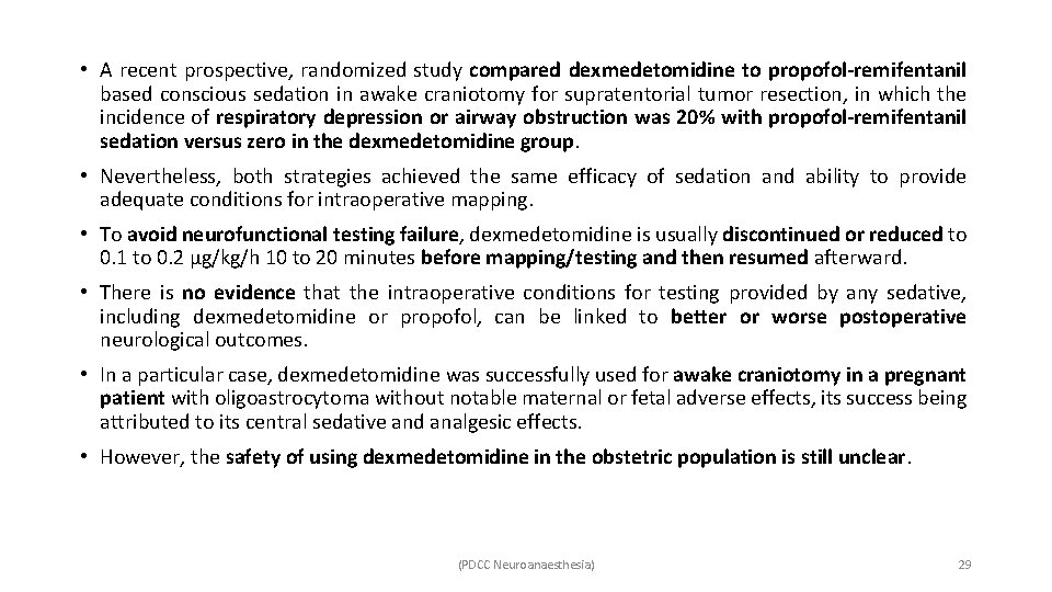
• A recent prospective, randomized study compared dexmedetomidine to propofol-remifentanil based conscious sedation in awake craniotomy for supratentorial tumor resection, in which the incidence of respiratory depression or airway obstruction was 20% with propofol-remifentanil sedation versus zero in the dexmedetomidine group. • Nevertheless, both strategies achieved the same efficacy of sedation and ability to provide adequate conditions for intraoperative mapping. • To avoid neurofunctional testing failure, dexmedetomidine is usually discontinued or reduced to 0. 1 to 0. 2 μg/kg/h 10 to 20 minutes before mapping/testing and then resumed afterward. • There is no evidence that the intraoperative conditions for testing provided by any sedative, including dexmedetomidine or propofol, can be linked to better or worse postoperative neurological outcomes. • In a particular case, dexmedetomidine was successfully used for awake craniotomy in a pregnant patient with oligoastrocytoma without notable maternal or fetal adverse effects, its success being attributed to its central sedative and analgesic effects. • However, the safety of using dexmedetomidine in the obstetric population is still unclear. (PDCC Neuroanaesthesia) 29
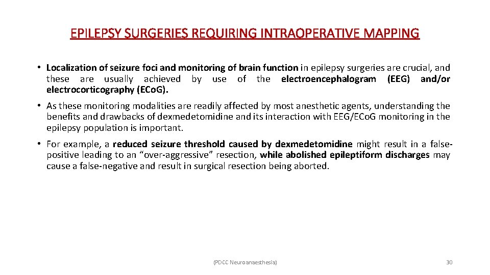
EPILEPSY SURGERIES REQUIRING INTRAOPERATIVE MAPPING • Localization of seizure foci and monitoring of brain function in epilepsy surgeries are crucial, and these are usually achieved by use of the electroencephalogram (EEG) and/or electrocorticography (ECo. G). • As these monitoring modalities are readily affected by most anesthetic agents, understanding the benefits and drawbacks of dexmedetomidine and its interaction with EEG/ECo. G monitoring in the epilepsy population is important. • For example, a reduced seizure threshold caused by dexmedetomidine might result in a falsepositive leading to an “over-aggressive” resection, while abolished epileptiform discharges may cause a false-negative and result in surgical resection being aborted. (PDCC Neuroanaesthesia) 30
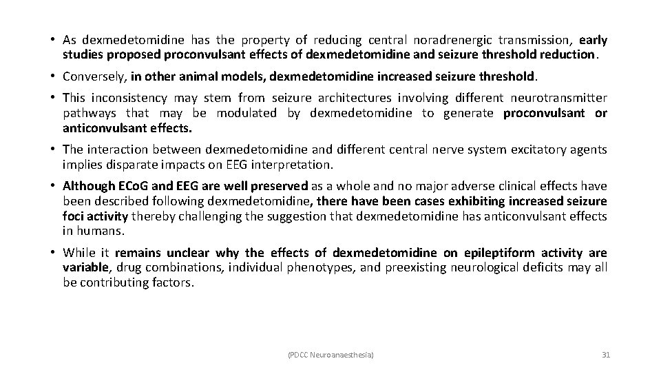
• As dexmedetomidine has the property of reducing central noradrenergic transmission, early studies proposed proconvulsant effects of dexmedetomidine and seizure threshold reduction. • Conversely, in other animal models, dexmedetomidine increased seizure threshold. • This inconsistency may stem from seizure architectures involving different neurotransmitter pathways that may be modulated by dexmedetomidine to generate proconvulsant or anticonvulsant effects. • The interaction between dexmedetomidine and different central nerve system excitatory agents implies disparate impacts on EEG interpretation. • Although ECo. G and EEG are well preserved as a whole and no major adverse clinical effects have been described following dexmedetomidine, there have been cases exhibiting increased seizure foci activity thereby challenging the suggestion that dexmedetomidine has anticonvulsant effects in humans. • While it remains unclear why the effects of dexmedetomidine on epileptiform activity are variable, drug combinations, individual phenotypes, and preexisting neurological deficits may all be contributing factors. (PDCC Neuroanaesthesia) 31
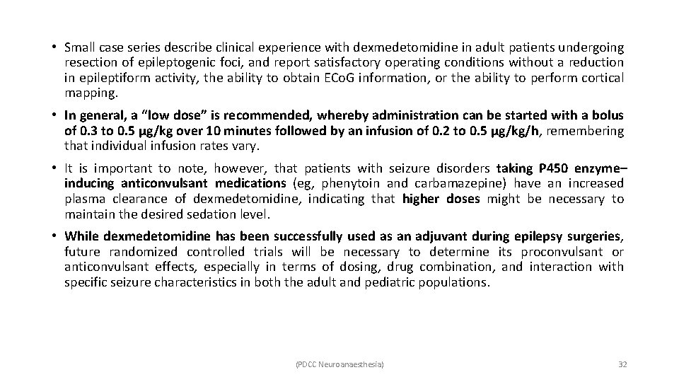
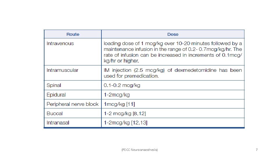
• Small case series describe clinical experience with dexmedetomidine in adult patients undergoing resection of epileptogenic foci, and report satisfactory operating conditions without a reduction in epileptiform activity, the ability to obtain ECo. G information, or the ability to perform cortical mapping. • In general, a “low dose” is recommended, whereby administration can be started with a bolus of 0. 3 to 0. 5 μg/kg over 10 minutes followed by an infusion of 0. 2 to 0. 5 μg/kg/h, remembering that individual infusion rates vary. • It is important to note, however, that patients with seizure disorders taking P 450 enzyme– inducing anticonvulsant medications (eg, phenytoin and carbamazepine) have an increased plasma clearance of dexmedetomidine, indicating that higher doses might be necessary to maintain the desired sedation level. • While dexmedetomidine has been successfully used as an adjuvant during epilepsy surgeries, future randomized controlled trials will be necessary to determine its proconvulsant or anticonvulsant effects, especially in terms of dosing, drug combination, and interaction with specific seizure characteristics in both the adult and pediatric populations. (PDCC Neuroanaesthesia) 32
STEREOTACTIC NEUROSURGERY/DEEP BRAIN STIMULATION • Stereotactic neurosurgeries such as deep brain stimulator (DBS) placement are treatments for patients with neurological movement disorders, for example, Parkinson disease (PD), Tourette Syndrome, or Obsessive-Compulsive Disorder. • The subthalamic nucleus, globus pallidus interna, and ventralis intermedius nucleus of the thalamus are the 3 common target areas. • The anesthetic considerations include an awake and cooperative patient, stable hemodynamics, maintenance of ventilation, airway reflexes and patency due to limited airway access because of a head frame, as well as minimizing anesthetic interference with microelectrode recordings (MER) and macrostimulation. • Propofol is commonly used as the sole sedative during DBS placement under MAC, and these cases may include scalp blocks and/or local anesthesia with a background infusion of short acting sedatives and narcotics (eg, remifentanil) during the procedure. • Propofol can decrease subthalamic nucleus activity and interfere with MER, although this interference can be terminated rapidly by stopping its administration. • This suppression is mediated by GABA inhibitory pathways in the subthalamic nucleus and globus pallidus that contain abundant GABAergic innervations. (PDCC Neuroanaesthesia) 33
• To avoid this, dexmedetomidine has been suggested as an alternative sedative agent. • It has been reported that dexmedetomidine (1. 5 μg/kg bolus over 20 min followed by 0. 2 to 0. 5 μg/kg/h infusion) effectively controlled propofol-induced dyskinesia during bilateral subthalamic nucleus DBS placement without adverse effect on MER. • Dexmedetomidine has been shown to possess other advantages in DBS surgeries – ü Easily arousable sedation ü Good patient cooperation ü No respiratory depression ü Anxiolytic ü Inhibit agitation and stress in prolonged surgery. ü Maintenance of hemodynamic stability. • A 3 -year retrospective study compared DBS surgeries performed with 0. 3 to 0. 8 μg/kg/h dexmedetomidine infusion titrating to Observer Assessment of Alertness and Sedation scale 4 versus patients without sedation, and found that dexmedetomidine not only protected electrophysiological mapping but also provided better hemodynamic stability. (PDCC Neuroanaesthesia) 34
• Lower infusion rates of dexmedetomidine are recommended for DBS surgery. • High-dose ( >0. 8 μg/kg/h) infusion should be avoided for several reasons – ü Oversedation (bispectral index< 80) may abolish MER by suppressing neuronal firing, decreasing background electrical activity and spike amplitudes, and thus delaying the commencement of MER or causing failure to guide electrode placement. ü Risk of respiratory suppression, especially in patients with obstructive sleep apnea. ü Clinically significant bradycardia. • Anxiety and failure to cooperate are more frequent in pediatric awake stereotactic neurosurgeries, thus hampering the procedure and increasing the risk of intracerebral bleeding. • A combination of dexmedetomidine and propofol infusion in pediatric DBS placement provided a safe, efficacious, and well-tolerated sedation with minimal respiratory depression. • The combination of three non-GABAergic agents (dexmedetomidine, ketamine, and opioids) in pediatric DBS surgery might be an optional anesthesia strategy to preserve MERs (PDCC Neuroanaesthesia) 35

SUMMARY (PDCC Neuroanaesthesia) 36
• The prevailing thought is that dexmedetomidine may be associated with – ü Less cerebral hemodynamic perturbation, ü Minimal neurophysiological monitoring interference, ü Greater ability to achieve cooperative sedation without respiratory compromise in awake procedures, and ü Acceptable pain control (alone or in combination with more potent analgesics). • Low-range to mid-range infusion doses of dexmedetomidine seem to exert the above benefits in perioperative neurosurgical care based on some current clinical evidence, although very notably there is yet no standard dexmedetomidine administration strategy for these indications. • Dexmedetomidine may result in severe bradycardia and hypotension and hence, should be used very cautiously in patients. • It is not a potent analgesic in its own right, but rather should be considered as an adjunct to a “true” analgesic, such as an opioid. remain. • It is unclear whether it is proconvulsant or anticonvulsant, or how it affects EPs in different cerebral pathologies, and there is a lack of clinical evidence regarding neuronal protection or toxicity following neuronal injury in both children and adults. • Further disease-based translational studies are required to understand both short-term and longterm neurological and neurocognitive outcomes after dexmedetomidine administration. (PDCC Neuroanaesthesia) 37
THANK YOU (PDCC Neuroanaesthesia) 38
- Slides: 38