Preparation of Smears and Simple Staining Lab 3
")
Preparation of Smears and Simple Staining: Lab ( 3 )

Introduction § Microbial Staining – giving color to microbes. • Because microbes are colorless and highly transparent structures. § • Staining – process in which microbes are stained.

Introduction -Stains § Stains/dyes - organic compounds which carries either positive charges or negative charges or both.
 Benzene Ring: As organic colorless")
* Stain: It is an organic compound containing: 1) Benzene Ring: As organic colorless solvent. 2) Chromophore: Chemical group that imparts color to benzene. 3) Auxchrome: Chemical group that conveys the property of ionization to the chromogen fibers or tissues.

* How Stains Work. The process of staining involve ion exchange reactions between the stain and the active sites at the surface or within the cell.

* Types Of Stains: - Simple Stain: Use only one stain and used to study the cell morphology, size and arrangements. - Negative Stain: That stain the background leaving the bacteria unstained. - Gram-stain: Use three stains and it used to differentiate between Gram positive and Gram negative bacteria. - Endospores Stain: Use two stains and used to study if the bacteria is spore forming or not. - Flagella Stain: Used to study if the bacteria have flagella or not.

*Classification of stains based on chemical behavior: - Acid Dye: The charge of this dye ions is negative, it stain the basic cell components. Eg: India Ink. - Basic Dye: The charge of this dye ions is positive, it stain the acidic cell components. Eg: Crystal Violet, Methylene Blue. - Neutral Dye: Complex salt of acid dye and base dye. Eg: Eosinate of Methylene Blue.
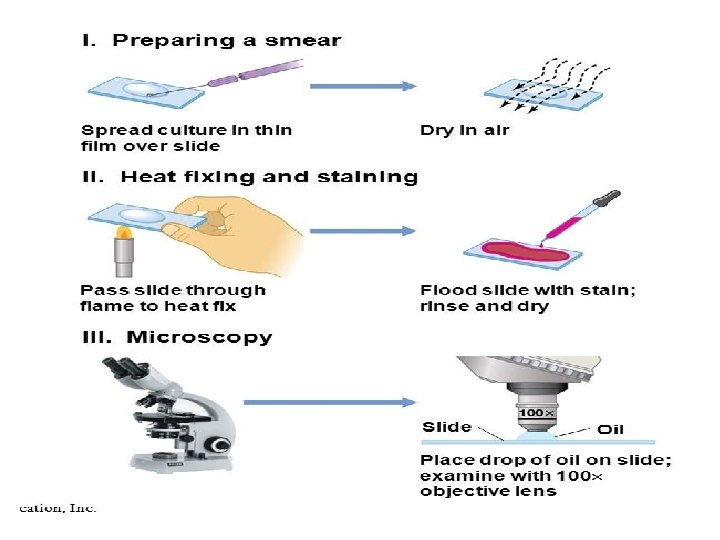
Preparation of Smears and Fixation 2 ) Staining")
Procedure of simple stain 1)Preparation of Smears and Fixation 2 ) Staining

1 - Fixation can be done by: 1 - Heat Fixation: By passing the slide through a Bunsen burner flame, but it may not kill all bacteria. 2 - Chemical Fixation: By embedding the slide with methyl alcohol 95% (Methanol) for 1 minute.
")
Differential Staining Lab ( 4 )

*Differential stain: It is used to describe staining processes which use more than one chemical stain. *Using multiple stains can better differentiate between different microorganisms or structures or cellular components of a single organism.

Gram Stain

*Gram Stain can be defined as: It is an empirical method of differentiating bacterial species into two large groups: a) Gram-positive b) Gram-negative based on the chemical and physical properties of their cell wall.
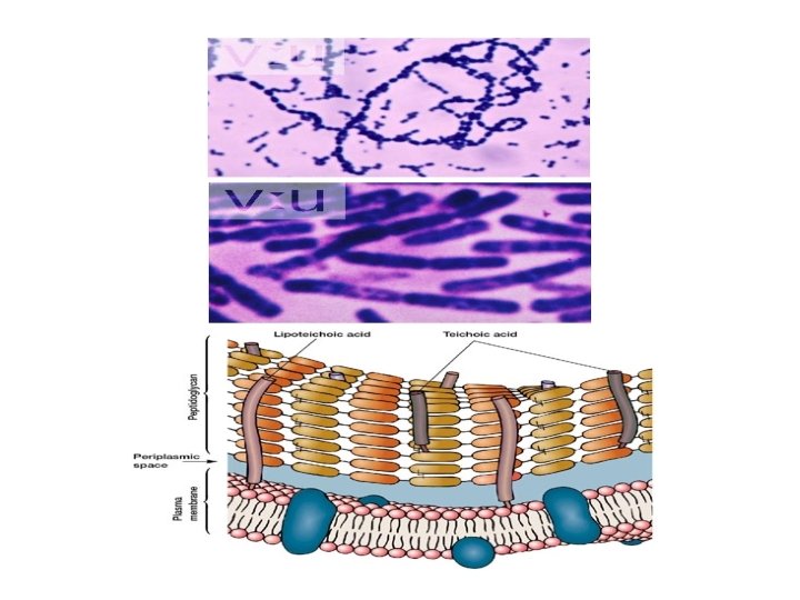
 Gram-positive bacteria: It have more peptidoglycan layers in their cell walls than Gram-negative")
a) Gram-positive bacteria: It have more peptidoglycan layers in their cell walls than Gram-negative and so are able to retain a Crystal violet dye during the Gram stain process. Gram-positive bacteria appear blue or violet under a microscope, whereas Gram-negative bacteria appear red or pink.
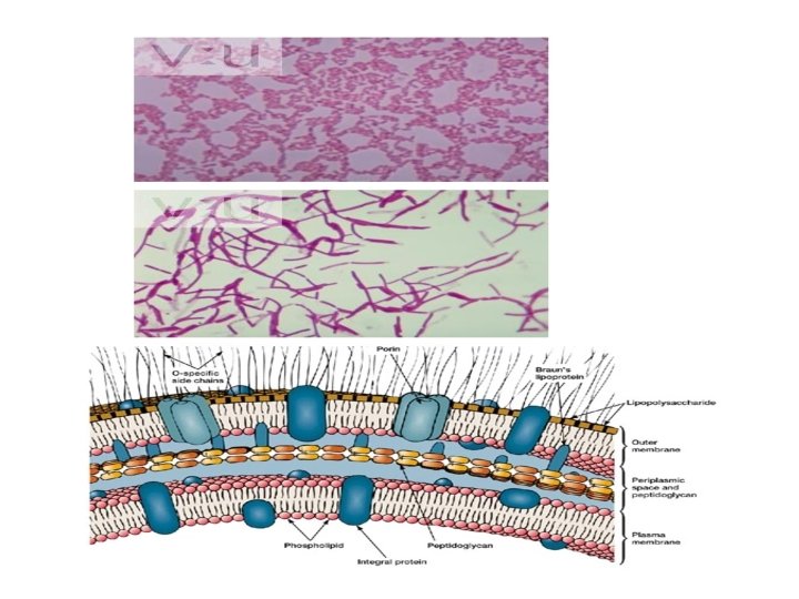
 Gram-negative bacteria: They are bacteria that do not retain Crystal violet dye in")
b) Gram-negative bacteria: They are bacteria that do not retain Crystal violet dye in the Gram staining protocol. Gram-positive bacteria will retain the crystal violet dye when washed in a decolorizing solution. In a Gram stain test, a counter stain (commonly safranine) is added after the crystal violet, coloring all Gram-negative bacteria a red or pink color. The test itself is useful in classifying two distinct types of bacteria based on structural differences in their cell walls.

Comparison Between Gram-Positive and Gram. Negative Bacterial Cell Wall.

Relative Differences Characteristics Gram-positive Gram-negative 1 - Cell Wall Composition 1 - Low in lipids (1 -4%). 2 - Peptidoglycan form (90%) 1 - High in lipids (11 -22%). 2 - Peptidoglycan form (5 -20%) 2 - Susceptibility to Penicillin More Susceptible Less Susceptible 3 - Inhibition by Basic Dye Marked Inhibition Less Inhibition 4 - Nutritional Requirements Many Species Relatively Complex Relatively Simple 5 - Resistance to More Resistant Physical Disruption Less Resistant

.")
*The Gram Stain requires four different solutions: 1 - The basic dye (Crystal Violet). 2 - Mordant solution (Iodine), it is a substance to increase the affinity or interaction between the cell and the dye. So the cell is more difficult to wash out the stain. 3 - Decolorizing agent (Ethanol). For removing the dye from stained cells. 4 - Counter stain (Safranin). To replace the basic dye if it is removed by decolorizing agent.

* The action of Gram stain on both G+ and G- bacteria Reaction and appearance of bacteria Solution Gram-positive Gram-negative 1 - Basic dye, Crystal Violet (CV). Cells stain violet 2 - Mordant solution, Iodine (I). CV-I complex formed within cells; cells remain violet 3 - Decolorizing agent, Alcohol 1 - Cell walls dehydrated, 2 - Shrinkage of pores occurs, 3 - Permeability decreases. 4 - CV-I complex cannot pass out of cells. So cells remain violet 1 - Lipid extracted from the cell walls. 2 - Porosity increases. 3 - CV-I complex is removed from cell 4 - Counter Stain, Safranin Cells not affected and remain violet Cells take up this stain and become red

Procedure & Principle of Gram Stain Prepare a smear, air dry ↓flame 3 times to fix Crystal violet, 1 min ↓wash Gram’s iodine, 1 min ↓wash Ethanol 95%, 0. 5 min ↓ wash Safranin, 1 min ↓ Primary stain Mordant Stain Decolonization Counterstain wash Dry slide with bibulous paper ↓ Observation (10× lens → 40× lens → oil-immersion lens )

Staphylococcus aureus

Escherichia coli

Bacillus subtilis
- Slides: 27