Prenatal and Perinatal History LOG 2 Learning objectives

Prenatal and Perinatal History LOG #2

Learning objectives for LOG 2 After completing this LOG, you should be able to 1. Describe the factors that lead to classification of a high-risk mother/delivery 2. Describe various methods of fetal assessment and monitoring 3. Describe the process of labour and delivery Prior to any delivery, it is imperative to identify those mothers and fetuses which are at a higher risk. By doing this, you can then be prepared for any treatment modality required. This module will guide students to explore the delivery process, methods of assessing the mother and fetus, as well as the potential complications of being ‘high-risk’.

High-Risk Pregnancy

High-Risk Pregnancy Prenatal history 1. Socioeconomic and demographic factors • Mother’s age • Income • Nutritional status

2. Obstetrical History Previous pregnancies – Anything pertinent to previous pregnancies • Miscarriages • Multiple births • Fetal/neonatal mortality • Dystocia
 Normal endogenous production of insulin increases")
3. Maternal Medical History Cardiac/pulmonary disease Diabetes (Pregestational) Normal endogenous production of insulin increases by 30% Associated with n Prematurity n Congenital anomalies n Stillbirth n Mild diabetes Macrosomic (large) infants and delayed lung maturation
 Increase risks")
Maternal Medical History n n Hypertension Infection (E. g. Rubella, ‘German Measles’) Increase risks of n Miscarriage n Congenital heart defects n Stillbirth n Cataracts n Prematurity n Mental retardation n IUGR n Deafness

Infection T – Toxoplasmosis O – Other R – Rubella C – Cytomegalovirus H – Herpes virus

4. Current Obstetric Status • • Absence of prenatal care Anomalies of the reproductive tract

5. Placental Problems n Increases incidence of premature labour and early separation from the placenta n Early separation reduces fetal/maternal gas exchange and may result in fetal asphyxia


Placenta Previa n Abnormal implantation of the placenta in the lower wall of the uterus n Can be diagnosed by ultrasound n Obstruction to fetal passage

Abruptio Placenta n Premature detachment of the placenta

Prolapse of the Umbilical cord n Premature expulsion of the cord before the infant is delivered

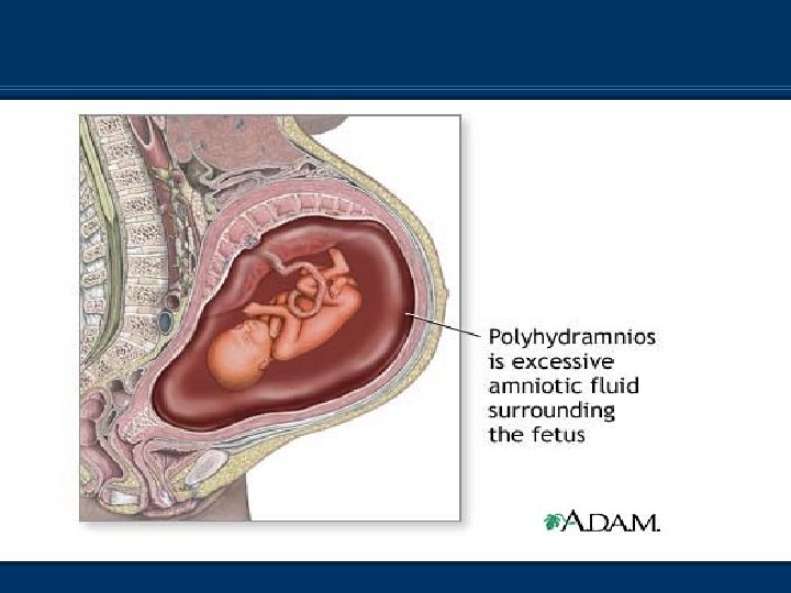
6. Amniotic Fluid Imbalance n Polyhydramnious > 2 litres of amniotic fluid, associated with impaired fetal swallowing (Caused by fetal malformations) n Associated with n Premature labour n Placental abruptio n Perinatal mortality n PROM – Overdistends uterus and increases chance of cord prolapse n

Treatment n Multiple amniocentesis n Prostaglandin synthesis inhibitors (Decrease amniotic fluid and act as a diuretic)

Amniotic Fluid Balance n Oligohydramnious n n n Deficiency in the amount of AF <300 mls Associated with -PROM -Post-term -Uteroplacental insufficiency -Use of prostoglandin inhibitors n Potential consequences n n n -Fetal deformities -Umbilical cord compression -Meconium passage -Fetal demise -Early in pregnancy, can cause underdeveloped lungs and limb deformities Treatment – Amnioinfusion
 n n n BP ≥ 140 mm. Hg or")
7. Pregnancy Induced Hypertension (PIH) n n n BP ≥ 140 mm. Hg or ≥ 90 mm. Hg after 20 wks’ gestation OR Rise of 30 mm. Hg systolic or 15 diastolic from prepregnant or 2 nd trimester measurements of BP

Pregnancy Induced Hypertension n Pre-Eclampsia – Hypertension plus proteinuria, edema, or both Severe Pre-Eclampsia – BP ≥ 160/110 plus proteinuria and edema Eclampsia – Hypertension plus seizures or coma related to underlying neurologic conditions
 n n If rupture occurs more than 24")
8. Premature Rupture of Membranes (PROM) n n If rupture occurs more than 24 hours prior to delivery More common with prematurity ↑ risk of fetal infection Decreases amniotic fluid and increases compression on cord

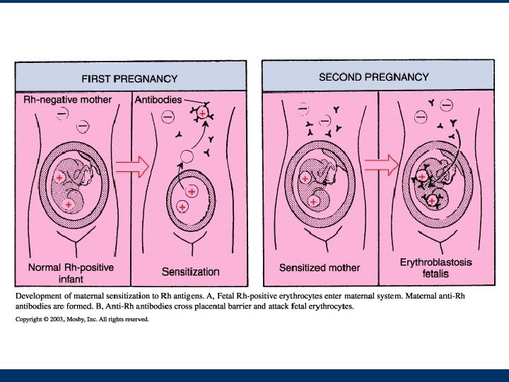
9. RH Isoimmunization or Erythroblastosis Fetalis n n A type of hemolytic anemia; results form fetal-maternal blood group incompatibility Rh factor when mother’s Rh is negative and infant is Rh positive Isoimmunization occurs after the first pregnancy May result in jaundice and severe anemia

10. Habits n n n Smoking Alcohol intake Drug use and abuse

11. Multiple Gestation n Twins/triplets IUGR Congenital anomalies

12. Postmaturity n n n n Placental function declines Greater than 42 weeks’ gestation Can cause maternal and fetal issues SGA, signs of wasting (placenta not able to supply nutrients, fetus can lose weight) Intra-uterine asphyxia, ? death Presentation n Dry, cracked skin; long nails; excessive hair n Meconium staining May require induction or caesarean section

13. Uteroplacental Insufficiency May result from PIH n May occur with post maturity n Inadequate blood flow to the placenta n Should be suspected with the 3 rd trimester – bleeding or oligohydromnios n May cause IUGR, chronic intrauterine asphyxia, meconium aspiration n Assessment of placental function a. Fetal Heart Rate (FHR) monitoring b. L/S ratio (Sample by amniocentesis) *Urinary estriol level are not done anymore n
 n n n Screened for at 24 - 28")
14. Gestational Diabetes Mellitus (GDM) n n n Screened for at 24 - 28 weeks’ gestation with glucose challenge test Abnormal glucose tolerance that is first recognized during pregnancy (3%) Poor blood sugar control will lead to macrosomia (Birth weight > 4000 g) With proper maternal glycemic control, pregnancies can proceed to full term and have normal deliveries

.")
Macrosomic infant. Excess of fat and organomegaly (liver and heart).
 n Highest infant mortality rate")
Consequences of Increased Fetal Risk Prematurity (<37 weeks’ gestation) n Highest infant mortality rate n Premature respiratory and digestive systems n Problems with thermoregulation and defense system n Poor tissue perfusion, incidence of intraventricular hemorrhage (brain) n Most common disorders § a. HMD (Hyaline Membrane Disease) § b. Asphyxia § c. Infection

Asphyxia n Impaired maternal blood flow to the placenta n Hypoxia, hypercarbia, and both resp. and met. acidosis IUGR n Diminished blood flow n Chronically hypoxic in-utero n Mec aspiration n Asphyxia n Mortality is higher in low birth wt vs. IUGR

Fetal Assessment

1. Ultrasound n n Uses sound wave to produce still or moving images (real -time) Rapid sequence of images, impression of movement Over abdomen or transvaginally with a transducer Can be used early on in pregnancy
")
Ultrasound Uses n n n n n Validates pregnancy (in the 1 st trimester) if used very early on Assesses fetal growth (in the 3 rd trimester) by the measurement of organs (Week 12 crown rump) – Gold standard to assess fetal age/growth Assess multiple gestation Assess fetal position Localize the placenta Assess congenital anomalies Amniotic fluid levels Fetal death Check for heart rate and respiratory effort Also to detect for incomplete miscarriage or ectopic pregnancy

3. Amniocentesis n n n Done in conjunction with ultrasound Performed ~18 weeks gestation Uses § § Lung maturity L/S ratio Genetic disorders alphafetoprotein Rh – Incompatibility bilirubin Renal maturity creatinine

4. Chorionic Villus Sampling • • • Performed at ~ 12 weeks’ gestation Alternative to amnio Obtain placental tissue for prenatal assessment (Sample of chorionic villus) With ultrasound, a catheter is inserted through the cervix and directed to the placenta Tissue aspirated Uses Chromasomal defects (More accurate than amnio) Risky – So early in gestation, can affect placental function Cannot detect neural tube defects
")
Fetal monitoring • • During labour and delivery or intrapartum Fetal Heart Monitoring (FHR) Before the rupture of membranes External monitor is used After the rupture of membranes Direct (usually the scalp) monitoring Heart rate and uterine contractions’ intensity, timing, and duration

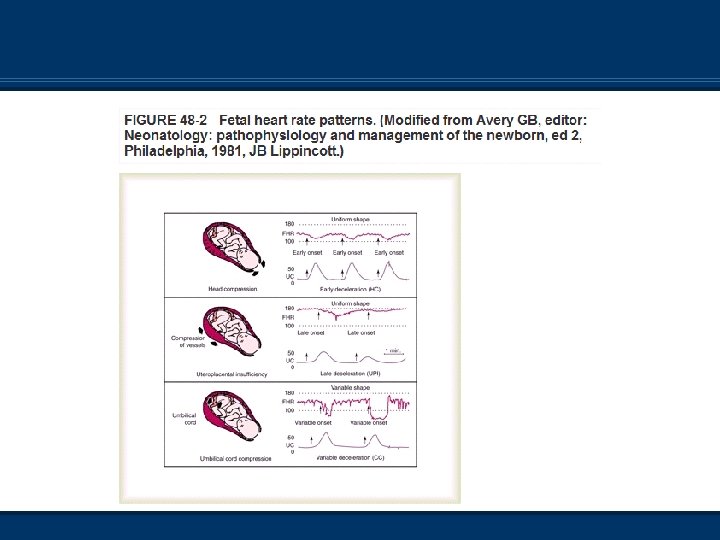
Establish a baseline • Normal is 120 - 160 bpm early in gestation 140 bpm • or 20 - 30 bpm • Bradycardia < 100 bpm most dangerous, cause asphyxia check scalp PH • Tachycardia > 180 bpm • Accelerations >160 bpm meds (sympathomemetics) and decelerations <120 bpm are measured • Three significant decelerations are 1. Early Type I 2. Late Type II 3. Variable Type III
 • FHR ’s (60 - 80) At the same time,")
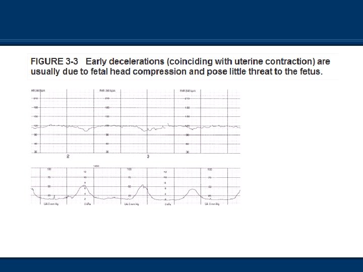
Early decelerations (type I) • FHR ’s (60 - 80) At the same time, uterine contractions occur and return to normal (120 - 160 beats/minute) caused by the compression of the fetal head against the cervix • Normal
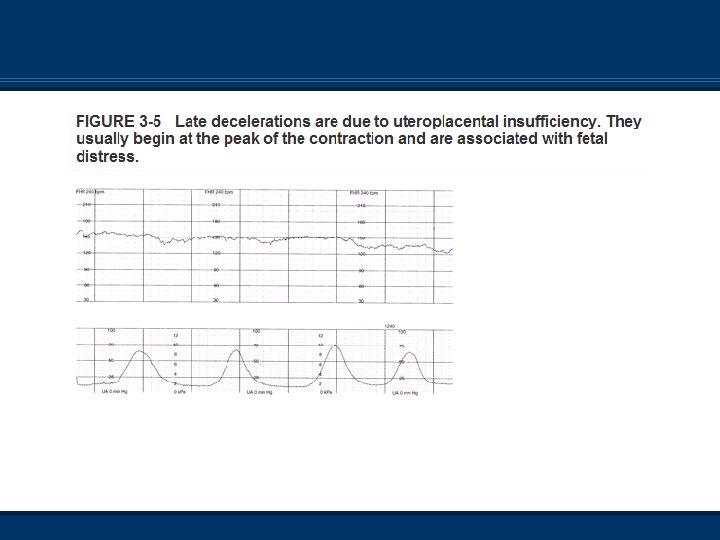
 • FHR ’s after the onset of a contraction and")
Late deceleration (type II) • FHR ’s after the onset of a contraction and returns to normal after the contraction has ended • Impaired maternal blood flow to the placenta • • UPI associated with fetal asphyxia and low Apgar scores • O 2 may help; attention to maternal hemodynamic status
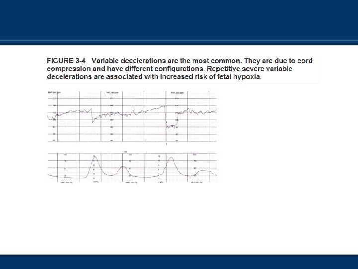
 • No relationship between FHR and contractions • Most common")
Variable decelerations (type III) • No relationship between FHR and contractions • Most common deceleration pattern caused by compression of the cord by fetal parts • Most fetuses can tolerate short periods of cord compression, longer = asphyxia
 • Used")
Fetal scalp monitoring • Analysis of fetal capillary blood (Usually the scalp) • Used to determine fetal well-being • p. H=7. 35 beginning of labour • p. H=7. 25 second stage of labour normal • p. H= 7. 2 -7. 24 slight asphyxia • p. H<7. 20= fetal distress ++ severe asphyxia
 2. If")
Two types of situations 1. Interference of placental blood flow (respiratory acidosis) 2. If interference is prolonged (lactic acidosis) *Fetal scalp blood sampling reserved for cases when the fetus has demonstrated distress, late decelerations, or is known to be at high risk for asphyxia

Labour and Delivery

Five Events 1. Membranes rupture 2. Dilatation of the cervix 3. Contraction of the uterus 4. Placental separation 5. Shrinking of the uterus

L&D Introduction n Labour occurs as a result of placental and fetal hormones n Progesterone decreases n Estrogen increases n Oxytocin stimulates uterine contractions n Relaxin ↑’s the flexibility of the pubic symphysis

True Labour n Regular intervals, produces pain n Localized back pain that intensifies with walking n Reliable indicator is dilatation of the cervix and the “show” (blood containing mucous)

False Labour or Braxton Hicks n Pain is felt in abdomen and is irregular n Does not intensify with walking; no show and no dilatation

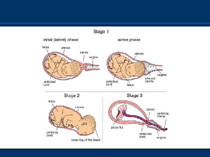
The Three Stages of Labour

Stage One – Dilatation n Onset of labour to complete dilation of the cervix n Thinning of cervix (effacement) % n Contractions 10 - 15 mins. apart, lasting 30 - 90 secs. n Amniotic sac ruptures n Regular contractions n 16 - 18 hrs. (primagravida) 7 - 12(multi)

Stage Two – Expulsion n 95% of all births are vertex n Aided by contraction of abdominal wall and diaphragm n Complete dilatation to delivery n Location of the head=stations or engagement n 20 minutes to 2 hours

Stage Three – Placental Delivery n Delivery of the placenta, expelled by uterine contractions n These contractions also constrict blood vessels, decreasing risk of hemorrhage n 5 to 45 minutes

Dystocia n Painful or difficult birth or difficult labour n Present when 1 st and 2 nd stages exceed 20 hrs Causes 1. Uterine dysfunction (abnormal contractions) 2. Excessive fetal size (hydrocephalus, macrosomia) 3. Any abnormality in shape or size of the birth canal 4. Abnormal fetal presentation (breech)
 • Complete breech (Both")
Abnormal Fetal Presentations Breech delivery • Frank breech (Buttocks first) • Complete breech (Both buttocks and lower extremities) • Footling breech (Lower extremities first) Breech deliveries = ↑ risk of trauma and asphyxia

Labour and Delivery Medication n 80 to 95% of births in the United States n Analgesics are pain-relieving drugs that help the mother relax n Anaesthetics are stronger painkillers that block sensation

Instrument Delivery Forceps

Vacuum Extraction

Induced Labour n Labour is started artificially by breaking the amnion and giving the mother a hormone that stimulates contractions n Used when continuing the pregnancy threatens the well- being of mother or baby n Contractions often longer, harder, and closer together
 Pharmacological prolongation of pregnancy Likelihood of neonatal survival/morbidity needs to be")
Tocolysis (Tocolytic therapy) Pharmacological prolongation of pregnancy Likelihood of neonatal survival/morbidity needs to be assessed before aggressive attempts to prolong pregnancy are initiated Indicated 1. Labour begins before 37 weeks, but it is generally difficult to justify risks to mom/fetus greater than 34 weeks 2. Placenta previa Preterm birth is leading cause of perinatal mortality

Factors that should be present before starting therapy 1. True labour 2. < 4 cm cervical dilation and < 50% effacement 3. Fetus 20 - 36 week gestation 4. No signs of fetal distress 5. No medical/obstetrical disorders that would contraindicate prolongation of pregnancy 6. Informed consent
 drugs used to relax smooth muscle contractions 2.")
Agents used 1. Beta sympathomimetic (adrenergic) drugs used to relax smooth muscle contractions 2. Anticonvulsant that stop uterine contractions (magnesium sulfate) 3. Prostoglandin synthesase (indomethacin) – Used to stop prostoglandin-induced labour 4. Calcium channel blockers These agents all have potential side effects that could be harmful to both mother and fetus
- Slides: 69