Preliminary Results using Shilla guided growing rods with


- Slides: 13
Preliminary Results using Shilla guided growing rods with sublaminar fixation in Early Onset Scoliosis Samuel R. Rosenfeld, M. D. Benjamin T. Smith, D. O. The Shilla technique utilizes apical deformity arthrodesis combined with guided cephalad and caudad growth in open pedicle screws. This is a one year review of two patients with severe early onset scoliosis treated with the Shilla technique incorporating sublamina fixation for guided growth. Pedicle screw fixation is used for stabilization / arthrodesis of the apical deformity. Cephalad and caudad fixation utilizes sublamina Songer cables at each level. The 5. 5 mm. stainless steel rods extend beyond the sublamina fixation for guided growth.
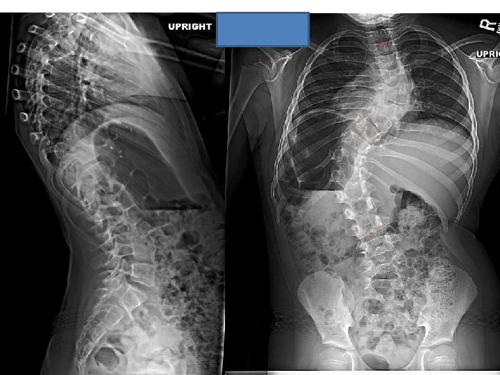
Case # 1 • M. S. is a 7 year 8 month old female with Prader-Willi syndrome. She had progressive scoliosis diagnosed at 1 year of age. She underwent MRI of the entire spinal cord demonstrating no abnormalities. She had no congenital deformities with normal spinal cord. Her neurological examination was significant for diffuse low tone with normal abdominal reflexes and absent Babinski. She began independent ambulation at 21 months of age. M. S. was treated with full time TLSO from 1 year of age until 5 years of age, then bending spinal orthosis. She had progressive scoliosis T 3 to T 9 R 61 degrees, T 9 to L 3 L 77 degrees with trunk decompensation.
• The surgical procedure consisted of posterior spinal arthrodesis from T 9 to T 12 with segmental spinal instrumentation from T 2 to L 3 in a guided growth construct. • One year post-op, the growth from T 1 to T 12 was 1. 4 cm. , and the growth from T 1 to S 1 was 3. 0 cm. based on Surgimap spine software (Nemeric, Inc. ).
Post-op
One year post-op
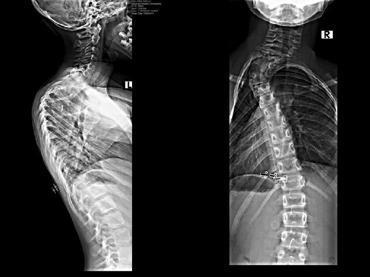
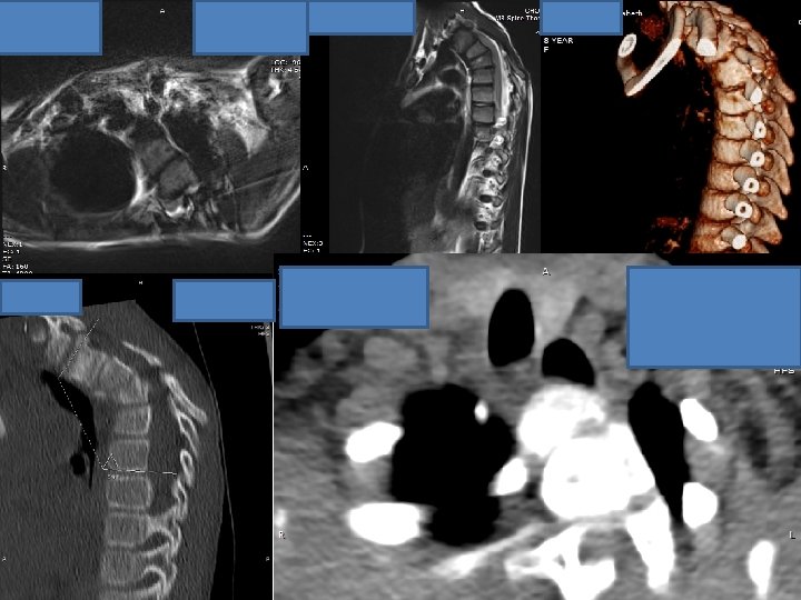
Case # 2 G. S. is a 8 year 10 month old female with a history of neuroblastoma left side of chest diagnosed in 2007. She completed chemotherapy followed by resection of the tumor in 2007. She developed progressive cervicothoracic kyphotic deformity. She was complaining of esophageal pressure with nausea. She had no neurological abnormalities, except for dysesthesias left axilla and left thorax. X rays, MRI, and computed tomography follow:
• The surgical procedure consisted of vertebral column resection with kyphectomy at T 4, posterior spinal arthrodesis from T 1 to T 6, and segmental spinal instrumentation from C 5 to T 10 in a guided growth construct. • One year post-op the growth from T 1 to T 12 was 0. 9 cm. , and the growth from T 1 to S 1 was 2. 6 cm. based on Surgimap spine software (Nemeric, Inc. ).
Post-op
One year post-op
. • The Shilla technique with sublamina fixation will allow guided growth without spontaneous arthrodesis. This technique should avoid implant complications reported with the Shilla procedure. This technique should allow improved deformity correction with fixation at all instrumented levels. • • References Skaggs, D. , Akbarnia, B. A. , Flynn, J. M. , et al. , A Classification of Growth Friendly Spine Implants. Journal of Pediatric Orthopedics. 2014; 34(3): 260 -274. Yang J. S. , Mc. Elroy, M. J. , Akbarnia, B. A. , et al. Growing Rods for Spinal Deformity: Characterizing Consensus and Variation in Current Use. J Pediatr Orthop. 2010; 30(3): 264 -7. Mc. Afee, P. , Lubicky, J, and Werner, F. , The Use of Segmental Spinal Instrumentation to Preserve Longitudinal Spinal Growth: An experimental study. J Bone Joint Surg Am, 1983; 65(7): 935 -42. Rinsky, L. A. , J. G. Gamble, and E. E. Bleck, Segmental Instrumentation without Fusion in Children with Progressive Scoliosis. J Pediatr Orthop, 1985; 5(6): 687 -90 Mc. Carthy, R. E. , Luhman, S. , Lencke, L. G. , The Shilla growth guidance technique for early onset spinal deformities at two year followup: a preliminary report. Spine, 2010 Mc. Carthy, R. E. , et al. , Shilla growing rods in a caprine animal model: a pilot study. Clin. Orthop. 2010; 468(3): 705 -710.