Pregnancy Parturition and Care of the Calf CowCalf


 • Sheath")


— – Not withstand much stretching – Ruptures due")


. •")


— –")


- Slides: 69
Pregnancy, Parturition and Care of the Calf Cow-Calf Operations Chapter 5
Order of Things • Estrus and Service of the Cow – Naturally or by artificial means – Followed by: Ovulation • Must precede: – Fertilization – Pregnancy – Production of a calf
Introduction • Where does fertilization occur? – Upper end of the oviduct. – Just below the infindibulum. • Fertilized egg moves from Fallopian tube to horn of uterus— – In about 3 -4 days. • Increase in size, differentiation into different cells.
Introduction • Zygote lies free in uterine cavity for— – As many as 30 days. • Then becomes implanted on uterine wall – Protects embryo – Transfer nutritive material – Transfer of waste products • Makes possible G&D; now called fetus.
Reproductive Organs of a Cow
Female Tract— Top View
Fetal Membranes • • Three separate structures. 1. Chorion 2. Amnion 3. Allantois
Chorion • Outer membrane • Lies close to mucous membrane of the uterus. • Surface quite large, extends into nonimpregnated horn. • Many blood vessels leading into the placenta.
Amnion • Innermost of the 3; begins at navel and surrounds fetus like a sack. • Contains liquid that protects fetus. – 6 -7 quarts of amniotic fluid. • During birth, lubricates vagina • Break during late stages of birth
Allantois • Large membranous sac between chorion and amnion – Contains fetal urine and other fluids – Fetal bladder (urachus) • All of the above membranes constitute— – Fetal membranes – Afterbirth
The Amniotic Sac
Placenta • Portion of the fetal membrane that unites the mother and fetus. • No direct vascular connection— – Blood vessels close together – Interchange materials – Not everywhere – Specialized locations called caruncles.
Umbilical Cord • Membranous portion that unites— – Fetus and placenta (navel) • Sheath composed of— – 2 umbilical arteries – 2 umbilical veins – Urachus • Arteries carry blood to placenta, veins carry it back. • Blood vessels contract forcefully at birth, when they are ruptured, preventing bleeding.
Umbilical Cord • UC that do not retract when ruptured— – May be avenue for bacterial infections – Navel Ill • Treatment with Iodine at birth – Will prevent.
Fetal Position at Birth
Normal Presentation of the Calf
Position of the Fetus in the Uterus • With growth, anterior end towards cervix— – Head toward the rear. • End of gestation, weight of fetus causes— – To rest on floor of abdomen • There is something that sits on left side-– The rumen stomach • Calf rests on cows right side.
Multiple Pregnancy • Seldom occurs; twins can occasionally • One fetus = in one horn of uterus. • Twins = one in each horn. – Fetal membranes can become fused in these – If of the opposite sex become fused, a common circulatory system, development of female reproductive organs is diminished. – Male hormones are dominant. • Therefore, heifers born twin to a male are— – Freemartins; < 1% are fertile, irreversible.
Signs of Pregnancy • Approximately 283 days average. • Angus shorter by 1 week, Brahman longer by 1 week. – Differences in birth weights? • NO visible signs during 1 st half of gestation. • Are some things that we can notice.
Signs of Pregnancy after 2 nd month • 1. Cessation of estrus or heat • 2. Noticeable enlargement of abdomen and udder. – Especially 1 st calf heifers. • 3. Pregnancy Testing – Palpation of reproductive tract by rectum – As young as 40 days can detect. – After 60 more reliable.
Embryonal Mortality • Every pregnancy doesn’t = live calf. • Infectious diseases account for most losses. • 1973 report said that— – 20. 3 % of 280, 215 cows had failure in birth.
Signs of Parturition • 1. Relaxation of pelvic ligament – Rump muscles drop inward – Sinking of tailhead/pinbones • 2. Enlargement of Vulva – Swollen or inflamed looking • 3. Distension of teats/Enlarged udder – Change in secretion from watery material to thick, milky colostrum
Signs of Parturition • Changes begin 3 -4 weeks before birth • Appear sooner— – In heifers than older cows.
Labor • First real sign of labor is what? – Noticeable uneasiness of animal – Several hours before calving • Cow will leave herd, isolated part • Frequently lies down and gets up— – In short intervals
Preliminary Contractions • These cause the uneasiness— – Are vital to the process – They move the fetus from lateral position on floor of abdomen to— – Longitudinal, upright position in front of the pelvic girdle. • Contractions bring about— – Dilation or enlargement of the cervix. – Cervix is practically obliterated. • Forms one continuous passageway.
Preliminary Contractions • Chorion (outer membrane)— – Not withstand much stretching – Ruptures due to increase in pressure. • Then, allantois and liquid contents— – Forced into vagina to appear at vulva. – See a semi-transparent baloon-like sac – First water bag.
Preliminary Contractions • Now fetus only in amniotic fluid – A lot of pressure on it • Too much pressure before in proper position or before genital passage are enlarged = difficult birth— – Longer water bag intact, the better.
Actual Birth • Rupture of first water bag, amniotic membrane appears with contained fetus— – Usually with the front feet. • With each labor contraction— – More and more of amniotic liquid is forced out to form another sac, the 2 nd water bag • Eventually bursts, (water breaking)— – This lubricates genital passage.
Actual Birth • Rupture of amniotic membrane— – Head and shoulders of fetus come through pelvic canal. • Cow will usually now stand up – Let influence of gravity take control.
Induced Parturition • Synthetic hormones – Used properly, excellent tools • Should not be done more than— – 7 to 10 days before normal gestation end. – Before that, inadequately developed lungs. – High mortality rate. – OXYTOCIN
Rendering Assistance • Do not give unless absolutely necessary. – Some rush in at the sign of a fetus. • Can cause injury to cow and calf in form of– Torn membranes and strained ligaments • If no progress after 2 hours, then aid. • Veterinarian should be called.
Rendering Assistance • On other hand, do not make mistake of allowing first-calf heifer to— – Labor until completely exhausted. • Large head or heavy shoulders— – May delay passage through pelvic cavity. • Posterior presentation— – Hips may cause the trouble.
Rendering Assistance • Traction should NEVER be used except when a cow labors, unless— – She is to the point of refusing to labor. • Mechanical calf-pullers— – Should only be used by experienced person – If not, usually fatal or cause permanent injury to cow or the calf.
Rendering Assistance • If no tension on umbilical cord— – Do not hasten process of birth. • Once head is out to the eyes— – Cord is pressed on floor of cow’s pelvis – Placental circulation is jeopardized. • Now, we can use traction in these types of situations.
Posterior Position of Calf • Delivery may often proceed without complications. • Assistance may be important if prolonged labor. • Calf death may occur due to rupture of navel cord and subsequent suffocation.
Posterior Position of Calf • With rear legs under the body. (breech position). • May correct by pushing forward and grasping legs one at a time. • As leg is drawn into birth canal, keep hock pointed towards the cows flank and hoof to the midline.
Posterior Position of Calf • With fetus in upsidedown position. • Can be caused by twisting of uterus or rotation of calf. • Delivery must NEVER be attempted in this position; professional assistance required.
Anterior Position of Calf • With rear legs extended beneath body. • Dog-sitting posture. • Very serious type of malpresentation. • If allowed to continue, fetal death may occur.
Anterior Position of Calf • With head and necked turned back over the body. • Secure legs with chains. • Push calf back into the body. • This often bring head into normal position.
Guidelines for Assisting in Difficult Deliveries
Correction of Simple Leg Flex • Anterior Presentation • Push calf forward & keep retained foot grasped in hand. • Carry foot outwards and then forward in an arc over pelvic rim. • More difficult—need a snare attached to fetlock to help extend the leg.
Correction of Hock Flex • Posterior position. • Push calf forward. • Hand grasps and cups the foot. • Draw back as hock is flexed.
Correction of Head & Neck Deviation • May require pulling the head and neck around with hand. • Push calf forward quickly and grab muzzle. • Pull head aligned with birth canal. • May use snare on lower jaw.
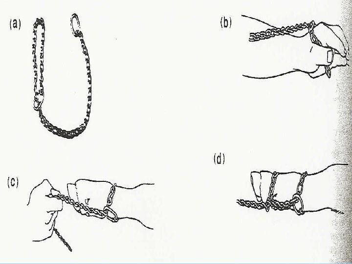
How to pull a calf with chains
Step 1 • Make a loop in the obstetrical chain by passing the chain through the oblong ring at the end of the calving chain.
Step 2 • Slip the loop over the gloved and welllubricated hand in order to allow for easy application and maneuverability in the birth canal or uterus.
Step 3 • Attach loop to one leg of calf and slit it up on the cannon bone 2 -3 inches above the dewclaws. • May need slight tension on the chain so it doesn’t slip off.
• Half-hitch the chain between dewclaw and the hoofhead. This can be made outside the cow on your hand. • Distributes the stress over 2 locations. • DO NOT apply single loop to both legs at same time. Step 4
Step 5 • Repeat steps 1 to 4 for the second leg.
Step 6 • Before applying handles to the chains, make sure that the chains will pull from the bottom sides of the legs (dewclaw side) • Ensures legs pulled straight and not at an angle.
Step 7 • Attach 2 handles to chains and pull GENTLY. • Make sure loop and half-hitch haven’t been pulled down on the leg.
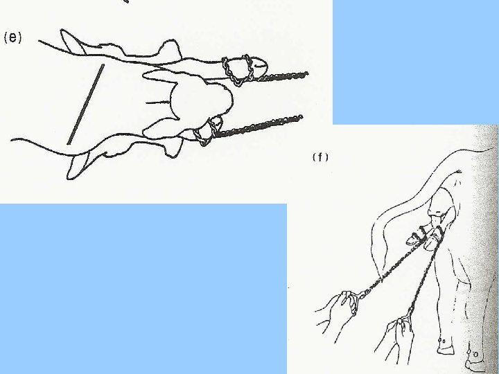
• Best to pull alternately on one leg then the other…. although can be done with pressure on both at same time. • Few inches at a time. • Shoulders and hips allowed through pelvic girdle one at a time. Step 8
Step 9 • Once legs exposed, pull head downward towards the cows hocks at 45° angle. • Head or shoulders can lacerate the uterus or cervix. • Slow traction is best. • Pressure dilates the cervix.
Step 10 • 1 or 2 people with manual strength should be able to pull a calf. • If too difficult, may use mechanical calfpullers. • Best here to seek experienced help. • Been demonstrated that leverage created by these pullers can pull over a ton of dead weight.
Birth by Caesarean Section • Must be done by a veterinarian or— – Almost certain to end in death for both • Large incision in left side just above flank – Go through skin, several membranes, muscle layers, and uterine wall. – Skilled surgeon usually; cow can be left in good condition with little/no infection. • Cow is immobilized by spinal anesthesia or a spinal block.
Care of the New-born Calf • All membranes cleaned from nose – To facilitate breathing. • Amniotic fluid removed from nostrils— – Prevent drowning. • Artificial respiration can be done if need. • Tickling the nostrils with straw or stem can stimulate onset of breathing.
Care of the New-born Calf • Umbilical cord is short (12 -15 inches)— – Always ruptures during birth – About time forequarter passes vulva • Tincture of Iodine or Formalin to the navel stump to destroy bacteria. • ASAP, Cow and calf left alone • Cow gets satisfaction from licking calf to dry it.
Care of the New-born Calf • Additionally, this may stimulate— – Calf’s circulatory system and cow’s milk letdown. • Usually, calf stands to nurse on own – Within few minutes, not necessary – Over 5 -6 hours, now a problem – Needs that colostrum – Protective antibodies
Care of the New-born Calf • Cow doesn’t claim a calf— – Can freeze colostrum, but must be warm. • Important to receive natural sunshine and exercise for health and growth.
Expulsion of the Placenta • After calf is born, outer fetal membranes— – And chorion still attached to uterus wall • At birth, exchange of nutrients— – Completely stops • Attachment between chorion and uterus is loosened by— – Contractions; then uterus returns to normal size – Freed membranes forced out through vulva.
Retention of the Placenta • Normally expelled 2 -6 hours after birth. – If over 24 h, abnormal condition may exist. • Cattle more susceptible than other species – Up to 20% of some cows in herds • Various cases; probability occur from— – Infection and inflammation of maternal cotyledons (where attaches) or from giving assistance during labor.
Retention of the Placenta • Retention is likely to come with failure of the uterine walls to contract promptly— – General weakness from animal; exhaustion • Cows thin or excessively fat— – Troubled by retained placenta more often • Deficiency in Vitamin A or carotene— – Linked to retained placentas
Retention of the Placenta • Also an ideal medium for— – Development of putrefying bacteria. • String-like portion hanging out of vulva— – Hindquarter of animal is – What else is there? – Bacteria and Feces
Dystocia • Applied to cases of prolonged and difficult birth, where death may or may not occur. – Aftereffects are usually on the cow. • Possible causes studied by MARC in Clay Center, Nebraska.
Causes of Dystocia and Effects • 1. Size of the Calf— – Most important; choice of sire breed, plane of nutrition, gestation length. • 2. Cow size— – Independent of cow breed or age, next in importance, small calves can’t birth as easily as larger cows.
Causes of Dystocia and Effects • 3. Cow age— – 2 year olds are only at approximately 75% of mature size; drop calves 90% as heavy as those of mature cows. Checking 1 st and 2 nd calf heifers every 3 -4 hours, round the clock, must. • 4. Cow Breed— – Associated with calf weight, shape, size and shape of cow’s pelvic opening.
End of Chapter 5