Pregnancy Honors Anatomy Physiology Chapter 28 Pregnancy covers


 • secreted by trophoblasts • target:")


 • • • True Labor:")
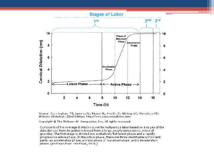


 the cervix to about 3 cm (1 in. ).")


 �last part of active labor: cervix")


- Slides: 68
Pregnancy Honors Anatomy & Physiology Chapter 28
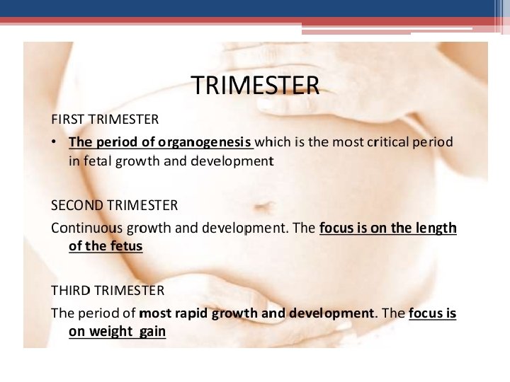
Pregnancy • covers from fertilization birth • period of time for this called gestation • use date of LMP (last menstrual period) to start count ▫ 280 days = full term ▫ fertilization 8 wks = embryonic period (embryo) ▫ 9 wks delivery conceptus called a fetus
Conception • once a month an ovum is released ▫ ovum- A female egg • secondary oocyte moves through the fallopian tube to the uterus ▫ where the baby develops during pregnancy • if not fertilized it disintegrates and is flushed away with menstruation
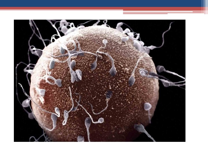
Fertilization • sperm‘s nucleus combines with nucleus of secondary oocyte 1 diploid cell called a zygote
Sperm • millions of sperm ejaculated • most either do not make it up vagina or are killed by the acidic environment • if female ovulatory, some sperm will make it thru endocervical canal (mucus clear, slippery) if not ovulatory: most sperm will be blocked • sperm that reach uterine cavity: ▫ whipped around by contractions of myometrium ▫ many phagocytosed
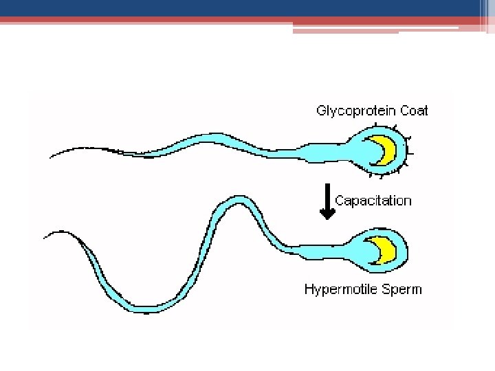
Sperm Capacitation • must be done w/in 2 – 10 hrs or cannot fertilize 1. enhances motility 2. membrane becomes more fragile ▫ membrane proteins removed by vaginal secretions
Acrosome Reaction • lysosomal enzymes released from acrosome • allows sperm head to penetrate zona pellucida & reach the plasma membrane of the secondary oocyte • membranes then fuse • sperm nucleus decondenses forming male pronucleus •
Polyspermy • entry of several sperm into ova • occurs in some animals , not in humans • after 1 sperm has entered Ca++ released from oocyte’s ER cytoplasm result in: 1. Cortical Reaction 2. activates 2 nd meiotic division
Cortical Reaction
Fertilization • secondary oocyte completes meiosis forming female pronucleus which joins with male pronucleus 2 n fertilized ovum (zygote)
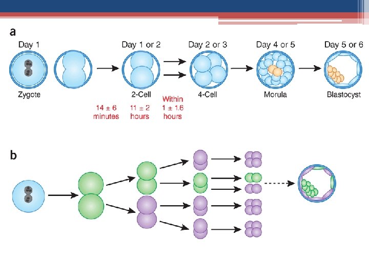
Embryonic Development • immediately after zygote formed chromosomes duplicate & cell prepares for mitosis ▫ 1 st mitotic division ~36 hrs ▫ 2 identical cells called blastomeres • initially mitosis repeated w/out cell growth developing very small cells with high surface-tovolume ratios • ~72 hrs: loose collection of cells forming a berryshaped cluster of 16 or more cells = morula
Morula Blastocyst • mitosis continues as embryo is moving through tube uterus • ~4 -5 days: fluid begins to collect in cavity: now called a blastocyst (2 parts) ▫ 1 layer flattened cells =trophoblast cells ▫ small cluster of 20 – 30 rounded cells = inner cell mass
Blastocyst • inner cell mass becomes the embryonic disc • Trophoblastic cells add adhesion molecules to plasma membrane eventually becomes placenta
Implantation: 4 Weeks • blastocyst initially floats around endometrial cavity 2 - 3 days after entering through tube ▫ nourishment from glycoproteins in uterine secretions • 6 – 7 days after ovulation implantation occurs ▫ E & P have prepared endometrium ▫ trophoblastic cells: adhesion molecules bind to the extracellular parts of the endometrial cells (collagen & others) ▫ https: //www. youtube. com/watch? v=ld. LWUpt 82 h I
Trophoblast • after implantation 2 layers: 1. Cytotrophoblast ▫ inner layer / retain plasma membranes 2. Syncytiotrophoblast ▫ outer layer/ lose membranes invade into endometrium blastocyst surrounded by blood from degraded maternal vessels
http: //www. as. wvu. edu/~s raylman/physiology/cleava ge_implant. html
https: //www. nlm. nih. gov/medli neplus/ency/anatomyvideos/00 0025. htm
h. CG • human chorionic gonadotropin (+preg. test) • secreted by trophoblasts • target: corpus luteum • action: 1. keep secretion of P & E ▫ ▫ so chorion is taking control from pituitary called “rescuing the corpus luteum” 2. protease activity ▫ acts as autocrine growth factor that promotes placental development
5 Weeks • is the start of the "embryonic period” : when all the baby's major systems and structures develop. • embryo's cells multiply and start to take on specific functions = differentiation • blood cells, kidney cells, & nerve cells start to develop. • brain, spinal cord, and heart begin to develop. • GI tract starts to form.
5 Weeks • during this time in the first trimester that the baby is most at risk for damage from things that may cause birth defects. • This includes certain medicines, illegal drug use, heavy alcohol use, infections such as rubella, and other factors.
6 Week Embryo
6 – 7 Weeks • arm and leg buds start to grow. • brain forms into 5 different areas ▫ some cranial nerves are visible. • eyes and ears begin to form • tissue grows that will become spine & other bones. • heart continues to grow and now beats at a regular rhythm.
Development of the Heart http: //www. indiana. ed u/~anat 550/cvanim/
8 Weeks • arms & legs longer • hands & feet begin to form, look like little paddles • brain continues to grow • lungs start to form
1 st Trimester
12 -14 Weeks • eyelids close, will not reopen until about the 28 th week • face is well-formed • limbs long and thin. • nails appear on the fingers & toes • genitals appear • liver is making red blood cells • head is very large -- about half of baby's size. • can now make a fist. • tooth buds appear for baby teeth.
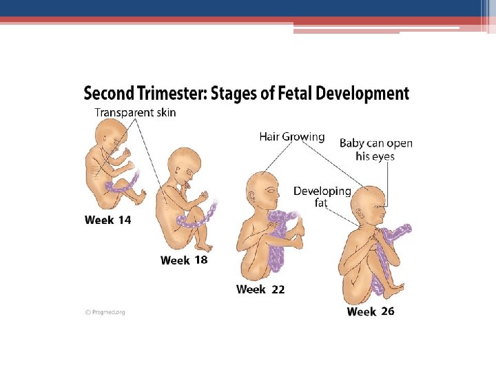
15 – 18 Weeks • skin is almost transparent • fine hair called lanugo develops on head • muscle tissue & bones keep developing, bones become harder • baby begins to move and stretch. • liver and pancreas produce secretions •
Weeks 19 - 21 • baby can hear • baby is more active, continues to move & float around • mother may feel a fluttering in the lower abdomen= quickening (when mom can feel baby's first movements) • by the end of this time, baby can swallow ▫ swallows amniotic fluid
Week 22 • lanugo hair covers entire body • meconium, baby's first bowel movement, is made in the intestinal tract • eyebrows & lashes appear • baby is more active with increased muscle development • mother can feel the baby moving • baby's heartbeat can be heard with stethoscope • nails grow to the end of fingers
Lanuga
Week 23 - 25 • bone marrow begins to make blood cells • lower airways of the baby's lungs begin to develop ▫ baby inhales amniotic fluid • baby begins to store fat
Week 26 • • • eyebrows & eyelashes are well-formed all parts of baby's eyes are developed startle response intact footprints & fingerprints forming air sacs form in baby's lungs ▫ lungs are still not ready to work outside the womb
Weeks 27 - 30 • brain grows rapidly • nervous system is developed enough to control some body functions • eyelids can open and close • the respiratory system, while immature, produces surfactant ▫ helps the air sacs fill with air
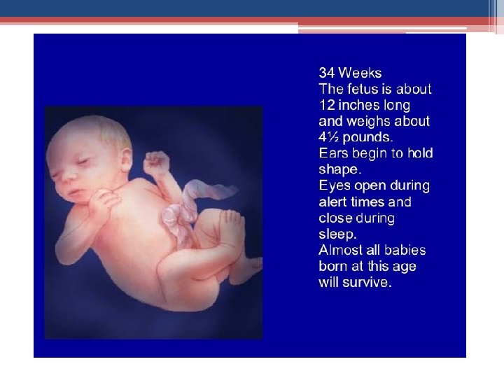
Weeks 31 - 34 • baby grows quickly & gains fat • rhythmic breathing occurs ▫ baby's lungs are not fully mature • bones are fully developed ▫ still soft • begins storing: ▫ iron ▫ calcium ▫ phosphorus
35 – 37 Weeks • weighs about 5 1/2 lbs • baby keeps gaining weight ▫ will probably not get much longer • skin is not as wrinkled as fat forms under the skin • definite sleeping patterns • heart & blood vessels are complete • muscles & bones are fully developed
38 - 40 Weeks • • • lanugo gone except for upper arms & shoulders fingernails may extend beyond fingertips small breast buds present on both sexes head hair is now coarse & thicker 40 th week of pregnancy it has been 38 weeks since conception
Labor & Delivery • 1 st Stage of Labor ▫ Early Labor ▫ Active Labor ▫ Transition • 2 nd Stage of Labor • 3 rd Stage of Labor
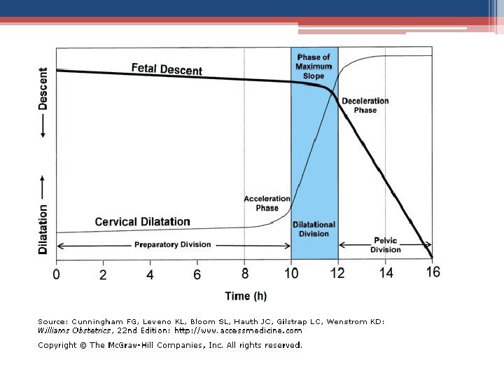
True vs False Labor: Williams Obstetrics (22 nd edition) • • • True Labor: Contractions occur at regular intervals Intensity gradually increases Discomfort is in the back and abdomen. *Cervix dilates. Discomfort is not stopped by sedation • “False Labor” • Contractions are irregular • Intensity remains the same • No cervical dilatation • Discomfort relieved by sedation
Length of first stage labor in healthy nulliparous and multiparous childbearing women adapted from Albers L. (2007) bold = nullips, italics = multips Mean (hrs) 95 th percentile (hrs) Friedman (1978) 4. 1 8. 5 Kilpatrick & Laros (1989) 8. 1 16. 6 Albers, Schiff & Gorwoda (1996) 7. 7 19. 4 Albers (1999) 7. 7 17. 5 Friedman (1978) 2. 4 7. 0 Kilpatrick & Laros (1989) 5. 7 12. 5 Albers (1996) 5. 7 13. 7 Albers (1999) 5. 6 13. 8
Early Labor • often longest part of birthing process ▫ sometimes lasting 2 to 3 days • uterine contractions: ▫ mild to moderate (you can talk while they are happening) and last about 30 to 45 seconds. ▫ often irregular (5 to 20 minutes apart) and may even stop for a while
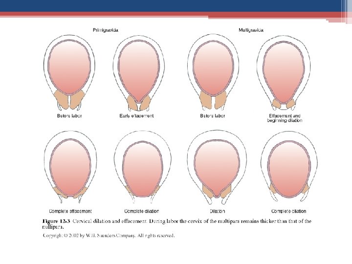
Early Labor • open (dilate) the cervix to about 3 cm (1 in. ). 1 st time mothers can have many hours of early labor without the cervix dilating ▫ dilation progresses 1 cm/2 hr in “true” labor • cervix has to efface (thin) ▫ starts 2 – 3 cm paper thin(by end of active phase)
Presentation
Passenger: Station • Engagement ▫ aka “dropping” or “lightening” ▫ @ the level of ischial spines = 0 station ▫ above ischial spines �-5 to -1 �-5 = unengaged ▫ below ischial spines �+1 to +5 �+5 = crowning
Active Labor • starts when the cervix is about 3 cm (1. 2 in. ) to 4 cm (1. 6 in. ) dilated • is complete when the cervix is fully effaced and dilated and the baby is ready to be pushed out
Active Labor • compared with early labor contractions during active labor are more intense & more frequent (every 2 to 3 minutes) and longer-lasting (50 to 70 seconds)
Transition (few min, to a few hours) �last part of active labor: cervix dilates from 8 to a full 10 centimeters �contractions usually very strong �coming every two and a half to three minutes �lasting a minute or more
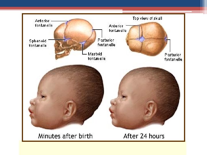
The Passenger Fontanelles and Sutures
Second Stage of Labor • 10 cm to birth of baby • pushing or expulsion • contraction pattern may alter • up to 2 hrs for 1 st shorter for others
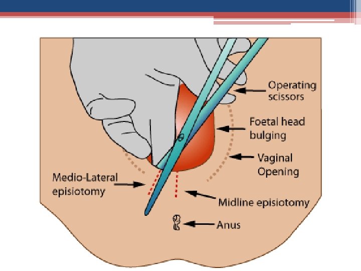
Birth • perineal management ▫ stretching tissues ▫ episiotomy if necessary
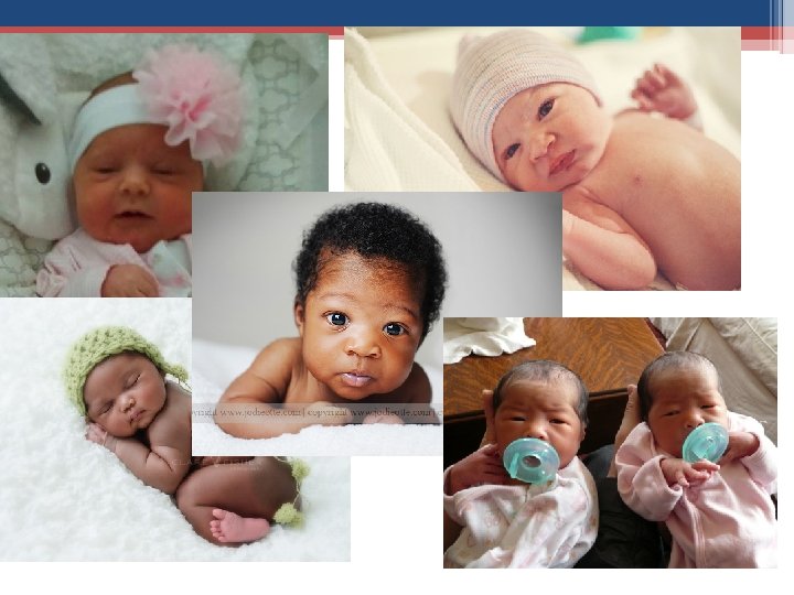
• Apgar score is a score given at one, five and ten minutes after the birth of a child. A score of 7 -9 is normal.
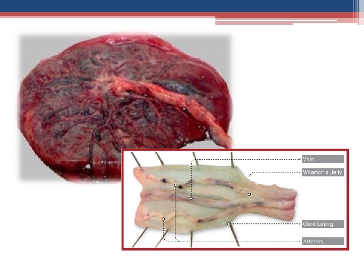
Third Stage of Labor • the placenta • 5 to 30 minutes…. or more • signs of placental separation: small gush of blood • inspection
Pathways of Placement of Anesthetics for Labor Pain Eltzschig H et al. N Engl J Med 2003; 348: 319 -332
Labor and Delivery • http: //www. webmd. com/baby/video/eddlemanwhat-expect-during-labor • http: //www. dailymotion. com/video/x 12 exwi_la bor-and-birth-babycenter-video_news