PreDissection Drawing Activity Pathway of Blood Through Heart

Pre-Dissection Drawing Activity Pathway of Blood Through Heart

Bell Work 2 -21 -2018 1. Name the four chambers of the heart. 2. The first heart sound (s 1) is what valves closing 3. The second heart sound (s 2) is what valves closing? 4. What structure divides the heart into right and left halves?

Standard Create an infographic to identify gross heart anatomy and physiology and related cardiac conduction and circulatory pathways.
")
Objective n Learn blood flow of the heart (circulatory pathway)

Video n https: //www. youtube. com/watch? v=7 Xaf td. E_h 60
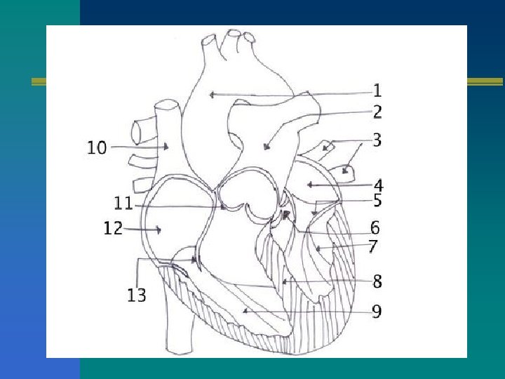


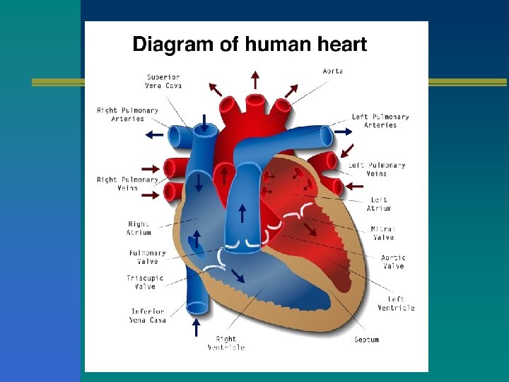
RIGHT SIDE OF THE HEART n DEOXYGENATED n PULMONARY BLOOD n SUPERIOR VENA CAVA n INFERIOR VENA CAVA n RIGHT ATRIUM n TRICUSPID VALVE n RIGHT VENTRICLE SEMILUNAR VALVE n PULMONARY ARTERIES n RIGHT AND LEFT LUNGS

LEFT SIDE OF THE HEART n OXYGENATED BLOOD n PULMONARY VEINS n LEFT ATRIUM n BICUSPID (MITRAL) VALVE n Left VENTRICLE n AORTIC SEMILUNAR VALVE n AORTA

PHYSIOLOGY OF THE HEART The heart is a double pump. When the heart beats… Right Heart Deoxygenated blood flows into heart from vena cava right atrium tricuspid valve right ventricle pulmonary semilunar valve pulmonary artery lungs (for oxygen) Left Heart Oxygenated blood flows from lungs via pulmonary veins left atrium mitral valve left ventricle aortic semilunar valve aorta general circulation (to deliver oxygen)

Supplies: n 3 sheets paper n pencil n 3 fine tip colored markers (blue, red, green) n Optional: highlighter (pink or orange)

The heart is a double pump. Right Heart Pump Left Heart Pump

The heart is a double pump. Right Heart Pump Left Heart Pump
 blood. Right Heart Pump")
The right heart pumps venous (deoxygenated) blood. Right Heart Pump
 blood. Left Heart Pump")
The left heart pumps arterial (oxygenated) blood. Left Heart Pump

Righ t hea rt Drawing the right heart.

Step 1: Draw outline of heart. Rt. side Lf. side apex

Step 2: Draw myocardium & septum. Identify chambers. Rt. side Lf. side LA RA RV myocardium LV septum apex

Step 3: Draw both superior & inferior vena cavae Rt. side superior vena cava inferior vena cava Lf. side LA RA RV LV septum apex

Step 4: Draw tricuspid valve. Rt. side superior vena cava Lf. side LA RA tricuspid valve inferior vena cava RV LV septum apex

Step 5: Draw pulmonary artery. Rt. side superior vena cava Lf. side pulmonary artery LA tricuspid valve inferior vena cava RA RV LV septum apex

Step 6: Draw pulmonary valve. Lf. side Rt. side pulmonary artery superior vena cava tricuspid valve inferior vena cava LA RA pulmonary valve RV LV septum apex

Step 7: Outline great vessels & chambers with blue marker. Rt. side Lf. side pulmonary artery superior vena cava tricuspid valve inferior vena cava LA RA pulmonary valve RV septum LV apex

Step 8: Outline valves in green. Color septum & myocardium in pink. Rt. side superior vena cava tricuspid valve inferior vena cava Lf. side pulmonary artery LA RA pulmonary valve RV septum LV apex

Step 9: Indicate pathway of blood through right heart with arrows. Rt. side superior vena cava tricuspid valve inferior vena cava Lf. side pulmonary artery LA RA pulmonary valve RV septum LV apex
 When you hear lub dub, that is the")
Bell Work 2 -22 -18 1) When you hear lub dub, that is the heart beating. The Lub is the first sound. What valves are closing? 2) When you hear the second sound (the dub), what valves are closing? 3) Blood flows in one direction in the heart. When the blood dumps into the right atrium, what Vein did it come from?

RIGHT SIDE OF THE HEART n DEOXYGENATED n PULMONARY BLOOD n SUPERIOR VENA CAVA n INFERIOR VENA CAVA n RIGHT ATRIUM n TRICUSPID VALVE n RIGHT VENTRICLE SEMILUNAR VALVE n PULMONARY ARTERIES n RIGHT AND LEFT LUNGS

LEFT SIDE OF THE HEART n OXYGENATED BLOOD n PULMONARY VEINS n LEFT ATRIUM n BICUSPID (MITRAL) VALVE n Left VENTRICLE n AORTIC SEMILUNAR VALVE n AORTA

PHYSIOLOGY OF THE HEART The heart is a double pump. When the heart beats… Right Heart Deoxygenated blood flows into heart from vena cava right atrium tricuspid valve right ventricle pulmonary semilunar valve pulmonary artery lungs (for oxygen) Left Heart Oxygenated blood flows from lungs via pulmonary veins left atrium mitral valve left ventricle aortic semilunar valve aorta general circulation (to deliver oxygen)

Standard Create an infographic to identify gross heart anatomy and physiology and related cardiac conduction and circulatory pathways.
")
Objective n Learn blood flow of the heart (circulatory pathway)

Left hear t Drawing the left heart.

Step 1: Draw outline of heart. Rt. side Lf. side apex

Step 2: Draw the myocardium and septum Rt. side Lf. side LA RA RV myocardium LV septum apex

Step 3: Draw mitral valve. Rt. side Lf. side LA RA myocardium mitral valve RV septum LV apex

Step 4: Draw aorta. Rt. side Lf. side aorta LA RA myocardium septum mitral valve RV LV apex

Step 5: Draw aortic valve. Rt. side aortic valve Lf. side aorta LA RA myocardium septum mitral valve RV LV apex

Step 6: Draw pulmonary veins. Rt. side pulmonary veins aorta Lf. side pulmonary veins LA RA aortic valve myocardium septum mitral valve RV LV apex

Step 7: Outline great vessels & chambers with red marker. Rt. side Lf. side pulmonary veins aorta pulmonary veins LA aortic valve RA myocardium RV septum mitral valve LV apex

Step 7: Outline valves in green. Color septum & myocardium in pink. Lf. side Rt. side pulmonary veins aorta pulmonary veins LA aortic valve RA myocardium RV septum mitral valve LV apex

Step 7: Indicate pathway of blood through left heart with arrows. Lf. side Rt. side pulmonary veins aorta pulmonary veins LA aortic valve RA myocardium RV septum mitral valve LV apex

Final Step: Draw entire heart.

Blood Flow review video n http: //www. registerednursern. com/how- to-remember-blood-flow-of-the-heart/
- Slides: 44