
Precision XRay Inc The Global Standard for PreClinical
Precision X-Ray Inc. The Global Standard for Pre-Clinical Specimen X-ray Irradiation and Imaging Systems X-Ray Irradiation and Imaging Research Systems! Headquarters: North Branford, CT, USA Incorporated: 2003 FOCUS : X-Ray Irradiation for Radiation Based Research Key Product Lines: • Standard Products : X-RAD 160, 225, 320, 350 k. V: Fixed Beam X-Ray Irradiation Systems • X-RAD 320 : Workhorse of the Industry – 100’s Worldwide Introduced Since 2015 • NEW : X-RAD Sm. ART – 3 D Image Guided Small Animal Radiation Therapy System • NEW: X-RAD 160, 225 and 320 XL/XLi family – Designed for High Throughput Irradiation and 2 D Multimodal Imaging. Largest Irradiation Chambers • NEW : Opti. MAX - Optical and X-Ray Multimodal Imaging Modules for X-RAD XL/XLi Systems
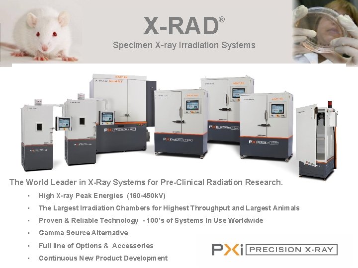
X-RAD ® Specimen X-ray Irradiation Systems The World Leader in X-Ray Systems for Pre-Clinical Radiation Research. • High X-ray Peak Energies (160 -450 k. V) • The Largest Irradiation Chambers for Highest Throughput and Largest Animals • Proven & Reliable Technology - 100’s of Systems In Use Worldwide • Gamma Source Alternative • Full line of Options & Accessories • Continuous New Product Development

Precision X-Ray Inc. The Global Standard for Pre-Clinical Specimen X-ray Irradiation and Imaging Systems X-Ray Irradiation and Imaging Research Systems! • X-RAD Customer List in CA 2018 • USC, Los Angeles, CA ( X-RAD 320) • University of California San Diego, CA (X-RAD 320) • SRI International, Menlo Park, CA (X-RAD 320) • Cedar Sinai Medical Center, Los Angeles, CA (X-RAD • Lawrence Berkeley National Labs, Berkeley, CA (XSm. ART) RAD 320) • VA Northern California Healthcare, Mc. Clellan, CA (X- • UCLA, Los Angeles, CA (X-RAD Sm. ART) RAD i. R 160) • Stanford University, Stanford, CA (X-RAD Sm. ART) • University of Southern California, Los Angeles, CA (X- • UC Irvine – Irvine CA ( X-RAD 320) RAD 320 ix) • City of Hope, Los Angeles, CA (X-RAD 320) • Stanford University, Stanford, CA (X-RAD 320) • Bayer, San Francisco, CA (X-RAD 320) nd • University of California, Berkeley, CA (X-RAD 320) 2 • Pfizer, La Jolla CA, (X-RAD 225) system • University of California San Diego, La Jolla, CA (X • University of California, Berkeley, CA (X-RAD 320) RAD 225 XL) • Henkel, Irvine, CA (X-RAD i. R-225) • City of Hope, Los Angeles, CA (X-RAD Sm. ART) • University of California – Riverside, CA (X- • VA Palo Alto, CA (X-RAD 320) • Merck & Co, Palo Alto, CA (X-RAD 320) • USC, Los Angeles, CA (X-RAD i. R-160) • UCSD La. Jolla CA ( X-Rad Sm. ART)
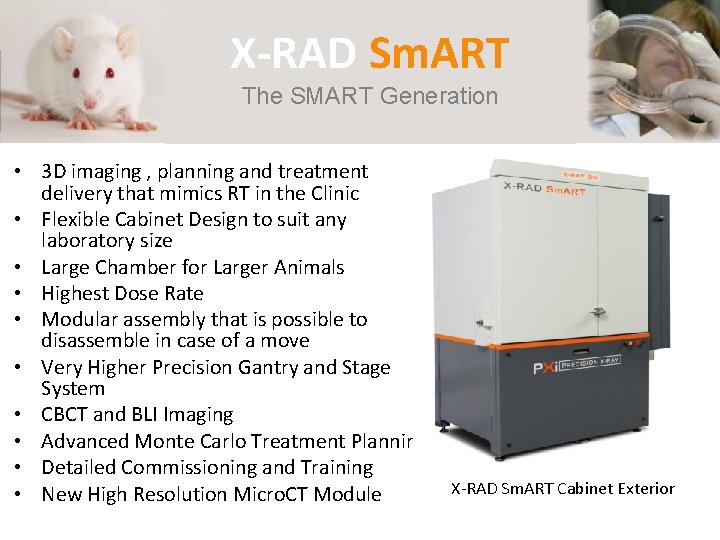
X-RAD Sm. ART X-RAD 225 Cx Small Animal IGRT The SMART Generation • 3 D imaging , planning and treatment delivery that mimics RT in the Clinic • Flexible Cabinet Design to suit any laboratory size • Large Chamber for Larger Animals • Highest Dose Rate • Modular assembly that is possible to disassemble in case of a move • Very Higher Precision Gantry and Stage System • CBCT and BLI Imaging • Advanced Monte Carlo Treatment Planning • Detailed Commissioning and Training • New High Resolution Micro. CT Module X-RAD Sm. ART Cabinet Exterior
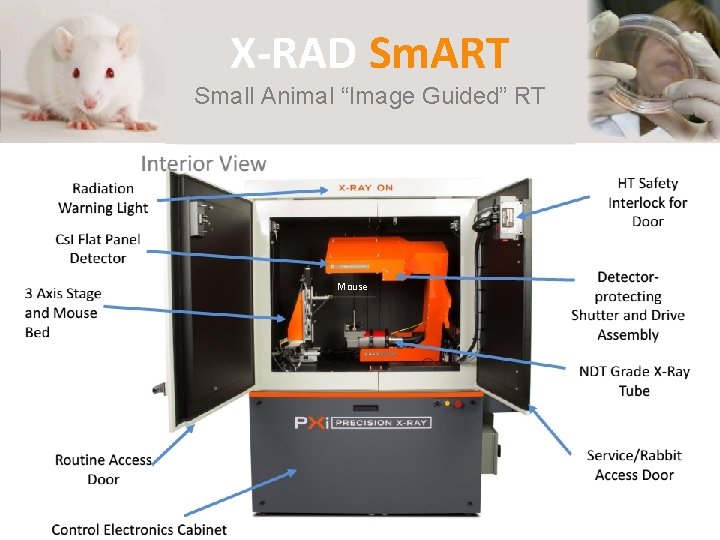
X-RAD Sm. ART Small Animal “Image Guided” RT X-Rad 225 Cx: Interior View Radiation Warning Light HT Safety Interlock for Door NDT X-ray Tube (5225 k. Vp) Load-bearing Gantry Ring Cs. I: Tl a-Si: H Detector Routine Access Door Control Electronics Cabinet Mouse U-arm and Drive Assembly Detectorprotecting Shutter and Drive Assembly Service Access Door
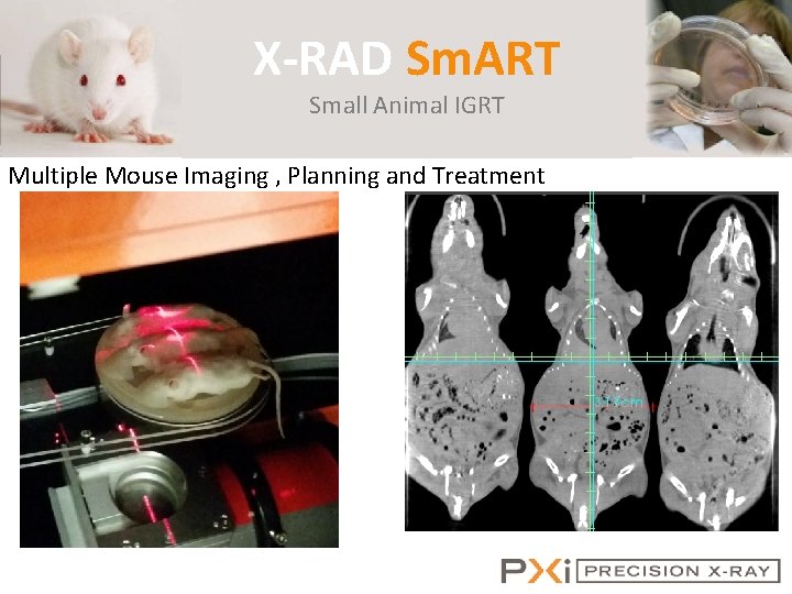
X-RAD Sm. ART Small Animal IGRT Multiple Mouse Imaging , Planning and Treatment
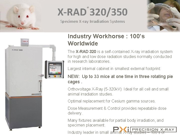
X-RAD 320/350 ® Specimen X-ray Irradiation Systems Industry Workhorse : 100’s Worldwide The X-RAD 320 is a self-contained X-ray irradiation system for high and low dose radiation studies normally conducted in research laboratories. Largest internal cabinet in smallest external footprint: NEW: Up to 33 mice at one time in three rotating pie cages. Orthovoltage X-Ray (5 -320 k. V): Ideal for all cell and small animal irradiation studies. Optimal replacement for Cesium gamma sources. Dose Measurement & Control provides repeatable dose delivery. Many fixtures available for partial body irradiation, and specimen placement. Industry leader in small animal X-ray studies – 100’s of
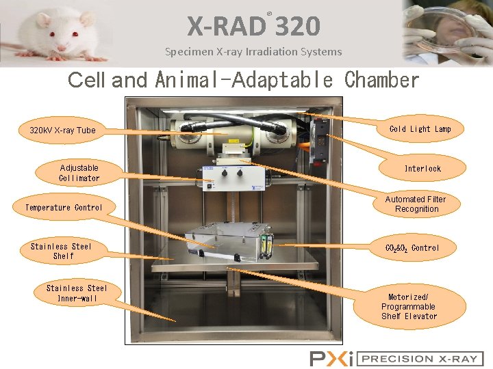
X-RAD 320 ® Specimen X-ray Irradiation Systems Cell and Animal-Adaptable Chamber 320 k. V X-ray Tube Adjustable Collimator Temperature Control Stainless Steel Shelf Stainless Steel Inner-wall Cold Light Lamp Interlock Automated Filter Recognition CO 2&O 2 Control Motorized/ Programmable Shelf Elevator
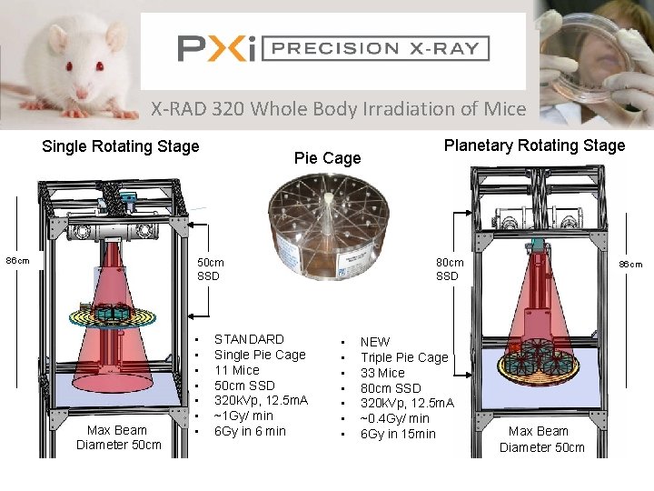
Precision X-Ray Inc. X-RAD 320 Whole Body Irradiation of Mice Single Rotating Stage 86 cm Pie Cage 50 cm SSD Max Beam Diameter 50 cm • • STANDARD Single Pie Cage 11 Mice 50 cm SSD 320 k. Vp, 12. 5 m. A ~1 Gy/ min 6 Gy in 6 min Planetary Rotating Stage 80 cm SSD • • NEW Triple Pie Cage 33 Mice 80 cm SSD 320 k. Vp, 12. 5 m. A ~0. 4 Gy/ min 6 Gy in 15 min 86 cm Max Beam Diameter 50 cm
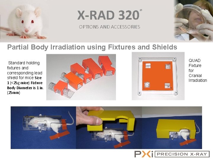
X-RAD 320 ® OPTIONS AND ACCESSORIES Partial Body Irradiation using Fixtures and Shields Standard holding fixtures and corresponding lead shield for mice Size 1 (<25 g mice) Fixture Body Diameter is 1 in. (25 mm) QUAD Fixture for Cranial Irradiation
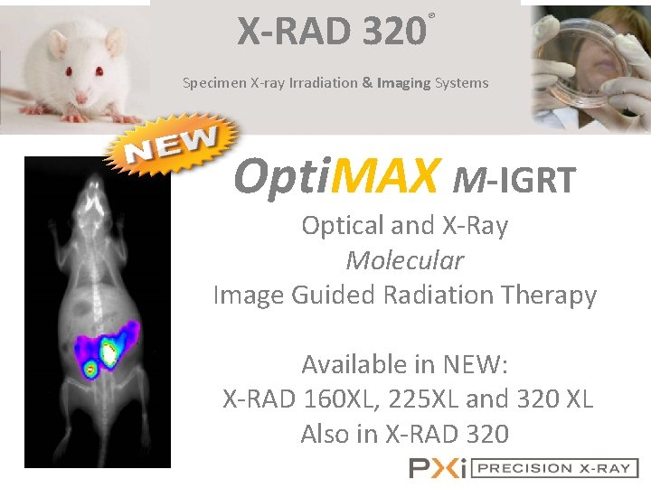
X-RAD 320 ® Specimen X-ray Irradiation & Imaging Systems Opti. MAX M-IGRT Optical and X-Ray Molecular Image Guided Radiation Therapy Available in NEW: X-RAD 160 XL, 225 XL and 320 XL X-RAD 320/350 Also in X-RAD 320
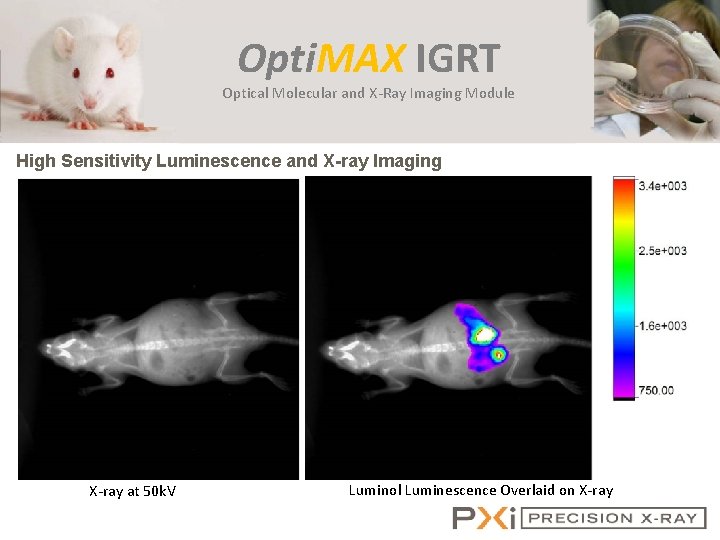
Opti. MAX IGRT Optical Molecular and X-Ray Imaging Module High Sensitivity Luminescence and X-ray Imaging X-ray at 50 k. V Luminol Luminescence Overlaid on X-ray
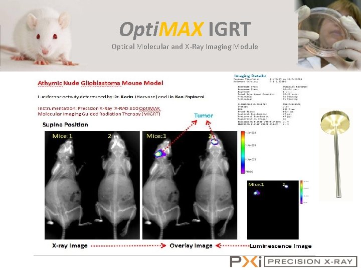
Opti. MAX IGRT Optical Molecular and X-Ray Imaging Module
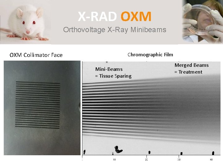
X-RAD OXM Orthovoltage X-Ray Minibeams OXM Collimator Face Chromographic Film Mini-Beams = Tissue Sparing Merged Beams = Treatment
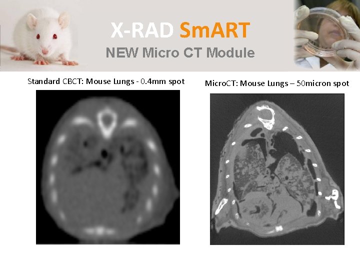
X-RAD Sm. ART NEW Micro CT Module Standard CBCT: Mouse Lungs - 0. 4 mm spot Micro. CT: Mouse Lungs – 50 micron spot
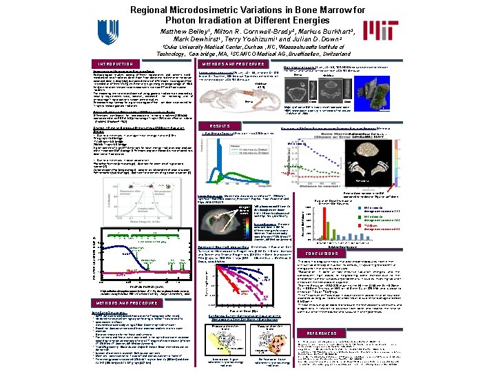
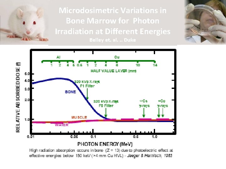
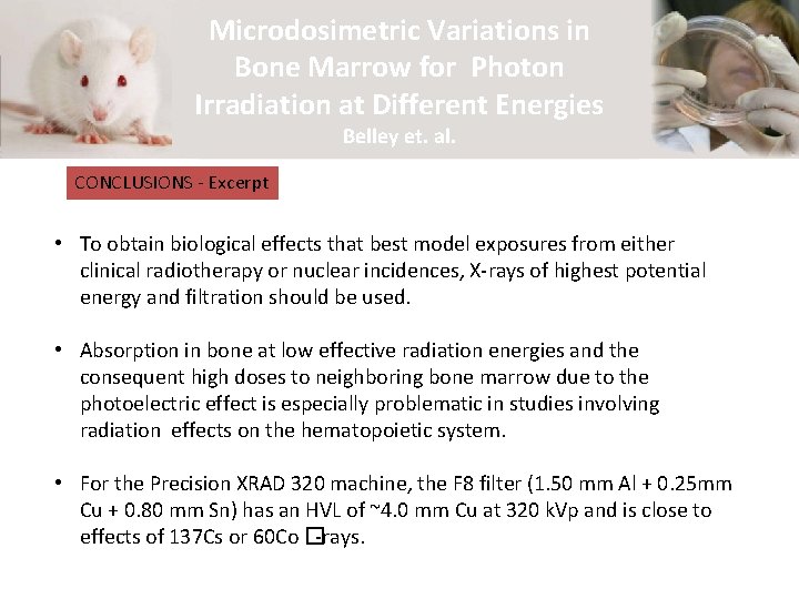
Regional Microdosimetric Variations in Bone Marrow for Photon Irradiation at Different Energies Matthew Belley 1, Milton R. Cornwall-Brady 2, Markus Burkhart 3, Mark Dewhirst 1, Terry Yoshizumi 1 and Julian D. Down 2 1 Duke University Medical Center, Durham, NC, 2 Massachusetts Institute of Technology, Cambridge, MA, 3 SCANCO Medical AG, Bruettisellen, Switzerland INTRODUCTION METHODS AND PROCEDURE Importance of Uniformity of Radiation Dose Radiobiological studies among different laboratories and centers need standardization of radiation dose. Apart from dosimetry to determine the actual absorbed dose in Gray (Gy), due consideration of differences in biological effect is needed at different energies. Relatively high energies (megavoltage of >500 Ke. V) are the most relevant to both exposures in clinical RT and from nuclear incidents. The escalating restrictions and costs of using gamma irradiators as imposed by the increasing use of security requirements have, however, prompted orthovoltage X-ray machines in experimental studies. Reasonable equivalency for a given biological effect per dose is warranted for X-rays to replace gamma-irradiators. High resolution scans (1 µm, µCT 50, SCANCO Medical) were performed on the femur and lumbar vertebrae from a C 57 BL/6 mouse Lower resolution scan (25 µm, µCT 50, e. Xplore CT 120 Micro CT Scanner, GE Medical Systems) performed on the whole body of a C 57 BL/6 mouse Femur Scout Majority of active BM is in cavities of trabecular bone. ~50% hematopoietic activity in vertebrae of the mouse – Colvin et al. , 2004. Relative Biological Effectiveness (RBE) of X-rays vs. -rays Differences well-known for hematopoietic lethality in rodents (LD 50/30): approximated at an RBE of 0. 8 (orthovoltage X-rays ~20% more effective in dose - Sinclair & Blackwell, 1962) Change in Relative Biological Effectiveness (RBE) with Radiation Energy • Due to differences in average linear energy transfer (LET): Vertebrae (L 2 -5) RESULTS 1. 0 mm Variations in BM Dose According to the Different Radiation Energies (Vertebra) X-Ray Energy Spectra (Precision Xrad 320 Machine) Radiation Dose Contours – 1 mm Cu HVL = 85 ke. V -rays ~ 0. 4 ke. V/ m -rays ~ 0. 8 ke. V/ m 200 k. Vp X-rays ~3. 5 ke. V/ m Higher content of high LET delta rays for lower energy irradiation may produce more irreparable DNA damage. Differences may be enhanced with protracted low dose rate or fractionation. 60 Co 137 Co HVL = 1 mm Cu • Due to differences in dose absorption: Photoelectric effect (orthovoltage) – dominant for materials of high atomic number (Z) Compton scattering (megavoltage) – absorption independent of atomic number Pair formation (supravoltage) - dominant for materials of high atomic number (Z) HVL = 4 mm Cu 137 Cs -rays Vertebra Relative dose increase in the BM compared to the dose at “Equilibrium” depth Lower Resolution - Monte Carlo Simulation with Micro-CT – 225 k. Vp Xrays from XRad 225 Cx machine, Precision X-Ray Inc – from Chow et al. Med Phys. 2010; 37: 5322 -9. 360° photon arc and 0. 3 mm Cu filter through mouse thorax. HVL = 1. 0 mm. Cu (Newton et al. Med Phys. 2011; 38: 6754 -62) Future Analysis - Estimate 2 4 5 0. 5 1 5. 0 4. 0 3. 0 10 2 4 6 HALF VALUE LAYER (mm) 14 BONE 2. 0 320 k. Vp X-rays F 8 Filter 137 Cs -rays 60 Co -rays Agrees with Existing Biological Data - Differences in Radiation Cell Survival for Hematopoietic Progenitors (CFU-C) in Bone Marrow and Spleen and Stromal Progenitors (CFU-F) in Bone Marrow with 60 Co -rays vs. 200 k. Vp X-rays (HVL = 1. 05 mm. Cu) – Beekman & Down, unpublished 1 MUSCLE 1. 0 WATER 0. 01 0. 05 0. 1 0. 5 1. 0 PHOTON ENERGY (Me. V) High radiation absorption occurs in bone (Z = 13) due to photoelectric effect at effective energies below 150 ke. V (>4 mm Cu HVL) - Jaeger & Harnisch, 1983 METHODS AND PROCEDURE 137 Cs Cs- -rays Average 137 dose increase = 0. 98 20% 0% • GATE Software Used (Geant 4 Application for Tomographic Emission) • Validated the physics settings by performing a “planar” simulation of a • bone/spleen interface Simulated a mouse body using a 2. 5 cm diameter cylinder of water • Placed the Scanco vertebra and femur phantom models into this water • phantom 5 micron resolution for the femur and vertebra • The vertebra and femur were positioned in the cylindrical water phantom according to the actual anatomy of micro-CT images of whole mouse (e. Xplore CT 120 Micro CT Scanner, GE Medical Systems) Xrad-320 geometry - 50 cm Source-Object Distance. Beam entered mouse on dorsal side Number of particles simulated: 10 e 9 source particles Materials – bone was set to “rib bone” and marrow was set to “spleen” Three energy beams simulated: 320 k. Vp X-rays at 1 mm Cu (85 ke. V) and 4 mm Cu HVL (150 ke. V) and Cs-137 -rays (662 ke. V). BM CFU-F X Spleen CFU-C 0. 001 0. 0001 0 1 2 3 4 5 X 6 7 • To obtain biological effects that best model exposures from either clinical radiotherapy or nuclear incidences, X-rays of highest potential energy and filtration should be used. Absorption in bone at low effective radiation energies and the consequent high doses to neighboring bone marrow due to the photoelectric effect is especially problematic in studies involving radiation effects on the hematopoietic system. For the Precision XRAD 320 machine, the F 8 filter (1. 50 mm Al + 0. 25 mm Cu + 0. 80 mm Sn) has an HVL of ~4. 0 mm Cu at 320 k. Vp and is close to effects of 137 Cs or 60 Co -rays. The “inconvenient” decrease in dose-rate with added filtration has to be accepted as long as it does not allow repair of sub-lethal damage at below 0. 5 Gy/min. These effects may be exploited to locate the hematopoietic stem cells and progenitors in relation to distance from bone and resolve the relative contribution of the endosteal and vascular niche hypotheses. • X 0. 01 Relative Dose Increase CONCLUSIONS • 0. 1 • BM CFUC 8 9 10 Radiation Dose (Gy) M onte-Carlo Simulations • • 40% 80% 0. 94 1. 01 1. 08 1. 15 1. 22 1. 29 1. 36 1. 43 1. 50 1. 57 320 k. Vp X-rays F 1 Filter Surviving Fraction RELATIVE ABSORBED DOSE (f) 1 absorbed dose in BM for different energies for whole skeleton –from whole body scan (e. Xplore CT 120 Micro CT Scanner, GE Medical Systems) Cu Al 60% HVL = 1 mm Cu Average dose increase = 1. 33 F 1 HVL = 4 mm Cu F 8 Average dose increase = 1. 08 100% Percent BM Volume x 3 higher dose to bones Radiation Dose Increase at Different BM Volumes • Can Adress Current Controversy on Location of the Hematopoietic Stem Cell Niche in Bone Marrow Endosteal Stem Cell Niche Vascular Stem Cell Niche HSC REFERENCES Ellis RE. Some experiments relating to dose in a model of trabecular bone. Br J Radiol. 1966; 39: 211 -5. Bone Osteoblast Next to bone - Higher radiosensitivity at low energy irradiation Gengozian N, Taylor T, Jameson H, Lee ET, Good RA, Epstein RB. Radiation-induced hemopoietic death in mice as a function of photon energy and dose rate. Radiat Res. 1986; 105: 320 -7. Endothelial Cell Far from bone - Lower radiosensitivity at low energy irradiation Jaeger SS, Harnisch BD. Corrected f factors for photons from 10 ke. V to 2 Me. V. Med Phys. 1983; 10: 714 -6. King SD, Spiers FW. Photoelectron enhancement of the absorbed dose from X rays to human bone marrow: experimental and theoretical studies. Br J Radiol. 1985; 58: 345 -56. Sinclair WK. The relative biological effectiveness of 22 -Mevp x-rays, cobalt-60 gamma rays, and 200 -Kvcp x-rays. V. Absorbed dose to the bone marrow in the rat and the mouse. Radiat Res. 1962; 16: 369 -83 Colvin GA, Lambert JF, Abedi M, Hsieh CC, Carlson JE, Stewart FM, Quesenberry PJ. Murine marrow cellularity and the concept of stem cell competition: geographic and quantitative determinants in stem cell biology. Leukemia. 2004 ; 18: 575 -83.
Microdosimetric Variations in Bone Marrow for Photon Irradiation at Different Energies Belley et. al. . . Duke
Microdosimetric Variations in Bone Marrow for Photon Irradiation at Different Energies Belley et. al. CONCLUSIONS - Excerpt • To obtain biological effects that best model exposures from either clinical radiotherapy or nuclear incidences, X-rays of highest potential energy and filtration should be used. • Absorption in bone at low effective radiation energies and the consequent high doses to neighboring bone marrow due to the photoelectric effect is especially problematic in studies involving radiation effects on the hematopoietic system. • For the Precision XRAD 320 machine, the F 8 filter (1. 50 mm Al + 0. 25 mm Cu + 0. 80 mm Sn) has an HVL of ~4. 0 mm Cu at 320 k. Vp and is close to effects of 137 Cs or 60 Co �-rays.
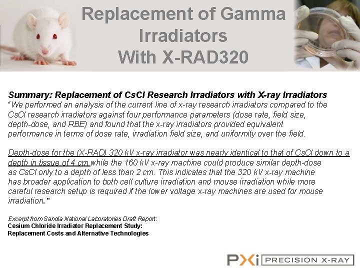
Replacement of Gamma Irradiators With X-RAD 320 Summary: Replacement of Cs. Cl Research Irradiators with X-ray Irradiators “We performed an analysis of the current line of x-ray research irradiators compared to the Cs. Cl research irradiators against four performance parameters (dose rate, field size, depth-dose, and RBE) and found that the x-ray irradiators provided equivalent performance in terms of dose rate, irradiation field size, and uniformity over the field. Depth-dose for the (X-RAD) 320 k. V x-ray irradiator was nearly identical to that of Cs. Cl down to a depth in tissue of 4 cm while the 160 k. V x-ray machine could produce similar depth-dose as Cs. Cl only to a depth of less than 2 cm. This indicates that the 320 k. V x-ray machine has broader application to both cell culture irradiation and mouse irradiation while more careful research setup is required if the lower voltage x-ray machines are used for mouse irradiation. ” Excerpt from Sandia National Laboratories Draft Report: Cesium Chloride Irradiator Replacement Study: Replacement Costs and Alternative Technologies
- Slides: 20