PRACTICAL SKILLS LAB PRACTICAL SKILLS In papers 2

PRACTICAL SKILLS
, you will be")
LAB PRACTICAL SKILLS • In papers 2 and 3 (esp. 3), you will be expected to demonstrate mastery of collecting, organizing, and displaying data • Plotting points • Creating a table • Measuring • Drawing

PLOTTING POINTS • Independent var. : x-axis • Variable being manipulated (ex: temperature, concentration, p. H) • Dependent (response) var. : y-axis • Variable being measured (ex: reaction rate, products formed) D I

PLOTTING POINTS • Always plot points with a small x or small dot inscribed in a circle • Always connect data points with a ruler • Different data sets should use different points OR different lines (solid, dashed, dotted)

CREATING A TABLE • • ALWAYS USE A RULER!!! Tables should be drawn in pen only Units go on column headings First column = always independent
 103 • Meter (m) 1 • Centimeter (cm)")
UNITS OF MEASUREMENT • Kilometer (km) 103 • Meter (m) 1 • Centimeter (cm) 10 -2 • Millimeter (mm) 10 -3 • Micrometer (µm) 10 -6 • Nanometer (nm) 10 -9 • Picometer (pm) 10 -12

UNITS OF MEASUREMENT Important for measuring cells! • BIG to SMALL: decimal to the right • SMALL to BIG: decimal to the left 1. Convert 10 μm to mm 2. Convert 10 µm to nm

DRAWING CELLS Do’s Don’ts • • • pencil only Clear, continuous lines Accurate proportions Tissues completely enclosed by lines Label all tissues Correctly identified parts Representative portions or cells Scale bar Drawing different tissues: Shape, Size, and Shading Textbook versions Individual cells on low power Nucleus as a solid blob on high power


VISUALIZING ANIMAL AND PLANT CELLS • Every plant and animal cell is surrounded by a very thin cell surface membrane (aka plasma membrane) • All have a nucleus which is very large and stains very darkly (especially chromatin) • The nucleolus located within the nucleus stains even more deeply (variable amount, can be ~1 -5)

VISUALIZING ANIMAL AND PLANT CELLS • The most numerous organelles seen with light microscopes are usually mitochondria (single mitochondrion) • Golgi apparatus can only be seen with silver containing stains

PLANT CELLS ONLY • Usually larger than animal cells • All surrounded by cell wall • Linked to neighboring cells by plasmodesmata (single plasmodesma)

PLANT CELLS ONLY • Large central vacuole, surrounded by tonoplast • Chloroplasts: show grana (single granum) at high magnification

LIGHT MICROSCOPES •

MAGNIFICATION I M × A

CHECK YOUR UNDERSTANDING This length of the displayed lymphocyte is 36 mm. The actual length of the lymphocyte is 6µm. What is the magnification?

CHECK YOUR UNDERSTANDING •

CHECK YOUR UNDERSTANDING •

MEASURING CELLS

MEASURING CELLS • Cells and organelles can be measured with a microscope by means of an eyepiece graticule • This is a transparent scale with 100 divisions that is placed in the eyepiece so it can be seen at the same time and the sample on the slide

USING AN EYEPIECE GRATICULE • This figure shows the scale over a human cheek epithelial cell. • The cell lies between 40 and 60 on the scale, therefore we say it measures 20 eyepiece units in diameter • We will not know the actual size of the cell until the eyepiece graticule scale is calibrated

CALIBRATING AN EYEPIECE GRATICULE SCALE What is • To calibrate the eypiece graticule scale, a miniature transparent ruler called a stage micrometer scale is placed on the microscope stage and is brought into focus • This scale may be etched onto a glass slide or printed on transparent film • It commonly has subdivisions of 0. 1 and 0. 01 mm • The images can then be super imposed to calibrate the graticule the value of 1 epu? 8 cm ? ? cm

CALIBRATING AN EYEPIECE GRATICULE SCALE •

CALIBRATING AN EYEPIECE GRATICULE SCALE •

FIND THE SIZE OF THE NUCLEUS
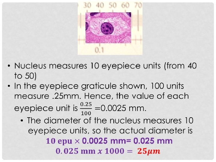

SOLUTION •
- Slides: 30