Practical Hematology Lab Normal Cell Maturation Blood Cells

Practical Hematology Lab Normal Cell Maturation

Blood Cells Maturation • Blood cells go through several stages of development; progression from one stage to the next is not rapid, so frequently the cell being studied may be between stages. • When this occurs the cell is generally given the name of the mature stage). • As a cell transforms from the primitive blast stage to the mature form found in the blood, there are changes in the cytoplasm, nucleus, and cell size.

• Normally, all three of these changes occurs gradually and simultaneously. • In some disease states, however, changes will take place at different rates
 because")
Cytoplasmic Maturation • The immature cytoplasm usually stain a deep blue color (basophilic) because of the high content of RNA present as the cell matures, there is a gradual loss of cytoplasmic RNA and therefore a lessening of blue color. • In some cells (for example, the myelogenous cells), granules appear in the cytoplasm as the cell matures. At first, these granules are few and nonspecific. As the cell matures further, these granules increase in number and take on specific characteristics and functions. • The amount of cytoplasm in relationship to the rest of the cell usually increases as the cell matures.

Nuclear Maturation • The nucleus of the immature cell is round oval and is large in proportion to the rest of the cell. As the cell matures the nucleus decreases in relative size and may or may not take on various shapes (depending on the cell type). • The nuclear chromatin transforms from a fine, delicate pattern to become more coarse and clumped on the mature form, and the staining properties change from a reddish purple to a bluish purple. • Nucleoli present in early stages of cell development usually disappear gradually as the cell ages.

Cell Size • As a cell matures, it usually becomes smaller in size. (for the new student, this change may be difficult to detect. • The normal mature red blood cells or small lymphocytes are generally of relatively constant size and may be used as a guide for comparison) the student should know the relative size of each cell type.

Identification of cells In identifying a cell we should think of the following terms: ØWhat is the size of the cell? • Small • Medium • Large ØWhat are the characteristics of the cytoplasm? • Granular or nongranular; specific or nonspecific granules. • Color (staining properties). • Relative amount.

ØWhat are the characteristics of the nucleus? • Shape. • Relative size. The size of the nucleus compared to the amount of cytoplasm is expressed as the nuclear to cytoplasmic ratio (N/C ratio). • Chromatin pattern: smooth or coarse. • Presence of or absence of nucleoli; number and size.

Erythrocyte Maturation § 4 mitotic divisions between pronormoblast and orthochromic normoblast stage. § Thus giving rise 16 RBCs. § But not all of the 16 will be good RBCs, some are bad and will be destroyed, these destroyed cells is called ineffective erythropoiesis. § 2 – 7 days for pronormoblast to mature into orthochromic normoblast § 1 more day to extrude the nucleus from the orthochromic normoblast. § Reticulocyte further matures for 2 – 3 days in bone marrow before it is released into the peripheral blood. § Red cell has life span of 120 days in peripheral blood.

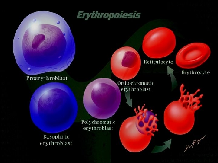
Erythrocyte Maturation The overall trend in RBC maturation is large, pale nucleus to darker, smaller nucleus to loss of nucleus; increase in cytoplasm; gradual decrease in size; cytoplasm from intensely blue (full of RNA) to grayish (mixture of RNA and hemoglobin) to reddish (full of hemoglobin, no RNA). Identify the following cells. Maturation
 • 14 -19 µm • Large nucleus 12 Microns")
Erythrocyte Maturation 1. Pronormobast (Rubriblast) • 14 -19 µm • Large nucleus 12 Microns occupies 3/4 th of the cell volume. • Nucleus has fine stippled reticulum & many nucleoli. • Cytoplasm is scant and basophilic (deep violet-blue staining). • Has no Hemoglobin.
 • Smaller in size. 12 -17 µm •")
2. Early Normoblast , Basophilic (Prorubriblast) • Smaller in size. 12 -17 µm • Shows active Mitosis. • Slightly smaller nucleus with slight chromatin condensation • Increased cytoplasm and intensely blue (RNA abundance); no granules and no nucleoli present.
 • • 12 -15 µm Shows active Mitosis.")
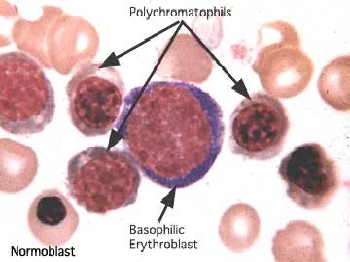
3. Intermediate normoblast, Polychromatophilic Normoblast (Rubricyte) • • 12 -15 µm Shows active Mitosis. Lighter, grayish cytoplasm. Increased Hemoglobin content in the cytoplasm hence, cytoplasm is Polychromatophilic • Color of the cytoplasm is due to coloring by both acidic and basic components of the stain. • Basophilia is from staining of ribosomes and acidophilia from hemoglobin. • The nucleus is condensed and intensely basophilic with moderately coarse heterochromatin granules.
 • • 8 -12 µm Dark, opaque nucleus;")
4. Late Normoblast Orthochromatophilic Normoblast (Metarubricyte) • • 8 -12 µm Dark, opaque nucleus; gray-red cytoplasm (trace blue) Cytoplasm has an eosinophilic appearance. The nucleus has become pyknotic (a homogenous blueblack mass with no structure) and there is abundant acidophilic hemoglobin. • Appears like a “Cartwheel” • In some instances you can detect the nucleus in the process of extrusion.
 • Nucleus has been extruded; cytoplasm is reddish-pale blue.")
Reticulocyte • (7 -10 µm) • Nucleus has been extruded; cytoplasm is reddish-pale blue. RNA is still present. • The penultimate stage cell. • Has a fine network of reticulum like a heavy wreath or as clumps of dots • This is the remnant of the basophilic cytoplasm, comprising RNA. • In the Neonates, Count is 2 – 6/Cu. mm. • Falls to <1 in the first week of life.

Mature Erythrocyte • Biconcave disc. • No nucleus. • About One-third filled with Hemoglobin.

Factors regulating erythropoiesis • • • Single most important regulator: “tissue oxygenation” Burst promoting activity Erythropoietin Iron Vitamins: • Vitamin B 12 • Folic acid • Miscellaneous

Erythropoietin • • • A hormone produced by the Kidney. A circulating Glycoprotein Nowadays available as Synthetic Epoietin Acts mainly on CFU – E. Increases the number of: • Nucleated precursors in the marrow. • Reticulocytes & Mature Erythrocytes in the blood.

Vitamins • B 12: Cyanocobalamine & Folic Acid: • Is also called Extrinsic Factor of Castle. • Needs the Intrinsic Factor from the Gastric juice for absorption from Small Intestine. • Deficiency causes Pernicious (When IF is missing) or Megaloblastic Anemia. • Stimulates Erythropoiesis • Is found in meat & diary products.

Iron • Essential for the synthesis of Hemoglobin. • Deficiency causes Microcytic, Hypochromic Anemia. • The MCV, Color Index & MCH are low.

Regulation of Erythropoiesis Stimulates CFU – E Proerythroblasts ERYTHROPOIETIN Mature Erythrocytes s se De a e r c Tissue Oxygenation Factors decreasing: Hypovolemian Anemian Poor blood flown Pulmonary Diseasen An example of a Negative feed back mechanism
 As its name suggests, EPO stimulates growth of Erythrocytes, and its function")
Erythropoietin (Epo) As its name suggests, EPO stimulates growth of Erythrocytes, and its function include: § Activates stem cells of bone marrow to differentiate into pronormoblasts. § Shortening cell cycle. § Decrease maturation time. § Increases rate of mitosis and maturation process. § Increases rate of hemoglobin production. § Causes increased rate of reticulocyte release into the peripheral blood, (normally the reticulocyte when it is released to the peripheral blood it need only one day to mature to RBC, but here they will be released prematurely into peripheral blood, thus they need more than one day to mature RBC.

EPO receptors §Found on surface on bone marrow erythroid progenitor and precursor cells. §The highest number of EPO receptors is seen on the CFU -E and the pronormoblasts. §The number of EPO receptors per cell gradually decreases during erythroid cell differentiation, and studies have shown that the reticulocyte and mature erythrocyte do not contain EPO receptors

EPO Receptor Divided into extracellular, transmembrane, and cytoplasmic domains. The cytoplasmic domain has terminal that contains both positive and negative growth-regulatory domains. After EPO binding, EPOR homodimarizes and JAK-2 phosphorylates itself, so EPOR and other proteins like STAT-5 initiate the cascade of proliferative signals.

Myeloid Maturation myeloblast promyelocyte metamyelocyte MATURATION band neutrophil

Maturation and Morphology of Immature Granulocytes

1. Myeloblast • Cells in the BM proliferation pool take 24 -48 hours for a single cell cycle • The first and earliest granulocyte • Is a large cell (15 μm) • High nucleus to cytoplasm (N: C) ratio (5: 1) • Round or oval nucleus with loose light staining euchromatin • 1 -2 nucleoli • Has minimal light blue cytoplasm • Contains no cytoplasmic granules

1. Myeloblast • • Comprises 1% of the nucleated cells in the bone marrow Cytochemical staining shows presence of myeloperoxidase which is required for intracellular kills Killing function is the first to be operational in the neutrophil cell line Myeloblast is incapable of motility, adhesion and phagocytosis and is therefore nonfunctional
 High N: C")
2. Promyelocyte • • • Larger than a myeloblast (20 μm) High N: C ratio (3: 1) Loose chromatin with nucleoli Dark blue cytoplasm Contains large nonspecific cytoplasmic granules • Containing myeloperoxidase (MPO) • Comprises 3 -4% of nucleated bone marrow cells
 High N: C ratio")
3. Neutrophilic Myelocyte • • Medium cell size (12 μm) High N: C ratio (3: 1) Round, oval, or slightly indented nucleus with darker Blue heterochromatin Last stage of cell division Has active RNA, therefore, the cytoplasm is blue Comprises 10% of bone marrow nucleated cells

3. Neutrophilic Myelocyte • Production and accumulation of secondery granules is characteristic of the myelocyte • Myelocytes demonstrate morphologic variability as this development stage lasts from 4 -5 days and cause alterations in the staining characteristics of the cell • Contains MPO and secondary granules containing leukocyte alkaline phosphatase • The cell acquires some motility

Granulopoiesis Notice change in granules color
 •")
4. Neutrophilic Metamyelocyte • 10 -15 μm • N: C ratio (2: 1) • Last mononuclear stage, no mitosis • Nucleus is kidney or horseshoe shaped, and has condensed heterochromatin • Has a prominent Golgi apparatus – clear area located at the indentation site of the nucleus • Cytoplasm is similar to the mature cell • Comprises 18% of bone marrow cells • Not seen in normal PB • Not fully functional, part of the maturation component of the marrow
 • N:")
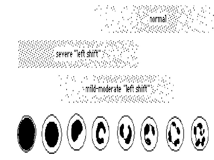
5. Band Neutrophile Same size as a mature neutrophil (10 -12 μm) • N: C ratio has reversed (1: 2) • Nucleus is band- or sausage-shaped without segmentation • Cytoplasm is filled with small neutrophilic granules • Last immature stage • Comprises 11% of bone marrow cells and 0 -3% of peripheral WBCs Stored in the bone marrow and released when there is an increased demand for neutrophils

Band Neutrophile • The band is a transitional form that exists in both the PB and the BM and considered part of both the maturation and storage pools • Represents the “almost mature” neutrophil having full motility, active adhesion properties, and some phagocytic ability • Membrane maturity shows changes in cytoskeleton, surface charge and presence of receptors for complement

Morphology of Mature Granulocytes

Neutrophils • This cell’s nucleus continues to indent until thin strands of membrane and heterochromatin form into segments, hence it is also called a “seg” • Polymorphonuclear means “many-shaped nucleus”, describing the varied nuclear shapes • Cell is completely functional and spend time in the storage pool of the BM as well as marginating and circulating pools of the PB • N: C ratio is 1: 3, and the size is 10 -12 μm • Average nucleus contains 3 -5 segments connected by narrow filaments • Cytoplasm contains very small neutral granules

Neutrophils • Granules can become larger upon bacterial infection producing toxic granulation, which are numerous, large, basophilic granules • PMNs spend their life performing phagocytosis and pinocytosis • Phagocytosis involves larger material and can be observed with light microscopy, pinocytosis involves small material (liquids) and is observed with EM • Both of these function can be performed in the circulation of the blood stream or in the tissues • 50 -70% of circulating WBCs of PB

Neutrophil kinetics

2. Eosinophils • Average size is 13 μm • Nucleus is generally bilobed • Cytoplasm is bright red or orange which is due to large specific, secretory granules containing peroxidase, acid phosphatase, aryl sulfatase, betaglucuronidase, etc. that stain red with the eosin component of Wright’s stain Ø Makes up 3% of WBCs in the peripheral blood

3. Basophils • Is the smallest granulocyte at 10 μm • The nucleus is difficult to see due to heavy granulation • Cytoplasm contains large specific, secondary granules that ØNote: Tissue mast cells contain heparin and histamines, are similar to basophils but which stain purple with Wright’s are larger and have no developmental relationship stain. • These granules are water soluble with basophils. Mast cells have a mesenchymal and sometimes appear as holes in (connective tissue) origin and the cell if the cells are not fixed have granules containing well during staining. serotonin (basophilic • Makes up to 0. 5% of peripheral granules contain no serotonin). WBCs

4. Lymphopoiesis • The only human WBCs whose site of development is not just BM, but also tissues referred to as primary and secondary lymphoid organs • In humans, the primary lymphoid organs are thymus and bone marrow, the secondary organs include the spleen, Peyer’s patches of the GI tract, the Waldermyer ring of the tonsils and adenoids, the lymph nodes and modules scattered throughout the body • Lymphocytes circulate throughout the body in both PB and lymph which act as carrier streams to bring the lymphocytes to sites of activity

Lymphopoiesis • Lymphocytes migrate from thoracic duct through vessel endothelium to lymph nodes to blood stream and back. • Lymphocytes are categorized in a variety of ways and may be short-lived or long-lived cells • Lymphocytes may produce antibodies or lymphokines and have different surface charges, densities and antigen receptors. • Lymphocyte % in the PB varies, depending on age. • Children under the age of 4 have a higher proportion of lymphocytes in the PB than do adults • Lymphocytes are the second most common WBC of the PB making up 20 -40% of WBCs. • 20 -35% of circulating lymphocytes are B cells

Lymphopoiesis

A. Lymphoblast • Size : 10 to 18 µm • Cytoplasm : no granules present. Appears smooth. Moderate to dark blue. May stain deep blue at the periphery and a lighter blue near the nucleus. More abundant than in the myeloblast. • Nucleus : chromatin pattern is somewhat coarse. Round or oval in shape. • Generally contain one to two distinct nucleoli. • N/C ratio is 4: 1.

B. Prolymphocyte • Size: May be the same size as the lymphoblast or smaller. • Cytoplasm: Moderate to dark blue. • Usually nongranular, but may contain occasional azurophilic granules. More abundant than in the lymphoblast. • Nucleus: Round, oval, or slightly indented. Chromatin pattern is more clumped than in the lymphoblast. May contain one to two nucleoli.

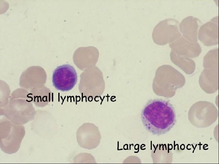
C. Small Lymphocyte • Size : 7 to 18 μm • N: C : 4: 1 • Chromatin : Oval nucleus with coarse lumpy chromatin with specific areas of clumping, a compact cell • Cytoplasm : Usually just a thin border, with few azurophilic, red granules.

D. Large Lymphocyte • Size : 9 to 12 μm • N: C : 3: 1 • Chromatin : Looser chromatin pattern, more transparent • Cytoplasm : Larger amount of cytoplasm, lighter in color • Distinguishing characteristic : Cytoplasm is more abundant with tendency for azurophilic granules.

Monocyte/Macrophage Maturation

5. Monopoiesis A. Monoblast • Size : 12 to 20 µm • Cytoplasm : moderately basophilic to blue-gray. Nongranular. • Nucleus : Ovoid or round in shape. Light blue-purple in color. Fine, lacey chromatin. One to two nucleoli. • N/C ratio is 4: 1 to 3: 1.
 • Size : 14 to 18 µm • Cytoplasm :")
B. Promonocyte (immature monocyte) • Size : 14 to 18 µm • Cytoplasm : Blue-gray. May contain fine dustlike azurophilic granules. Ground glass appearance. Moderate amount. • Nucleus : oval, may have a single fold or fissure. One to five nucleoli. Fine chromatin pattern. • N/C ratio is 3: 1 to 2: 1.

C. Monocyte • Size : 14 to 20 µm • Cytoplasm : Abundant. Blue-gray. Outline may be irregular because of the presence of pseudopods. Many fine azurophilic granules, giving a ground glass appearance. Vacuoles may some times be present. • Nucleus : Round, kidney shaped, or may show slight lobulation. It may be folded over on top of itself, thus showing brain-like convolution. No nucleoli are visible. Chromatin is fine and lacey (not clumped), arranged in skin-like strands.
 • Cytoplasm : varying shades of blue usually")
6. Megakaryocytopoiesis A. Megakaryoblast (stage 1) • Cytoplasm : varying shades of blue usually darker than the myeloblast. • May have small, blunt pseudopods. • Small to moderate amount. usually a narrow band around the nucleus. As the cell matures, the amount of cytoplasm increases. Usually nongranular.
 • Nucleus : Round, oval, or may be : kidney shaped.")
Megakaryoblast (stage 1) • Nucleus : Round, oval, or may be : kidney shaped. • Fine chromatin pattern. • Multiple nucleoli that generally stain blue. • N/C ratio is about 10: 1.
 • Cytoplasm : more abundant than previous stage. • Lass")
B. Promegakaryocyte (stage 2) • Cytoplasm : more abundant than previous stage. • Lass basophilic than in the blast. Granules begin to form in the Golgi region. • Nucleus : chromatin becomes more coarse. Multiple nucleoli are visible. Irregular in shape; may even show slight lobulation. • N/C ratio is 4: 1 to 7: 1 depending on the ploidy.
 • Cytoplasm : abundent. Pinkish blue in color. Very")
C. Granular Megakaryocyte (stage 3) • Cytoplasm : abundent. Pinkish blue in color. Very fine and diffusely granulat. Usually has an irregular peripheral border. • Nucleus : small in comparison to cell size. Multiple nuclei may be visible or the nucleus may show multilobulation. • Chromatin is coarser than in the previous stage. No nucleoli are visable. N/C ratio is 2: 1 to 1: 1.
 • Cytoplasm: contain coarse clumps of granules aggregating into")
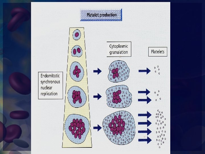
D. Mature Megakaryocyte (stage IV) • Cytoplasm: contain coarse clumps of granules aggregating into little bundles, which bud off from the periphery to become platelets. • Nucleus: multiple nuclei are present, or the nucleus is multilobulated. No nucleoli visible. N/C ratio is less than 1: 1.
 • Size: 1 to 4 µm in diameter. • Cytoplasm: light")
E. Platelet (Thrombocyte) • Size: 1 to 4 µm in diameter. • Cytoplasm: light blue to purple. Very granular. Consists of two parts: 1. The chromomere, which is granular and located centrally, and 2. The hyalomere, which surrounds the chromomere and is nongranular and clear to light blue.

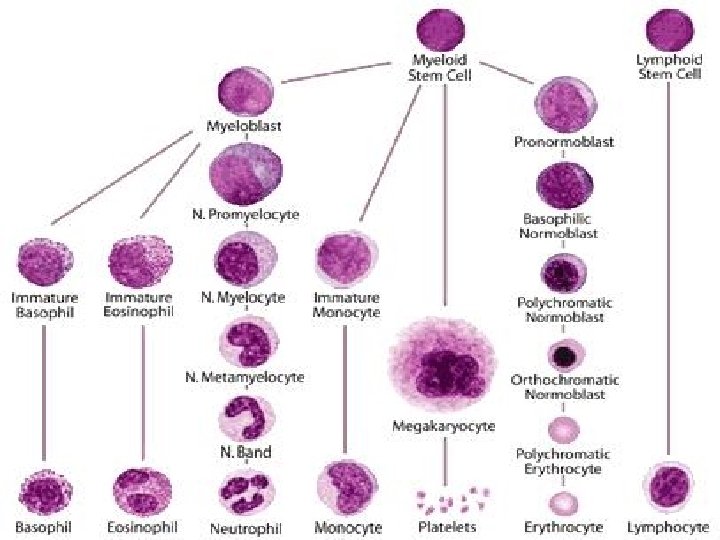
Hematopoiesis Just notice general trends, Don’t memorize
- Slides: 65