Practical Application of Diastolic Function Assessment ASEEACVI Diastology


- Slides: 34

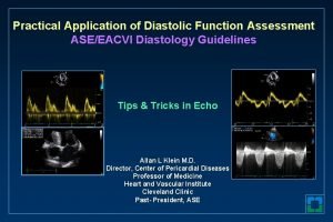
Practical Application of Diastolic Function Assessment ASE/EACVI Diastology Guidelines Tips & Tricks in Echo Allan L Klein M. D. Director, Center of Pericardial Diseases Professor of Medicine Heart and Vascular Institute Cleveland Clinic Past- President, ASE

Practical Application of Diastolic Function Assessment ASE Diastology Guidelines Update • Introduction • 2016 guidelines • Echo parameters • Myocardial pathology • Supplementary parameters 0718 -102

PRESIDENT’S MESSAGE 0718 -104

@ASE Connect Ongoing Dialogue “I had a discussion with a cardiologist who doesn’t believe in the assessment of diastolic function” “Diastology finally makes sense. . A big thanks to the writing group” 0718 -109

Epidemic of Indeterminate DF evaluations

Practical Application of Diastolic Function Assessment ASE Diastology Guidelines Update • Introduction • 2016 guidelines • Echo parameters • Myocardial pathology • Supplementary parameters 0718 -102

Key Diastology Parameters Nagueh et al. J Am Soc Echocardiogr 2016; 29: 277 -314 0718 -142

Criteria for Diagnosis of LV Diastolic Dysfunction (Algorithm 1) Nagueh et al. J Am Soc Echocardiogr 2016; 29: 277 -314 0718 -144

Estimation of LV Filling Pressures and Grading DF (Algorithm 2) Nagueh et al. J Am Soc Echocardiogr 2016; 29: 277 -314 0718 -145

Practical Application of Diastolic Function Assessment ASE Diastology Guidelines Update • Introduction • 2016 guidelines • Echo parameters • Myocardial pathology • Supplementary parameters 0718 -102

Mitral Inflow Measurements Appleton et al. , J Am Soc Echo 1997; 10: 271 -91 0718 -116

MV Inflow PW Doppler • Apical 4 chamber view • Color flow imaging for optimal alignment • SV size 1 – 3 mm placed at leaflet tips • Optimize spectral gain and wall filters • Sweep speed measure at 50 -100 mm/s

MV Inflow PW SV Placement Incorrect -towards Apex Correct - Leaflet Tips

MV Inflow PW Measurements • Measurements: peak E–wave, A–wave, E/A ratio, deceleration time • Limitations: sinus tachycardia, conduction system disease, arrhythmias, eccentric AI

Tissue Doppler Imaging s’ e’ a’ 0718 -125

PW DTI of Mitral Annular Velocities • Apical 4 chamber • SV (5– 10 mm) placed at or within 1 cm of mitral leaflet insertion sites • Optimize gain/filter settings and minimize the angle of incidence (<20 degrees) • Velocity scale 20 cm/s above below baseline, sweep speed 50 to 100 mm/s

PW DTI of Mitral Annular Velocities • Measure early (e’) diastolic velocities • Average septal and lateral velocities, calculate E/e’ • Limitations: e’ reduced with MV surgical rings (repair), prosthetic valves, annular calcification and mitral stenosis, e’ increased with > 2+MR

PW DTI SV Placement TDI e’ ? SV size 5 – 10 mm TDI e’ 5

Common mistake – don’t measure the first downward deflection IVC s’ IVC IVR e’ a’

LA Volume Index • LA volume measured using: • Simpson’s Method • Area-Length Method A 2 chamber view V=8 A 1 A 2/3 п. L • LAVI = LAV/Body Surface Area A 4 chamber view 0718 -131

LA Acquisition • LV and LA frequently lie in different planes • Avoid foreshortening LA • Base of the atrium should be at its largest size • LA length should be maximized

LV and LA frequently lie in different planes

LA Volume Measurement LA Volume Index (m. L/BSA)~ 32. 4 ml/m 2

LA Measurement Tips • Do not include LA appendage or pulmonary veins in LA tracings from A 4 C or A 2 C views • The long-axis lengths should be within 5 mm of each other

Peak TR Velocity • Diligence is necessary, scan from multiple windows • Agitated saline contrast is recommended if signal-tonoise ratio is poor • Measure full, well-defined envelopes • Consider PR signal for mean PAP

Peak TR Velocity Saline Enhanced

Practical Application of Diastolic Function Assessment ASE Diastology Guidelines Update • Introduction • 2016 guidelines • Echo parameters • Myocardial pathology • Supplementary parameters 0718 -102

How Do You Determine Diastolic Dysfunction Myocardial Pathology ( Algorithm 2) • Extensive cardiac history • Known CV disease as coronary artery disease • Wall motion • Pathologic LVH • Hypertensive CV Disease • Cardiomyopathy • Established Diagnosis of HFp. EF • If 3/4 positive parameters from Algorithm 1 • EF reduced • Specific Doppler signals 0718 -146

Practical Application of Diastolic Function Assessment ASE Diastology Guidelines Update • Introduction • 2016 guidelines • Echo parameters • Myocardial pathology • Supplementary parameters 0718 -102

Supplementary Measures A duration Mitral E-deceleration time <150 ms Ar - A duration difference > 30 ms Pulmonary venous S/D ratio <1 Ar duration LV global longitudinal strain Normal > 18% Left atrial reservoir strain Normal > 20%? Smiseth O. Journal of Echo 2018; 16: 55 -64 0718 -151

Special Populations • Atrial Fibrillation • Moderate to Severe MR • Hypertrophic Cardiomyopathy • Mitral Stenosis • Restrictive Cardiomyopathy • Sinus Tachycardia • Bundle Branch Block • Severe Aortic Insufficiency • Heart Transplant Patients • Cardiac causes of Pulmonary Hypertension

Practical Application of Diastolic Function Assessment ASE Diastology Guidelines Update Take Home Points • The “In” Parameters are Mitral E/A, TDI, LAVI and RVSP • Important to consider Clinical setting 2 D findings (LVH, LA volumes, LV EF and volumes) Evaluate technical quality of acquired signals • Who gets you into algorithm 2? • Supplementary parameters including LV and LA strain can help reclassify indeterminate DF • Report: LV filling pressures and grade of diastolic dysfunction 0718 -1108


Thank you kleina@ccf. org @Allan. LKlein. MD 1
 Diastology algorithm
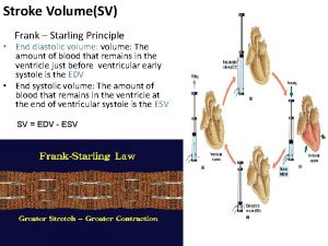
 What is stroke volume
 Preload stroke volume
 Diastolic notch
 Pansystolic murmur causes
 Still's murmur
 Csi 201
 Tapping sounds
 Ivrt echo
 Trunctus
 What is blood pressure
 What is systolic and diastolic pressure
 Practical application of calculus
 Charles law irl
 Practical situations examples
 Practical application of solar energy
 State five practical application of batteries
 Use of polynomials in daily life
 Stokes theorem physical significance
 Practical application
 Practical application of biotechnology
 Practical functional assessment
 Icass practical assessment task 2 2017 memorandum
 Common practical assessment criteria
 Grade 10 egd mechanical drawings
 Nus cs1101s
 Self assessment in job application process
 Lmia online pilot
 Stages in implementing portfolio assessment
 Define dynamic assessment
 Portfolio assessment matches assessment to teaching
 Real life example of inverse function
 Writing piecewise functions from a graph
 Rule application function
 Function of application software
