POXVIRUSES DR AMITA ARYA INTRODUCTION Family Poxviridae Subfamily

POXVIRUSES DR. AMITA ARYA.

INTRODUCTION • Family Poxviridae • Subfamily – Chordopoxvirinae - Entomopoxvirinae

CHORDOPOXVIRINAE

CHORDOPOXVIRINAE • Orthopox virus – Variola, Vaccinia - Cowpox, Monkeypox, Rabbitpox, Buffalopox - Camelpox, Mousepox • Parapox virus - orf, Paravaccinia (Ungulates) • Capripox virus – Sheep pox, Goat pox (Lumpy Skin disease) • Leporipox virus – Myxoma & Fibroma (rabbits, hares, squirerels) • Avipox virus - Fowl pox, Turkey pox, Pigeon pox, Canarypox virus • Suipox virus - Swinepox

HOOVES

UNCLASSIFIED VIRUSES • Molluscum Contagiosum virus • Tanapox • Yaba pox virus

VARIOLA VIRUS • Small Pox • Global eradication of Smallpox – 8 th May 1980 • Variola major – Classical smallpox • Variola minor – Alastrim

Vaccinia virus • Smallpox virus • Artificial virus • Vector for the development of recombinant vaccines • Genome can accommodate 25, 000 base pairs • Not a suitable vector for humans

MORPHOLOGY • Brick shaped •

PHYSICAL AND CHEMICAL PROPERTIES • Viable for months at room temperature • Susceptible – UV light, Formalin • Resistant - 50 % Glycerol, 1% Phenol, Ether • Enzymes ++ • Multiplication - Cytoplasm

ANTIGENIC STRUCTURE • Immunodiffusion – 20 antigens • Nucleoprotein antigen • LS antigen • Agglutinogen • Hemagglutinin

CULTIVATION AND HOST RANGE • Chick Embryo • Tissue culture • Animals

CHICK EMBRYO Vaccinia Pocks Variola Pocks

CEILING TEMPERATURE • Vaccinia – 41˚C • Variola major – 38˚ C • Variola minor – 37. 5˚ C

TISSUE CULTURE • Monkey kidney, He. La & Chick embryo cell lines • Vaccinia – Cytopathic effects (24 – 48 hours) - Plaque (Chick embryo) • Guarnieri bodies – eosinophilic inclusion bodies
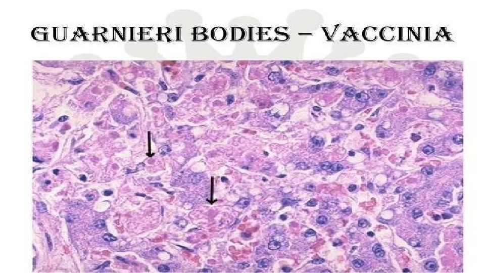

ANIMAL INOCULATION • Vaccinia virus – Monkeys, Calves, Sheep & Rabbits • Variola virus – Keratitis - Guarnieri bodies

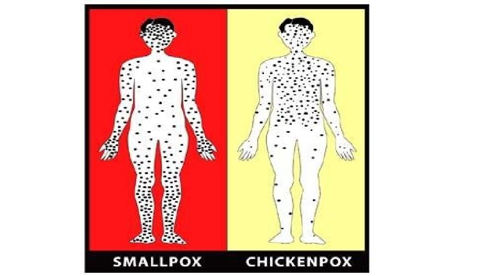
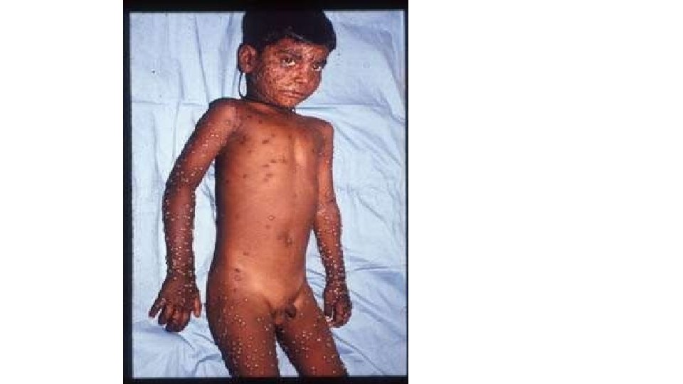
SMALL POX • No reservoir & carrier stage • Source – Patients • Route of spread – Inhalation • Incubation period – 12 days • Enanthems – Buccal mucosa • Exanthems – Skin lesions (Haemorrhagic, Flat, Ordinary or Modified) • Macule – Papule – Vesicle – Pustule - Scabbing

Haemorrhagic Exanthem Flat Exanthem

Ordinary Exanthem Modified Exanthem

LABORATORY DIAGNOSIS • Sample – Fluid from eruptive lesions - Blood • Antigen detection, Isolation of virus • Electron Microscopy • Paschen bodies

Milker’s Node • Paravaccinia virus • Lesions – small ulcerating nodules • Chick embryo – No growth • Bovine kidney cultures • Resembles orf virus morphologically

Orf virus • Sheep & Goats • Contagious Pustular Dermatitis • Single papulovesicular lesion with a central ulcer • Hands, forearm or face • Unrelated to Variola – vaccinia group

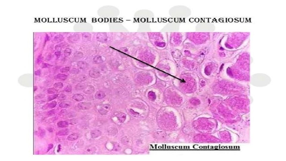
MOLLUSCUM CONTAGIOSUM • Sexually transmitted disease • Pink or pearly white wart like nodules on the skin • Molluscum bodies – large eosinophilic hyaline inclusion bodies • Virus cannot be grown in eggs, tissue culture or animals

GENITAL MOLLUSCUM CONTAGIOSUM

THANK U
- Slides: 29